Lupine Publishers Group
Lupine Publishers
Menu
ISSN: 2637-6636
Case Report(ISSN: 2637-6636) 
Erupted Complex Odontoma in Mandibula: Case Report Volume 1 - Issue 2
Alem Coşgun1*, Halenur Altan2 and Ahmet Altan3
- 1Department of Pedodontics, Mustafa Kemal University, Turkey
- 2Department of Pedodontics, Gaziosmanpaşa University, Turkey
- 3Department of Oral and MaxilloFacial Surgery, Gaziosmanpaşa University, Turkey
Received: March 16, 2018; Published: March 21, 2018
*Corresponding author: Alem Coşgun, Department of Paedodontics, Mustafa Kemal University, Turkey
DOI: 10.32474/IPDOAJ.2018.01.000107
Abstract
Odontomas are the most common odontogenic tumour. They are considered to be hamartous rather than neoplasms. They are consist of dentin, enamel and cementum. They are generally asymptomatic and discovered on routine radiographic examination. Disturbances in tooth eruption are one of the most common complications associated with odontomas. Eruption of odontoma in the oral cavity is rare. This case report presents an erupted complexodontoma in a 12-year-old girl long with its related clinical and radiological manifestations and the surgical management.
Keywords: Complexodontoma; Eruption; Surgical Management
Introduction
Odontogenic tumors were described by Broca in 1867 [1]. Odontomas are well differentiated benign mixed odontogenic tumors originating from epithelial and mesenchymal cells. Odontomas are not a real tumor; they are also defined as hamartomatous lesions [2,3]. Odontomas constitute 22% of all odontogenic tumors observed in jaws [4]. Although their etiology is not fully known, local trauma and infections are reported as possible factors. Furthermore, it has been indicated that inheritance and genetic mutation also play a role in etiology [5]. 2 types of odontoma were defined by the World Health Organization in 2005, these are complex and compound odontoma [6]. During the development of the tumor, anatomically similar structures to normal teeth called compound odontoma are formed with enamel and dentin accumulation. However, when the dental tissue forms create a simple irregular mass, this structure is defined as complex odontoma [2-6]. Compound type odontoma is almost 2 times more common than complex type odontoma. Compound odontomas are usually formed in the maxillary incisor and canine tooth region [7]. Complex odontomas are seen in the mandibula and the premolar- molar region by 70% [8].
Odontomas are usually noticed between the ages of 10 and 20 [9]. Since they are usually asymptomatic, the diagnosis of odontomasis made during routine radiographic examinations [10,11]. Furthermore, germ deficiency, malposition, diastema, impacted teeth, cyst formation, milk tooth retention, crowding, resorption or displacement of teeth are observed in the teeth adjacent to the relevant region depending on these tumors [712]. However, they may rarely cause swelling, pain, inflammation, regional adenopathy and enlargement of the bones. While a large size odontoma in mandibula may lead to inferior alveolar nerve injury, mandibular fracture or a large size bone cavity gap, an odontoma located in the maxilla may cause acute maxillary sinusitis [13].
Case Report
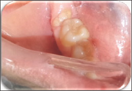
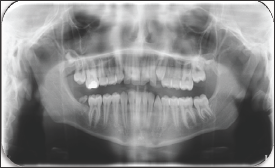
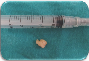
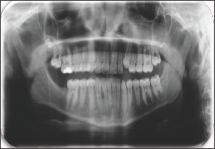
The case of erupted complex odontoma in mandibula was reported in this paper. The clinical and radiological examinations of a 12-year-old girl referred to the pediatric dentistry clinic with complaints of pain in the right mandibular region were performed. In the anamnesis taken from the patient, it was learned that she had no systemic disease. During her intraoral examination, a tooth-like tissue was observed inside of the gingiva in the right mandibular posterior region (Figure 1). No inflammation or ulceration was found in the mouth. During radiographic examination, it was observed that the patient's tooth number 47 was congenitally missing (Figure 2). The pre-diagnosis of complex odontoma was made after the clinical and radiological examinations. The patient was taken under an operation under local anesthesia and was operated with surgical procedures. As a result of the histopathological examination of the biopsy material obtained, the complex odontoma in opaque color with a dimension of 0.8x0.7x0.7 cm was confirmed (Figures 3-4).
Figure 1: Intraoral Appearance.

Figure 2: Pre-Operative Panoramic Film.

Figure 3: Odontoma (0.8X0.7X0.7 cm).

Figure 4: Post-Operative Panoramic Film.

Discussion
Odontomas may cause teeth to remain impacted and also lead to pathologies such as devitalization, malformation, malposition in adjacent teeth, and the congenital missing of the tooth [7]. Erupted odontomas may cause intraoral recurrent infections and pain and swelling caused by malocclusion [14]. In this case mentioned, in accordance with this information, the congenital missing of the lower-right 2nd molar tooth was identified with the presence of odontoma in the same region and led to the feeling of discomfort due to the erupted odontoma in the mouth. The treatment of odontomas is surgical excision [11]. The excision of odontoma with the surrounding soft tissue is the recommended treatment due to its cystic degeneration potential [15]. No recurrence is expected. Small-sized tumors without symptoms can be left untreated and can be followed [16]. All excised odontomas should be pathologically examined for microscopic examination and definitive diagnosis [17]. In this case report, the lesion was sent to the histopathologic examination after the surgical procedure and the diagnosis of complex odontoma was confirmed, supporting our pre-diagnosis. The majority of odontomas are present inside of the bone, but those that are present in extra oral localization have been rarely reported [18-20]. In this case reported, a rare erupted odontoma was observed in the case. Odontomas are usually noticed between the ages of 10 and 20 [9]. In this case reported, the incidence age of odontoma is consistent with the literature.
Conclusion
Although odontomas are benign tumoral tissues, they may cause pain, inflammation, infection, erythema, and ulcerous tissue formation when they are erupted in the mouth. Since their structure has no root and periodontal ligament, the eruption mechanisms of odontomas differ from healthy teeth. Unlike healthy teeth, the force required for the eruption of odontomas in the mouth is not associated with fibroblasts. Although there is no root formation in odontomas, eruption may occur depending on the increase in lesion dimensions and occlusal forces. In this case reported, we think that occlusal forces are effective in the eruption of odontoma. The early diagnosis of odontomas by radiographic examination will help prevent possible malocclusions.
References
- Reddy G, Reddy G, Sidhartha B, Sriharsha K, Koshy J, et al. (2014) large complex odontoma of mandible in a young boy: a rare and unusual case report. Case reports in dentistry.
- Baldawa RS, Khante KC, Kalburge JV, Kasat VO (2011) Orthodontic management of an impacted maxillary incisor due to odontoma. Contemporary clinical dentistry 2(1): 37-40.
- Yadav M, Godge P, Meghana S, Kulkarni SR (2012) Compound odontoma. Contemporary clinical dentistry 3(1): S13-S15.
- Aral İ L (1998) An erupted compound odontoma. The Journal of Dental, Atatürk University, Turkey.
- Owens B, Schuman N, Mincer H, Turner J, Oliver F (1997) Dental odontomas: a retrospective study of 104 cases. The Journal of clinical pediatric dentistry 21(3): 261-264.
- Morgan PR (2011) Odontogenic tumors: a review. Periodontology 2000 57(1): 160-76.
- Karaçay Ş, Gürbüzer B, Erkan M, Küçükodacı Z (2012) Mandibular Canine Transmigration Associated with Compaund Odontoma: Case Report. Turkiye Klinikleri Journal of Dental Sciences 18(1): 103.
- Avsever H, Kurt H, Suer TB, Ozturk HP, Piskin B (2015) The prevalence, anatomic locations and characteristics of the odontomas using panoramic radiographs. Journal of Oral and Maxillofacial Radiology 3(2): 49-53.
- Katz R (1989) An analysis of compound and complex odontomas. ASDC journal of dentistry for children 56(6): 445-449.
- Nelson BL, Thompson LD (2010) Compound odontoma. Head and neck pathology 4(4): 290-291.
- An SY, An CH, Choi KS (2012) Odontoma: a retrospective study of 73 cases. Imaging science in dentistry 42(2): 77-81.
- Salgado H, Mesquita P (2013) Compound odontoma-case report. Revista Portuguesa de Estomatologia, Medicina Dentária e Cirurgia Maxilofacial 54(3): 161-165.
- Casap N, Zeltser R, Abu Tair J, Shteyer A (2007) Removal of a large odontoma by sagittal split osteotomy. Journal of oral and maxillofacial surgery 64(12): 1833-1836.
- Avinash Tejasvi M, Balaji Babu B (2011) Erupted compound odontomas: a case report. Journal of dental research, dental clinics, dental prospects 5(1): 33-36.
- Amailuk P, Grubor D (2008) Erupted compound odontoma: case report of a 15-year-old Sudanese boy with a history of traditional dental mutilation. British Dental Journal 204(1): 11-14.
- Bora Ö, Özden B, Gündüz K, Burcu B, Şanal KO (2009) Impacted maxillary primary lateral tooth associated with compound odontoma: A case report. The Journal of Ondokuz Mayis 10(2).
- de Oliveira BH, Campos V, Marçal S (2001) Compound odontomadiagnosis and treatment: three case reports. Pediatric dentistry 23(2): 151-157.
- Özcan G, Şekerci AE, Ekizer A, Kara Ö (2015) Erupted Compound Odontoma: Two Case Reports. Turkiye Klinikleri Journal of Dental Sciences Cases 1(2): 126-30.
- Liu JK, Hsiao CK, Chen HA, Tsai MY (1997) Orthodontic correction of a mandibular first molar deeply impacted by an odontoma: a case report. Quintessence International 28(6): 381-385.
- Köymen R, Ortakoğlu K, Aydıntuğ YS, Günaydın Y (2001) An Erupted Complex Odontoma (A Case Report). Turkiye Klinikleri Journal of Dental Sciences 7(2): 101-104.
Editorial Manager:
Email:
pediatricdentistry@lupinepublishers.com

Top Editors
-

Mark E Smith
Bio chemistry
University of Texas Medical Branch, USA -

Lawrence A Presley
Department of Criminal Justice
Liberty University, USA -

Thomas W Miller
Department of Psychiatry
University of Kentucky, USA -

Gjumrakch Aliev
Department of Medicine
Gally International Biomedical Research & Consulting LLC, USA -

Christopher Bryant
Department of Urbanisation and Agricultural
Montreal university, USA -

Robert William Frare
Oral & Maxillofacial Pathology
New York University, USA -

Rudolph Modesto Navari
Gastroenterology and Hepatology
University of Alabama, UK -

Andrew Hague
Department of Medicine
Universities of Bradford, UK -

George Gregory Buttigieg
Maltese College of Obstetrics and Gynaecology, Europe -

Chen-Hsiung Yeh
Oncology
Circulogene Theranostics, England -
.png)
Emilio Bucio-Carrillo
Radiation Chemistry
National University of Mexico, USA -
.jpg)
Casey J Grenier
Analytical Chemistry
Wentworth Institute of Technology, USA -
Hany Atalah
Minimally Invasive Surgery
Mercer University school of Medicine, USA -

Abu-Hussein Muhamad
Pediatric Dentistry
University of Athens , Greece

The annual scholar awards from Lupine Publishers honor a selected number Read More...


