Lupine Publishers Group
Lupine Publishers
Menu
ISSN: 2637-6636
Case Report(ISSN: 2637-6636) 
Surgical Management of Massive Dentigerous Cyst in Mixed Dention- A Case Report Volume 7 - Issue 2
Santoshni Samal1*, Mukesh Kumar2, Prasanna Kumar Sahoo3 and Ratna Renu Baliar Singh4
1Post Graduate Student, Department of Pedodontics and Preventive Dentistry, SCB Dental College and Hospital, Utkal University, India
2Post Graduate Student, Department of Periodontics and Implantology, Kalka Dental College, India
3Department of Pedodontics and Preventive Dentistry, SCB Dental College and Hospital, Utkal University, India
4Professor and Head, Department Pedodontics and Preventive dentistry, SCB Dental College and Hospital, Utkal University, India
Received: January 14, 2022; Published: February 01, 2022
*Corresponding author: Santoshni Samal, Post Graduate Student, Department of Pedodontics and Preventive Dentistry, SCB Dental College and Hospital, India
DOI: 10.32474/IPDOAJ.2022.07.000260
Abstract
A dentigerous cyst is an odontogenic cyst thought to be of developmental origin associated with the crown of an unerupted tooth. Such cysts remain initially completely asymptomatic unless when infected. It is detected clinically when becomes large and associated with swelling of the face. The purpose of this case report was to describe the diagnosis and management of a dentigerous cyst in an 8-year-old boy. The chosen treatment was cyst enucleation and tooth extraction.
Keywords: Dentigerous cyst; enucleation; Mixed dentition
Abbreviation: DC: Dentigerous Cyst
Introduction
Dentigerous cysts(DC) are the second most common type of odontogenic cysts associated with the crowns of impacted or unerupted permanent teeth [1]. It frequently occurs during the second and third decades of life and is more common in male patients [2]. The occurrence of dentigerous cysts in children has been reported very little in the dental literature. It is often detected during the routine radiographic examination and detected clinically when it is large (>2 cm) in diameter [3]. Histologically, dentigerous cysts lined with nonkeratinized stratified squamous epithelium and consist of a fibrous wall containing variable amounts of myxoid tissue and odontogenic remnants. The epithelial-connective tissue interface is typically flattened but becomes highly irregular when associated with inflammation [4]. The complications associated with dentigerous cyst include pathologic bone fracture, loss of a permanent tooth, bone deformation, and development of squamous cell carcinoma [5]. Among the treatment options proposed for dentigerous cysts, two techniques can be emphasized: total enucleation for small lesions and marsupialization for decompression of large volume cysts; or a combination of both [6]. Some small cysts can be treated by enucleation and extraction of the involved tooth [6]. The extensive DCs often block the eruption of teeth, displace these teeth, and destroy the bone. Motamedi et al. show that extensive DCs (involving three or more teeth) primarily occurs between the ages of 10–19 years [7] In this report, we presented a case of an extensive DC associated with maxillary right first premolar and second deciduous molar in an 8-year-old boy.
Case Report
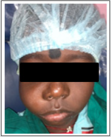
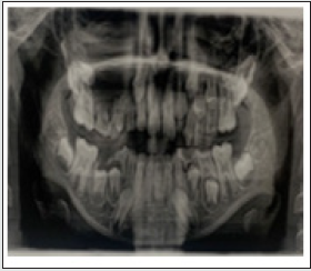
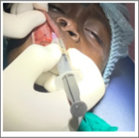
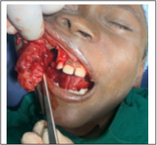
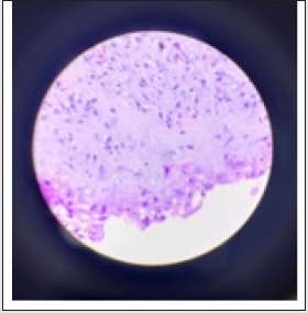
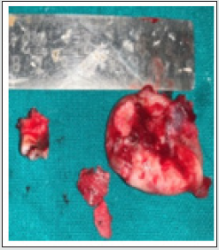
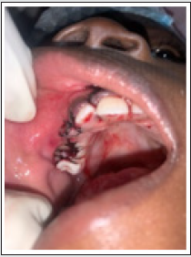
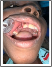
An eight-year-old boy was reported to the department with the chief complaint of swelling of the right upper cheeks for 1 month. The swelling was initially small and gradually increased in size. There was no relevant medical history. There was no history of associated trauma. On extraoral examination, the swelling was 4x 4 cm extending from the lateral border of the nose to lateral canthus of eyes mediolaterally and from zygomatic bone to corner of mouth vertically obliterating the nasolabial fold (Figure 1). The swelling was slightly hard and tender on palpation. On intraoral examination, there was the obliteration of the buccal vestibule of the right side, and 55 was carious and there were no other associated findings. Panoramic radiograph shows radiolucency extending from lateral of 13 to medial of 16 and second premolar and canine was impacted and displaced laterally (Figure 2). Since the compliance was poor, surgical treatment was planned. On the day of surgery, informed consent was taken. After adequate local anesthesia (1:1000 adrenaline) (Figure 3), surgical enucleation was done (Figure 4) followed by extraction of 1st premolar and second deciduous molar. Enucleation of the 2nd premolar was also done to prevent recurrence and a silk 3.0 suture was placed (Figure 5). The cyst was 2.5 x 3 cm in size (Figure 6) and sent for Histopathological examination which showed variable amounts of myxoid tissue and odontogenic remnants along with inflammatory cells (Figure 7). The patient was reviewed after 7 days (Figure 8) and under follow-up.
Discussion
DC arises from an extraordinary expansion of the dental follicle of an unerupted tooth. DC can be suspected if the follicular space on the radiograph is more than 5 mm [8]. In our case, panoramic radiographs clearly show radiolucent lesion associated with one premolar and deciduous carious molar. Because of its extensive large size, it displaced canine and second premolar also. According to Benn and Altini [9], three possible mechanisms exist for the histogenesis of the dentigerous cyst. Developmental dentigerous cyst forms from the dental follicle and becomes secondarily inflamed and associated with the non-vital tooth. The second type develops from the radicular cyst, and the third type is due to peri-apical inflammation from non-vital deciduous tooth or another source, which spreads to involve follicle of a permanent successor, as a result of inflammatory exudate, dentigerous cyst formation occurs as seen in our case is of the third type [9]. The treatment of DCs is generally dependent on cyst sizes, sites, patients’ age, the dentition involved, and involvement of vital structures [10]. Marsupialization reduces the size of the cyst by relieving the hydraulic pressure, which may be responsible for cyst enlargement or displacing the involved tooth and preserving the involved permanent tooth [11]. The major disadvantage of marsupialization is that pathologic tissue is left in situ, without a thorough histologic examination. Enucleation should be preferred in case of a small lesion and where the possibility of recurrence is there. In our case, the cystic sac was surrounding the unerupted premolar and was firmly attached to it [12]. So, it was decided to do enucleation of the cyst followed by extraction of carious deciduous molar and impacted premolar. A thorough diagnosis and removal of cyst along with granulation tissues should be done to prevent recurrence and to provide the best care to child patients.
Conclusion
Management of odontogenic lesions, such as dentigerous cysts in children, presents a unique challenge to general and pediatric dentists. Coupled with the need for appropriate behavior management and the delicate balance between the primary and the developing permanent dentitions, warrant a highly meticulous approach on the part of every clinician involved in providing dental care to such child patients.
References
- Daley TD, Wysocki GP (1995) The small dentigerous cyst: A diagnostic dilemma. Oral Surg Oral Med Oral Pathol Oral Radiol Endod 79: 77-81.
- Bodner L, Woldenberg Y, Bar-Ziv J (2003) Radiographic features of large cysts lesions of the jaws in children. Pediatr Radiol 33: 3-6.
- Tuzum MS (1997) Marsupialization of a cyst lesion to allow tooth eruption: A case report. Quintessence Int 28: 283-284.
- Motamedi MH, Talesh KT (2005) Management of extensive Dentigerous cysts. Br Dent J 198: 203-206.
- Perez DM, Molare MV (1996) Conservative treatment of dentigerous cysts in children. A report of 4 cases. J Indian Soc Pedod Prev Dent 14: 49-51.
- Kalaskar RR, Tiku A, Damle SG (2007) Dentigerous cysts of anterior maxilla in a young child: A case report. J Indian Soc Pedod Prev Dent 25: 187-190.
- Motamedi MH, Talesh KT (2005) Management of extensive dentigerous cysts. Br Dent J 198: 203-206.
- Ko KS, Dover DG, Jordan RC (1999) Bilateral dentigerous cysts — report of an unusual case and review of the literature. J Can Dent Assoc 65: 49-51.
- Benn A, Altini M (1996) Dentigerous cysts of infl ammatory origin. A clinicopathologic study. Oral Surg Oral Med Oral Pathol Oral Radiol Endod 81: 203-209.
- Sands T, Tocchio C (1998) Multiple dentigerous cysts in a child. Oral Health 88: 27-29.
- Jones TA, Perry RJ, Wake MJ (2003) Marsupialization of a large unilateral mandibular dentigerous cyst in a 6-year-old boy a case report. Dent Update 30: 557-561.
- Peterson LJ, Ellis E III, Hupp JR (1998) Contemporary Oral and Maxillofacial Surgery. 3rd Ed. St Louis, MO; Mosby, USA p. 540.
Editorial Manager:
Email:
pediatricdentistry@lupinepublishers.com

Top Editors
-

Mark E Smith
Bio chemistry
University of Texas Medical Branch, USA -

Lawrence A Presley
Department of Criminal Justice
Liberty University, USA -

Thomas W Miller
Department of Psychiatry
University of Kentucky, USA -

Gjumrakch Aliev
Department of Medicine
Gally International Biomedical Research & Consulting LLC, USA -

Christopher Bryant
Department of Urbanisation and Agricultural
Montreal university, USA -

Robert William Frare
Oral & Maxillofacial Pathology
New York University, USA -

Rudolph Modesto Navari
Gastroenterology and Hepatology
University of Alabama, UK -

Andrew Hague
Department of Medicine
Universities of Bradford, UK -

George Gregory Buttigieg
Maltese College of Obstetrics and Gynaecology, Europe -

Chen-Hsiung Yeh
Oncology
Circulogene Theranostics, England -
.png)
Emilio Bucio-Carrillo
Radiation Chemistry
National University of Mexico, USA -
.jpg)
Casey J Grenier
Analytical Chemistry
Wentworth Institute of Technology, USA -
Hany Atalah
Minimally Invasive Surgery
Mercer University school of Medicine, USA -

Abu-Hussein Muhamad
Pediatric Dentistry
University of Athens , Greece

The annual scholar awards from Lupine Publishers honor a selected number Read More...


