Lupine Publishers Group
Lupine Publishers
Menu
ISSN: 2637-6636
Case Report(ISSN: 2637-6636) 
Surgical Management of Impacted Inverted Mesiodens With cyst Excision Volume 6 - Issue 5
Gaurav Gupta1*, DK Gupta2, Priyanka Gupta3, Parth Shah4 and Neelja Gupta5
1MDS, Associate Professor, Department of Paediatric and Preventive Dentistry, Jaipur Dental College Senior Consultant, Wisdom Dental Clinics, India
2MDS in Oral and Maxillofacial Surgery, Senior consultant at Wisdom Dental Clinic, India
3MDS in Paediatric and Preventive Dentistry, Senior Demonstrator, Department of Paediatric and Preventive Dentistry, RUHS College of Dental Science, India
4MDS, Department of Paediatric and Preventive Dentistry, Senior consultant at Saanchi Pediatric Hospital, Surat, India
5BDS, Department of Cosmetic Dentistry, Senior consultant at Wisdom Dental Clinic, India
Received:September 29, 2021; Published: October 11, 2021
*Corresponding author: Gaurav Gupta, MDS, Associate Professor, Department of Paediatric and Preventive Dentistry, Jaipur Dental College Senior Consultant, Wisdom Dental Clinics, India
DOI: 10.32474/IPDOAJ.2021.06.000249
Abstract
Impacted tooth is most frequently diagnosed during the routine dental check-up, commonly associated teeth are third molars, but impacted inverted mesiodens are considered to be one of the infrequent findings in dentistry. Here with, a rare case is reported which is suspected of inverted mesiodens describing its surgical intervention along with cyst removal which was detected in radiographic examination and also showing importance of radiograph in diagnosis of infrequent cases in dentistry.
Keywords:Inverted mesiodens; palatal impaction; supernumerary
Introduction
Supernumerary teeth are also known as hypodontia which is defined as an extra number of teeth as compared to normal dentition formula of oral cavity [1]. The prevalence percentage of supernumerary teeth ranges from 0.10% to 3.6% in permanent dentition while 0.02% to 1.9% in case of primary dentition [2]. The supernumerary tooth cited in premaxillary region is termed as mesiodens. Usually, it has conical crown with single root which may be single or multiple. It might be one of the cause for creating eruption disturbances of incisors. Largely, (55.2%) of mesiodens found are in vertical position, whereas (37.6%) are inverted and remaining 7% are horizontally positioned [3]. The cause might be due to complex interaction of genetic as well as environmental factors [1]. Inverted mesiodens etiology still remains unknown. Various complications are found associated with inverted mesiodens such as adjacent teeth eruption disturbance, rotation and displacement of central incisors, diastema evolution and cyst formation. Therefore, initial stage detection and timely surgical management of inverted mesiodens is important to prevent such consequences. In existing literatures, there is no such specified time for its removal [4]. In pediatric dentistry, numerous anomalies in tooth size, shape, number and eruption of teeth are encountered. Some of the anomalies are detected during the routine dental checkup associated with erupted teeth while others may go unnoticed and remain impacted as they are sometimes asymptomatic. Here is such a case which is suspected for inverted mesiodens and was found palatally impacted, upon radiographic examination and also describing about its surgical management.
Case Report
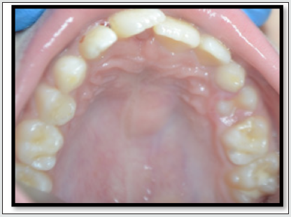
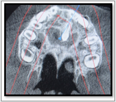
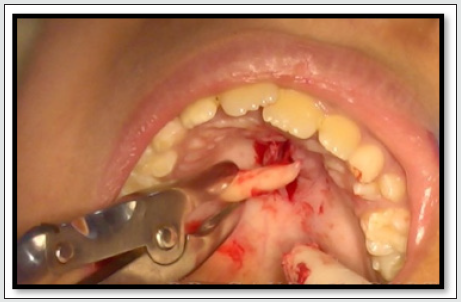
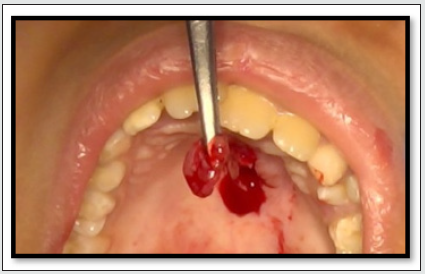
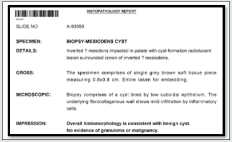
A 9-year-old male patient reported to the clinic with chief complain of pain in upper front tooth region. There was no significant medical history, patient presented with good general health. On clinical examination palatal swelling was found (Figure 1). Radiographic examination through CBCT was done to evaluate any underlying infection or hard tissue defect which surprisingly disclosed the presence of impacted mesiodens, with radiolucent area surrounding the crown of the tooth. Based upon the clinical and radiological findings supernumerary tooth was provisionally diagnosed to be like inverted mesiodens followed with pathology (Figure 2). Due to pressure created by impacted tooth the roots of the permanent maxillary central incisors were tilted, thereby necessitating the removal of impacted mesiodens. Routine pre-operative procedure was carried out with informed consent to patient’s family regarding the surgical procedure. Local anaesthesia was administered, an incision was made at the alveolar crest towards midline. The flap was then raised with minimal bone removal to visualize the impacted tooth, it was luxated with care so not to hamper permanent teeth and impacted tooth was carefully extracted (Figure 3). Conservative surgical enucleation of associated lesion was done with suture placement after the gentle curettage and saline irrigation (Figure 4). The patient was prescribed with antibiotic and anti-inflammatory coverage along with homecare instructions including oral hygiene measures. The excised soft tissue specimen was sent for histopathological examination to know any linked malignancy which gave final impression of benign cyst with no evidence of malignancy (Figure 5). The healing was uneventful, patient was recalled for follow-up phase and post-operative results were as expected. Clinical and radiological controls were timely scheduled.
Discussion
Morphologically, the mesiodens appear to be as a rudimentary tooth with smaller size and conical crown as compared to normal teeth. Sometimes, it may mimic as natural tooth. Root is usually globular in shape and fully developed. Mesiodens are frequently found between maxillary central incisors, particularly on palatine side, along with sagittal mid-plane, which gives it this name [5]. According to the literature they shown case reported the presence of impacted tooth among the adjacent maxillary central incisors and found to be in inverted position, adding up with the statistics that 80% to 90% of all supernumerary teeth are found in maxilla [6]. Various authors have stated, the inverted mesiodens might disrupt the erupting permanent incisor thereby creating malocclusion. Few authors recommend for the early removal of supernumerary teeth, particularly those which are inverted as they are unlikely to erupt [1,7]. In present case it seems impacted mesiodens caused the roots of neighboring permanent incisors to tilt thus forming malocclusion. The supernumerary teeth erupting in palate causes early loss, ectopic eruption, diastema, crowding, root resorption of adjacent teeth and also leads to development of cyst which might be dentigerous/primordial or seen incursion into nasal cavity [8,9]. As a result of histopathology the associated lesion with impacted inverted mesiodens was benign cyst. It gave the possibility of development of dentigerous cyst, moreover because cyst was surrounding the crown of impacted tooth. Furthermore, the inverted position of the mentioned tooth leaves no doubt for the surgical intervention since there was no such possibility of its eruption other than causing complications. The diagnosis of mesiodens is done by radiographs, Cone Beam Computed Tomography (CBCT) has proved to be advanced technique to detect exact location as well as any hard or soft tissue pathology associated with tooth, thus avoiding surgical complications, and decreasing iatrogenic damages to permanent tooth [10]. In this case report too CBCT played major role in locating the exact position and linked pathology with impacted mesiodens. Some clinicians recommend for postponement of surgical management till the root formation of associated adjacent permanent incisors [11]. The case presented with early diagnosis and surgical management for more favorable prognosis with minimal complications.
Conclusion
This case presents with highlights about the major role of radiography in diagnosing rare cases of inverted mesiodens and successful surgical removal of cyst along with impacted supernumerary teeth, thus minimizing its complications and enabling better prognosis. Thereby creating a need for clinicians to make timely diagnosis and treatment plan for such rare developmental anomalies.
References
- Primosch RE (1981) Anterior supernumerary teeth – Assessment and surgical intervention in children. Pediatr Dent 3: 204-215.
- Jafri S, Pannu PK, Galhotra V, Dogra G (2011) Management of an inverted impacted mesiodens associated with a partially erupted supplemental tooth – A case report. Indian J Dent 2: 40-43.
- Desai V, Baghla P, Sharma R, Gaurav I (2014) Inverted impacted mesiodens: A case series. J Adv Med Dent Sci Res 2: 135-140.
- Onauma Angwaravong, Navavit Sirikajorndechakul, Pattaramon Rattanapan (2011) Dental Management of Inverted Mesiodens: Review of the Literature and Report of Two Cases. KDJ 14(1): 55-58.
- Giancotti A, Grazzini F, De Dominicis F, Romanini G, Arcuri C (2002) Multidisciplinary evaluation and clinical management of mesiodens. J Clin Pediatr Dent 26(3): 233–237.
- Ghogre P, Singh VD (2014) Management of an impacted inverted mesiodens associated with a large circumferential type of dentigerous cyst: a rare case report with one-year follow-up. Int J Case Rep Images 5(1): 80-85.
- Nazif MM, Ruffalo RC, Zullo T (1983) Impacted supernumerary teeth: A survey of 50 cases. J Am Dent Assoc 106: 201-204.
- Dalledone M, Tassi-Junior PA, Souza JF, Losso EM (2015) Mesiodens surgery at deciduous and permanent dentition. RSBO 12(1): 94-97.
- Anil S, Beena VT , Raji MA, Sreela LS, Vijayakumar T (1992) Morphometric analysis of premaxillary supernumerary teeth. J Indian Dent Assoc 63(8): 337-341.
- Hattab FN, Yassin OM, Rawashdeh MA (1994) Supernumerary teeth: Report of three cases and review of literature. ASDC J Dent Child 61: 382-393.
- Henry RJ, Post AC (1989) A labially positioned mesiodens: Case report. Pediatr Dent 11: 59-63.
Editorial Manager:
Email:
pediatricdentistry@lupinepublishers.com

Top Editors
-

Mark E Smith
Bio chemistry
University of Texas Medical Branch, USA -

Lawrence A Presley
Department of Criminal Justice
Liberty University, USA -

Thomas W Miller
Department of Psychiatry
University of Kentucky, USA -

Gjumrakch Aliev
Department of Medicine
Gally International Biomedical Research & Consulting LLC, USA -

Christopher Bryant
Department of Urbanisation and Agricultural
Montreal university, USA -

Robert William Frare
Oral & Maxillofacial Pathology
New York University, USA -

Rudolph Modesto Navari
Gastroenterology and Hepatology
University of Alabama, UK -

Andrew Hague
Department of Medicine
Universities of Bradford, UK -

George Gregory Buttigieg
Maltese College of Obstetrics and Gynaecology, Europe -

Chen-Hsiung Yeh
Oncology
Circulogene Theranostics, England -
.png)
Emilio Bucio-Carrillo
Radiation Chemistry
National University of Mexico, USA -
.jpg)
Casey J Grenier
Analytical Chemistry
Wentworth Institute of Technology, USA -
Hany Atalah
Minimally Invasive Surgery
Mercer University school of Medicine, USA -

Abu-Hussein Muhamad
Pediatric Dentistry
University of Athens , Greece

The annual scholar awards from Lupine Publishers honor a selected number Read More...


