Lupine Publishers Group
Lupine Publishers
Menu
ISSN: 2637-6636
Research Article(ISSN: 2637-6636) 
Reliability of Construction of Gonial Angle in Mixed Indian Population: OPG or Lateral Cephalogram – A Pilot Study Volume 6 - Issue 2
Sukhbir Singh Chopra1*, Rajat Mitra2, Nanda Kishore Sahoo2 and Ashish Kamboj1
- 1Department of Orthodontics & Dentofacial Orthopedics, Maharashtra University of Health Sciences, India
- 2Department of Orthodontics & Dentofacial Orthopedics, Delhi University, India/li>
Received:June 08, 2021 Published: June 16, 2021
*Corresponding author: Sukhbir Singh Chopra, Department of Orthodontics & Dentofacial Orthopedics,Maharashtra University of Health Sciences, India
DOI: 10.32474/IPDOAJ.2021.06.000234
Abstract
Aims: The aim of the present study is to compare the measurement of gonial angles and vertical ramal length (both right and left) from OPG with the gonial angle and vertical ramal length measured from lateral cephalograms in a mixed Indian Population with Class I, II & III malocclusion. Settings and Design: The retrospective cross‑sectional diagnostic study was conducted after obtaining due approval from the institutional ethical committee.
Methods and Material: Gonial angle and ramal length was measured from 57 OPG and lateral cephalogram of orthodontic patients (26 females and 31 males). The patients were a heterogeneous group comprising of individuals from pan India.
Statistical analysis used: The analyses of descriptive statistics, reliability, analysis of variance and correlation was conducted with SPSS 20.0, IBM. The means based on malocclusion classes were compared using one-way analysis of variance (ANOVA) followed by comparison of correlation values using Pearson correlation. Gender differences were also compared using T-test. Post Hoc analysis could not be carried out due no statistically significant difference found through ANOVA.
Results: There was no statistical difference in the measurements of gonial angle made from OPG as compared to the same measurements made from a lateral cephalogram.
Conclusions: In addition to the conventional method of measuring the gonial angle and ramal length, OPG may also be used for their accurate measurement.
Keywords: OPG; lateral cephalogram; gonial angle; ramal length
Introduction
Diagnosis, formulation, and execution of treatment plan are the steps involved in successful management of malocclusions. Diagnosis defines the problem and lays the foundation for the treatment objectives. Treatment plan is execution of the treatment objectives. Proper determination and treatment planning are dependent on information obtained from clinical examination, study models, and the applicable radiographs. Lateral cephalograms and Orthopantomograms (OPG) are important radiographic tools for treatment planning and are regularly used for orthodontic patients. OPGs may be utilized for assessing the supporting bone, screening for cysts, neoplasms, ankylosed teeth, eruption path of teeth and asymmetry of mandible and supernumerary or missing teeth [1,2]. The angle formed by the junction of the posterior and lower borders of the mandible is called the gonial angle. The radiographic gonial angle measurements aid in ascertaining growth patterns of facial skeleton, mandibular rotation, facial asymmetry, age estimation in forensic odontology, decisions on extractions in Class II and orthognathic surgery in Class III skeletal base [3-6]. Lateral cephalograms are usually used for measuring this angle. However, superimposed images on a lateral cephalogram adversely affect reliability of measurements of the gonial angle and are of utmost importance while planning orthognathic surgery [7]. The right and left gonial angles can be measured individually without superimposition in an OPG; hence the measurement may be more accurate than lateral cephalometry [2].
Aim
The aim of the present study is to compare the measurement of gonial angles and vertical ramal length (both right and left) from OPG with the gonial angle and vertical ramal length measured from lateral cephalograms in a mixed Indian Population with Class I, II and III malocclusion.
Materials and Methods
The retrospective cross‑sectional diagnostic study was conducted after obtaining due approval from the institutional ethical committee. Data was mined from 57 orthodontic patients (26 females and 31 males). Good quality radiographs from the departmental archives of the Orthodontic Department of a tertiary care Government Dental Institution providing care at no cost to the patients were used. Records of patients in permanent dentition upto at least second permanent molar were included. Records of subjects with a history of craniofacial syndromes or tooth extraction and those in mixed dentition were excluded from the study. The patients were a heterogeneous group comprising of individuals from pan India. All radiographs were examined according to the standard radiographic procedures. OPGs and cephalometric radiographs were acquired with a New Tom (Verona, Italy) radiographic unit, using a standardized technique. Gonial angles were recorded in OPG by drawing a tangent to the lower border of the mandible and a tangent to the distal border of the ramus and condyle on both sides. In lateral cephalograms, the mean of gonial angles in the superimposed projections was calculated. The lines were traced on tracing paper using 0.5 mm 2H pencil led. A protractor with 1° accuracy was used to measure the angles. All measurements were made by two senior orthodontists. The data obtained were inserted in an Excel spreadsheet for further analysis.
Error Assessment
To assess the reproducibility of measurements, fifteen OPGs and lateral cephalograms were randomly selected and re‑traced after two weeks of the initial tracings. There was no difference in any measurement more than 0.5°. The intraclass correlation coefficient was found to be >0.90, thus indicating a high level of reproducibility of both measurements [3-8].
Statistical Analysis
The analyses of descriptive statistics, reliability, analysis of variance and correlation was conducted with SPSS 20.0, IBM. The means based on malocclusion classes were compared using oneway analysis of variance (ANOVA) followed by comparison of correlation values using Pearson correlation. Gender differences were also compared using t-test. Post Hoc analysis could not be carried out due no statistically significant difference found through ANOVA.
Results
The study group comprised of 57 subjects (31 males, 26 females). The group comprised of patients from 13 years to 26 year. The average age of the entire study group was 16 years. The average age of the males was 15.25 and 16.88 for the females. There were 17 subjects of Class I, 33 of Class II and 07 of Class III.
Gonial Angle
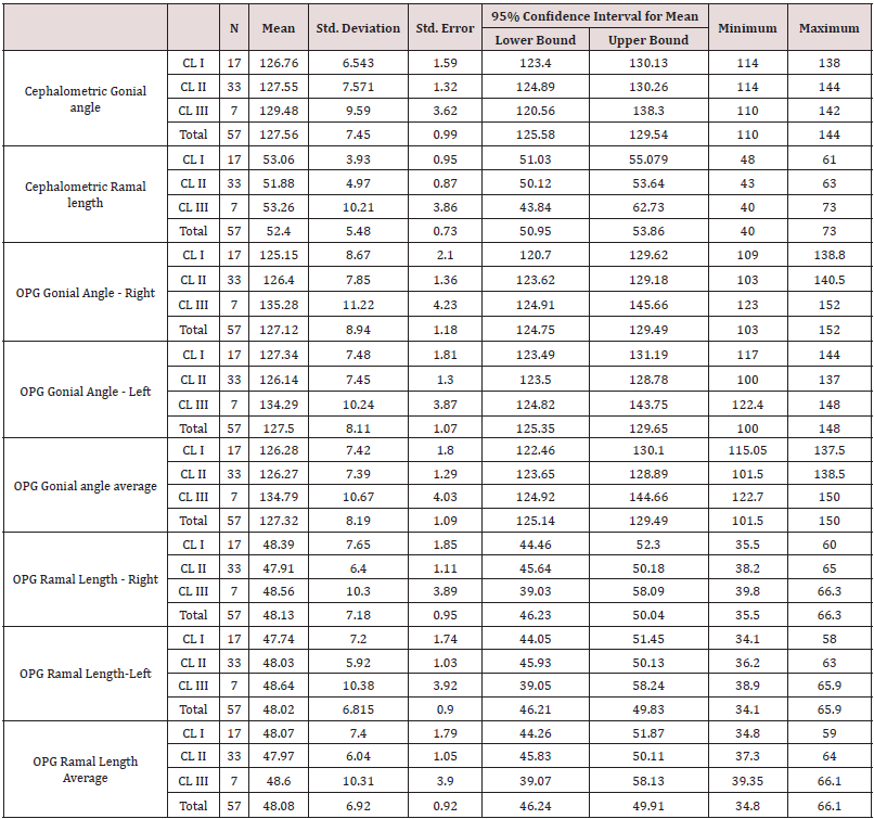
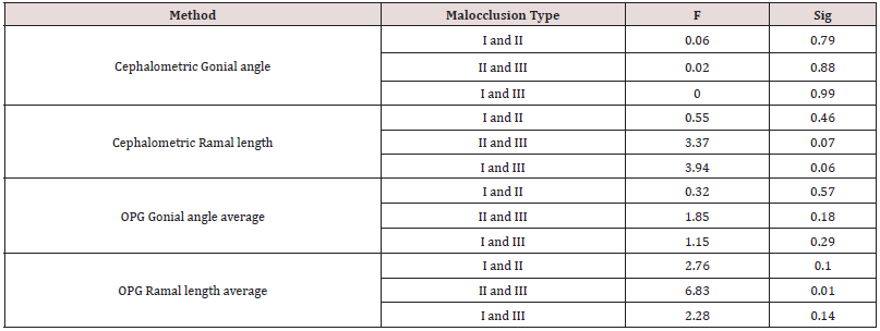
On the basis of malocclusion, the mean of gonial angles measured from the lateral cephalograms were (a) Class I: 126.76O+6.54O, (b) Class II: 127.57O+7.57O and (c) Class III:129.42O+9.58O. The mean of the average of right and left gonial angles measured from OPG were (a) Class I: 126.27O+7.42O, (b) Class II:126.26O+7.38O and (c) Class III: 134.78O+10.66O (Table 1). While no significant difference was found between the classes of cephalometric gonial angle (p=0.735), a significant statistical difference has been observed among the malocclusion classes of average gonial angle of OPG (p=0.33) (Table 2). This may be due to variances found in class III of gonial angles of OPG (Table 3 & Table 4). An independent sample t-test was conducted to analyse difference, if any to measure the gonial angle by both the methods for all Classes of malocclusion separately. It revealed no statistically significant difference (Table 5).
Table 1: Mean, standard deviation and standard error of OPG and cephalometric gonial angle values and ramal length in subjects distributed based on malocclusion.

Table 2: ANOVA comparing cephalometric gonial angle, OPG gonial angle right, OPG gonial angle left, OPG gonial angle total, cephalometric ramal length, OPG ramal length right, OPG ramal length left and OPG ramal length average.

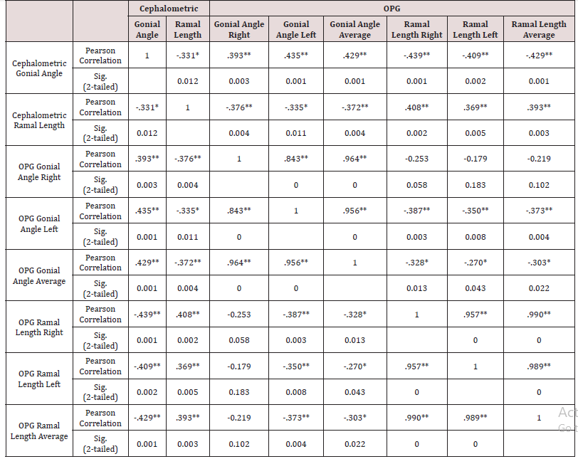
N=57 Correlation is significant at 0.05 level (2-tailed). Correlation is significant at 0.01 level (2-tailed).
Table 5: Mean differences of cephalometric and OPG gonial angle, cephalometric ramal length, OPG gonial angle and average ramal length average across malocclusion classes.

Ramal Length
On the basis of malocclusion, the mean of vertical ramal length measured from the lateral cephalograms were (a) Class I: 53.05mm+3.62mm, (b) Class II: 51.87mm+4.96mm, (c) Class III: 53.28mm+10.20mm respectively. The mean of the average of right and left vertical ramal length measured from OPG were (a) Class I: 48.06mm+7.40mm, (b) Class II: 47.96mm+6.03mm and (c) Class III: 48.60mm+10.30mm (Table 1).
No statistically significant difference was found between vertical ramal length measured by lateral cephalogram (p=0.70) and OPG (p=0.97) for all Class of malocclusion. However, a statistically significant difference was found between the means of Class II and Class III for the average vertical ramal length measured from OPG (p=0.01 (Table 4).
Gender
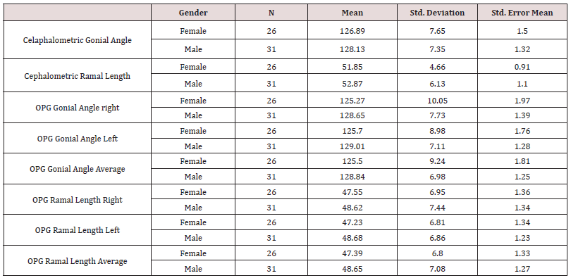
A t-Test was conducted for comparison of means of gonial angles and vertical ramal length on the basis of gender. The gonial angle measured from lateral cephalogram in females was 126.88O whereas in males it was 128.12O. Vertical ramal length measured from lateral cephalogram was 51.84mm in females and 52.87mm in males. The average vertical ramal length in females measured form OPG was 47.39mm and 48.65mm in males (Table 3). There was no statistically significant difference in gonial angles, and vertical ramal length values measured from lateral cephalogram (p=0.53) and OPG (p=0.49) found with respect to gender. Pearson correlation was also applied to examine the correlation between gonial angle and ramal length measured from cephalogram and OPG. A moderate to high correlation has been found on both methods of radiographs (Table 5).
Discussion
The gonial angle is an indicator of the mandibular form and shape and hence an important diagnostic parameter in planning treatment, especially in planning therapeutic extractions for orthodontic treatment and orthognathic surgery. The present study aimed at assessing the reliability of OPG in measuring the right and the left gonial angles by comparing the measured angles with the gonial angles determined using lateral cephalograms in patients with different types of malocclusion (Class I, II &III) , to view the feasibility of enhanced application of OPG as a diagnostic tool in orthodontic practice. Gonial angle is formed by a tangent to the lower border of the mandible and a tangent touching the posterior border of the ramus at two points, one at the condyle and one at the angle region [8]. The present study used the above method to measure the gonial angle in both OPG and lateral cephalogram. In the present study There was no significant difference between the various Classes of malocclusion of gonial angle measured from lateral cephalogram (p=0.735). A significant statistical difference was observed among the various Class of malocclusion of the mean of the average of right and left gonial angles measured from OPG (p=0.33). This may be due to variances along with found in Class III of gonial angles of OPG (Table 1), also number of Class III cases was relatively small. However, there was no statistically significant difference was found between both the methods i.e., lateral cephalogram and OPG for all malocclusion classes.
The results of the present study were in similar to the other studies [7,9-11]. Though none of the earlier studies have compared all three Class of malocclusion in the same study. Mattila et al. [2] measured gonial angle using OPG and lateral cephalograms, followed by comparing with values found using dry skulls. They concluded that the measurements made using the OPG were more accurate. Hence OPG can be considered reliable for measuring the gonial angle, particularly in cases where the outlines of asymmetry or when the two sides are not clearly visible on a lateral cephalogram, as the right and the left gonial angles can be accurately visualized in OPG. A comparison of gonial angle of Class I patients using OPG and lateral cephalograms by Shahabi et al. [12] concluded that OPG could be used for determining the gonial angle as accurately as a lateral cephalogram. This was similar to the present study which used a study sample comprising of Class I, II and III patients. However, the present study was in variance with a study by Araki et al. [13] which compared the gonial angles measured using 49 OPG with the gonial angle estimated using lateral cephalometric radiographs taken from 2 dry mandibles and found that the gonial angle measurements were slightly smaller on the OPG than on the lateral cephalometric radiographs. The present study found no statistically significant gender difference in the gonial angle. Similar results were obtained by Dutra et al. [14].
Other studies [15-17] reported gonial angle of females was larger, which may be attributed to the impact of masticatory forces. Angles are not correctly reproduced in lateral cephalograms unless the angle plane is parallel to the film [18]. The gonial angle measured in a lateral cephalogram is geometrically an intermediate angle between the right and the left gonial angle. Arithmetically, it is the mean. Any distortion of the right and the left gonial angles is reflected in this angle. Angular values from OPG are more reliable, as the angular values in the posterior and the lateral aspects of the mandible are not influenced by the image distortion inherent to panoramic radiography [19], whereas Fischer-Brandies et al. [20] preferred only lateral cephalograms for determining the gonial angle. In the present study there was no significant difference found in the mean vertical ramal length between cephalogram and OPG as well as among the right and left gonial angles of OPG and thus post hoc analysis was not carried out (Table 2). This was in consonance with other similar studies [21-23]. However, the present study was unique as the study group comprised a record of Class I, II and III patients. Additionally, vertical ramal length was also measured among classes. While no statistically significant difference was found for both cephalometric (p=0.70) and OPG ramal length (p=0.97) for all malocclusion classes, a statistically significant difference was found between the means of Class II and III for the average ramal length of OPG (p=0.01). Kambylafkas et al. [24] in their study suggest that though OPG may be used to evaluate total ramal length, but there will be some underdiagnosis. Tronje et al. [25] concluded using a mathematical model that a properly oriented patient OPG can be used for vertical measurements.
Conclusion
In addition to the conventional method of measuring the gonial angle and vertical ramal length, OPG may also be used for accurately determining the gonial angle and vertical ramal length, as there are no significant differences in the measured values of gonial angle on lateral cephalogram and OPG. OPG has the advantage of easier and more accurate determination of both right and left gonial angles and vertical ramal length with mathematical average unlike arbitrary construction of overlapped anatomical structures as in a lateral cephalogram. Thus, the present study substantiates the possibility of enhancing the diagnostic utility of OPG in orthodontics.
References
- Graber TM (1967) Panoramic radiography in orthodontic diagnosis. Am J Orthod 53: 799-821.
- Mattila M, Könönen M, Mattila K (1995) Vertical asymmetry of the mandibular ramus and condylar heights measured with a new method from dental panoramic radiographs in patients with psoriatic arthritis. J Oral Rehabil 22: 741-745.
- Xiao D, Gao H, Ren Y (2011) Craniofacial morphological characteristics of Chinese adults with normal occlusion and different skeletal divergence. Eur J Orthod 33: 198-204.
- Nanda SK (1990) Growth patterns in subjects with long and short faces. Am J Orthod Dentofacial Orthop 98: 247-258.
- Tahmina K, Tanaka E, Tanne K (2000) Craniofacial morphology in orthodontically treated patients of class III malocclusion with stable and unstable treatment outcomes. Am J Orthod Dentofacial Orthop 117: 681-690.
- Upadhyay RB, Upadhyay J, Agrawal P, Rao NN (2012) Analysis of gonial angle in relation to age, gender and dentition status by radiological and anthropometric methods. J Forensic Dent Sci 4: 29-33.
- Larheim TA, Svanaes DB (1986) Reproducibility of rotational panoramic radiography: mandibular linear dimensions and angles. Am J Orthod Dentofacial Orthop 90: 45-51.
- Jensen E, Palling M (1954) The gonial angle. Am J Orthod 40: 120-133.
- Bhullar MK, Uppal AS, Kochhar GK, Chachra S, Kochhar AS (2014) Comparison of gonial angle determination from cephalograms and orthopantomogram. Indian J Dent 5: 123-126.
- Radhakrishnan PD, Ajith VV, Varma NKS (2017) Dilemma of gonial angle measurement: Panoramic radiograph or lateral cephalogram. Imaging Science in Dentistry 47: 93-97.
- Ganeiber T, Bugaighis I (2018) Assessment of the validity of orthopantomographs in the evaluation of mandibular steepness in Libya. J Orthodont Sci 7: 14.
- Shahabi M, Ramazanzadeh BA, Mokhber N (2009) Comparison between the external gonial angle in panoramic radiographs and lateral cephalograms of adult patients with Class I malocclusion. J Oral Sci 51: 425-429.
- Araki M, Kiyosaki T, Sato M, Kohinata K, Matsumoto K, et al. (2015) Comparative analysis of the gonial angle on lateral cephalometric radiographs and panoramic radiographs. J Oral Sci 57: 373-378.
- Dutra V, Yang J, Devlin H, Susin C (2004) Mandibular bone remodelling in adults: evaluation of panoramic radiographs. Dentomaxillofac Radiol 33: 323-328.
- Ghosh S, Vengal M, Pai KM, Abhishek K (2010) Remodeling of the antegonial angle region in the human mandible: a panoramic radiographic cross-sectional study. Med Oral Patol Oral Cir Bucal 15: 802-807.
- Huumonen S, Sipilä K, Haikola B, Tapio M, Söderholm AL, et al. (2010) Influence of edentulousness on gonial angle, ramus and condylar height. J Oral Rehabil 37: 34-38.
- Bhardwaj D, Kumar JS, Mohan M (2014) Radiographic evaluation of mandible to predict the gender and age. J Clin Diagn Res 8: 66-69.
- Slagsvold O, Pedersen K (1977) Gonial angle distortion in lateral head films: a methodologic study. Am J Orthod 71: 554-564.
- Nohadani N, Ruf S (2008) Assessment of vertical facial and dentoalveolar changes using panoramic radiography. Eur J Orthod 30: 262-268.
- Fischer-Brandies H, Fischer-Brandies E, Dielert E (1984) The mandibular angle in orthopantomogram. Radiologe 24: 547-549.
- Kumar SS, Thailavathy V, Srinivasan D, Loganathan D, Yamini J (2017) Comparison of orthopantomogram and lateral cephalogram for mandibular measurements. J Pharm Bioallied Sci 9(Suppl 1): S92-S95.
- Ongkosuwito EM, Dieleman M, Kuijpers-Jagtman AM, Neck JWV (2009) Linear mandibular measurements: comparison between orthopantomograms and lateral cephalograms. The Cleft palate-craniofacial journal 46: 147-153.
- Zangouei-Booshehri M, Aghili HA, Abasi M, Ezoddini-Ardakani F (2012) Agreement between panoramic and lateral cephalometric radiographs for measuring the gonial angle. Iran J Radiol 9(4): 178-182.
- Kambylafkas P, Murdock E, Gilda E, Tallents RH, Kyrkanides S (2006) Validity of panoramic radiographs for measuring mandibular asymmetry. Angle Orthod 76: 388-393.
- Tronje G, Welander U, McDavid WD, Morris CR (1981) Image distortion in rotational panoramic radiography. IV. Object morphology; outer contours. Acta Radiol Diagn (Stockh) 22: 689-696.
Editorial Manager:
Email:
pediatricdentistry@lupinepublishers.com

Top Editors
-

Mark E Smith
Bio chemistry
University of Texas Medical Branch, USA -

Lawrence A Presley
Department of Criminal Justice
Liberty University, USA -

Thomas W Miller
Department of Psychiatry
University of Kentucky, USA -

Gjumrakch Aliev
Department of Medicine
Gally International Biomedical Research & Consulting LLC, USA -

Christopher Bryant
Department of Urbanisation and Agricultural
Montreal university, USA -

Robert William Frare
Oral & Maxillofacial Pathology
New York University, USA -

Rudolph Modesto Navari
Gastroenterology and Hepatology
University of Alabama, UK -

Andrew Hague
Department of Medicine
Universities of Bradford, UK -

George Gregory Buttigieg
Maltese College of Obstetrics and Gynaecology, Europe -

Chen-Hsiung Yeh
Oncology
Circulogene Theranostics, England -
.png)
Emilio Bucio-Carrillo
Radiation Chemistry
National University of Mexico, USA -
.jpg)
Casey J Grenier
Analytical Chemistry
Wentworth Institute of Technology, USA -
Hany Atalah
Minimally Invasive Surgery
Mercer University school of Medicine, USA -

Abu-Hussein Muhamad
Pediatric Dentistry
University of Athens , Greece

The annual scholar awards from Lupine Publishers honor a selected number Read More...


