Lupine Publishers Group
Lupine Publishers
Menu
ISSN: 2637-6636
Review Article(ISSN: 2637-6636) 
Reciprocal Canine Distalisation Volume 5 - Issue 3
Sukhbir Singh Chopra1* and Ashish Kamboj2
- 1Professor, Department of Orthodontics, Armed Forces Medical College, India
- 2Assistant Professor, Department of Orthodontics, Armed Forces Medical College, India
Received:January 04, 2021; Published: January 12, 2021
*Corresponding author: Sukhbir Singh Chopra, Professor, Department of Orthodontics, Armed Forces Medical College, Pune, India
DOI: 10.32474/IPDOAJ.2021.05.000211
Abstract
Orthodontists have increasingly shown more interest in the problem of retracting permanent canines. This dental movement is necessary in space closure of the arch for resolving different orthodontic problems. Very often, the extraction of first bicuspids is necessary in cases where there is severe crowding due to a discrepancy in tooth size and jaw size. First bicuspid extraction followed by separate cuspid retraction may also be indicated in cases of dento-alveolar sagittal anomalies such as excessive dental protrusion of the upper and/or lower teeth. Separate canine retraction may be accomplished with elastic chains, NiTi compression springs, lacebacks, Poul Gjessing (PG) canine retraction spring, rare earth magnets or distraction osteogenesis. These tend to tax anchorage. Headgears or orthodontic implants may be used for anchorage enhancement. Turning the wheel back and improvising the age-old technique of moving teeth with open coil springs, the authors have devised this economical, intra-oral, biomechanically efficient anchorage conserving technique of canine Distalisation, with minimal increase in routine orthodontic armamentarium and patient co-operation, the reciprocal retraction of canines with the use of open coil springs. There is no published literature on reciprocal canine distalisation for an entire premolar width.
Keywords: Canine distalisation; open coil spring
Introduction
Over the year’s orthodontists have increasingly shown more interest in the problem of retracting permanent canines. This dental movement is necessary in space closure of the arch for resolving different orthodontic problems. Very often, the extraction of first bicuspids is necessary in cases where there is severe crowding due to a discrepancy in tooth size and jaw size. First bicuspid extraction followed by cuspid retraction is also indicated in cases of dento-alveolar sagittal anomalies such as excessive dental protrusion of the upper and/or lower teeth. Still in the case of basal sagittal malocclusions, such as a prognathic maxilla with a molar Class II relationship or a prognathic mandible with a Class III relationship, the extraction of bicuspids (the maxillary ones in the first case and the mandibular ones in the latter case) could be a suitable compromise solution followed by retraction of the anterior segment. Anchorage in orthodontics is one of the most important factors that determine the outcome of treatment. Salzmann [1] stated that “regardless of the skill one may possess in the mechanics of space closure following the extraction of the teeth, the teeth in the posterior buccal segment will be displaced mesially to some extent.” The extraction space may be closed by en-masse retraction using loops on a continuous arch wire (frictionless mechanics) or by elastic chains/Nickle Titanium (NiTi) coil springs (friction mechanics). Alternatively, separate canine retraction may be accomplished with elastic chains, NiTi compression springs, lacebacks, Poul Gjessing (PG) canine retraction spring, rare earth magnets or distraction osteogenesis. The aforementioned techniques tax anchorage. Hence, various anchorage enhancing techniques have been designed. Intraoral anchorage techniques have not always been successful. Although headgear has proved to be the best source of anchorage, the problems related to patient compliance [2], undesirable side effects on the maxillary complex, and the risk of injuries [3-5] have jeopardized success. Any dental anchorage would to a certain degree result in unwanted movement of the anchor teeth; devices have been developed that do not use teeth as the anchorage unit. Specifically designed orthodontic implants have been placed in various locations such as the alveolar bone [6], the retromolar region [7], the midpalatal region [8] and the lingual [9] and buccal [10] cortical plates. Orthodontic implants have the inherent disadvantage of adding to the cost of treatment. A recent meta-analysis on determinants for success rates of temporary anchorage devices in orthodontics by Dalessandri et al. [11] has concluded that though the reported success rates were greater than 80 per cent, the factors determining success rates were inconsistent between the studies analysed and this made conclusions difficult. Hence it can be inferred that orthodontic implants are not a failsafe. Ricketts [12] stated that a force between 115 and 150 gm is needed for retraction of canines. Storey and Smith [13] stated that a force of 100 to 200 gm would be necessary for canine retraction.
Aim
Turning the wheel back and improvising the age-old technique of moving teeth with open coil springs, the authors have utilized open coil springs to move canines distally into the premolar extraction space.
Technique
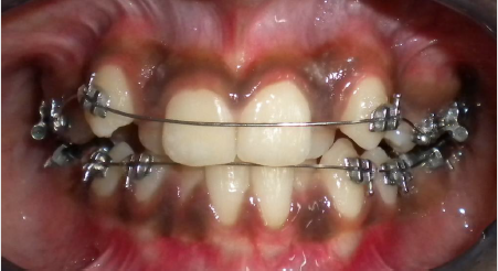
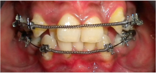
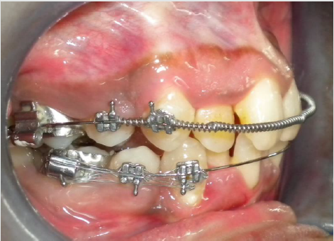
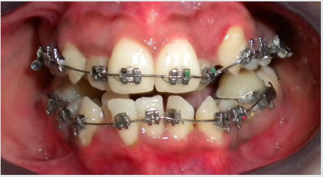
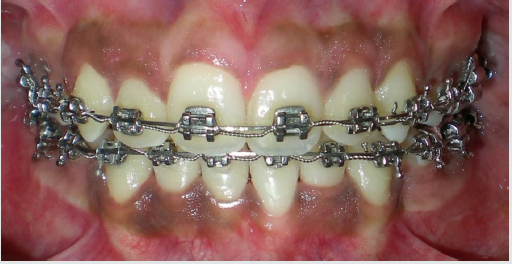
The first molars are banded/bonded with molar tubes followed by bonding of brackets to the second premolars and canines, the incisors are not bonded at this stage and 0.016 NiTi is ligated (Figure 1). The first premolars are extracted. On completion of leveling of the banded/bonded teeth, usually in 6-12 weeks, a 0.016” stainless steel (SS) arch wire is ligated with a NiTi open coil spring between the two canines (Figure 2). The length of the NiTi coil spring is calculated by measuring the distance from the mesial edge of the canine bracket on either side or adding 25% length to it. This delivers a force of approximately 150gms to the canines, as recommended in literature [12,13]. Initially the arch wire is away from the anterior teeth, subsequent activation at four-week intervals may be done by active cinching of the distal ends of the 0.016” SS arch wire. Usually by the second or third activation the arch wire begins to touch the incisors, at this stage small lengths of the NiTi open coil spring may be added to activate the appliance system (Figure 3). On completion of canine retraction, the anterior brackets may be bonded (Figure 4) and the remaining teeth are leveled and aligned (Figure 5).
Figure 5: Alignment upper and lower dentition in progress with optimal utilisation of extraction space.
Conclusion
The present technique is economical, intra-oral, biomechanically efficient anchorage conserving method of canine distalisation, with minimal increase in routine orthodontic armamentarium and patient co-operation. There is no published literature on reciprocal canine distalisation for an entire premolar width.
References
- Salzmann JA (1966) Principles of orthodontics. 2nd Ed, Lippincott, USA.
- Clemmer EJ, Hayes EW (1979) Patient cooperation in wearing headgear. Am J Orthod 75: 517-524.
- Samuels RH, Willner F, Knox J, Jones ML (1996) A national survey of orthodontic facebow injuries in the UK and Eire. Br J Orthod 23: 11-20.
- Holland GN, Wallace DA, Mondino BJ, Cole SH, Ryan SJ (1985) Severe ocular injuries from orthodontic headgear. Arch Ophthalmol 103: 649-651.
- Dickson G (1983) Contact dermatitis and cervical headgear. Br Dent J 155: 12.
- Kyung HM, Park HS, Bae SM, Sung VH, Bongkim IL (2003) Development of orthodontic microimplants for intraoral anchorage. J Clin Orthod 37: 321-328.
- Odman J, Lekholm U, Jemt T, Thilander B (1997) Osseointegrated implants as orthodontic anchorage in the treatment of partially edentulous adult patients. Eur J Orthod 31: 763-767.
- Roberts WE, Marshall KJ, Mozsary PG (1990) Rigid endosseous implant utilized as anchorage to protract molars and close an atrophic extraction site. Angle Orthod 60: 135-152.
- Block MS, Hoffman DR (1995) A new device for absolute anchorage for orthodontics. Am J Orthod Dentofacial Orthop 107: 251-258.
- Lee JS, Park HS, Kyung HM (2001) Micro-implant anchorage for lingual treatment of a skeletal Class II malocclusion. J Clin Orthod 35: 643-647.
- Dalessandri D, Salgarello S, Dalessandri M, Lazzaroni E, Piancino M, et al. (2013) Determinants for success rates of temporary anchorage devices in orthodontics: a meta-analysis (n>50). Eur J Orthod 36(3): 303-313.
- Ricketts RN (1976) Bioprogressive therapy as an answer to orthodontic needs part 2. Am J Ortho 70: 359 -397.
- Storey E, Smith R (1952) Force in orthodontics and its relation to tooth movement. Aust Dent J 56: 11-18.
Editorial Manager:
Email:
pediatricdentistry@lupinepublishers.com

Top Editors
-

Mark E Smith
Bio chemistry
University of Texas Medical Branch, USA -

Lawrence A Presley
Department of Criminal Justice
Liberty University, USA -

Thomas W Miller
Department of Psychiatry
University of Kentucky, USA -

Gjumrakch Aliev
Department of Medicine
Gally International Biomedical Research & Consulting LLC, USA -

Christopher Bryant
Department of Urbanisation and Agricultural
Montreal university, USA -

Robert William Frare
Oral & Maxillofacial Pathology
New York University, USA -

Rudolph Modesto Navari
Gastroenterology and Hepatology
University of Alabama, UK -

Andrew Hague
Department of Medicine
Universities of Bradford, UK -

George Gregory Buttigieg
Maltese College of Obstetrics and Gynaecology, Europe -

Chen-Hsiung Yeh
Oncology
Circulogene Theranostics, England -
.png)
Emilio Bucio-Carrillo
Radiation Chemistry
National University of Mexico, USA -
.jpg)
Casey J Grenier
Analytical Chemistry
Wentworth Institute of Technology, USA -
Hany Atalah
Minimally Invasive Surgery
Mercer University school of Medicine, USA -

Abu-Hussein Muhamad
Pediatric Dentistry
University of Athens , Greece

The annual scholar awards from Lupine Publishers honor a selected number Read More...


