Lupine Publishers Group
Lupine Publishers
Menu
ISSN: 2637-6636
Case Report(ISSN: 2637-6636) 
Management of a Dentigerous Cyst in a 6-Year-Old Child – A Case Report Volume 7 - Issue 2
Shilpa S Naik, Shreya Khodke and Shreya K*
School of Dentistry, DY Patil University, India
Received: January 14, 2022; Published: January 28, 2022
*Corresponding author: Shreya K, School of Dentistry, DY Patil University, Nerul, India
DOI: 10.32474/IPDOAJ.2022.07.000259
Abstract
Dentigerous cysts are epithelial in origin and most common odontogenic cysts. They are usually asymptomatic and hence diagnosed on radiological examination. The standard treatment for these cysts is enucleation and extraction of the affected teeth. This is a case report of a 6-year-old female patient with dentigerous cyst associated with a primary molar. The cyst was enucleated and unerupted premolars were removed from the lower left region. The patient was given a fixed functional band and loop postsurgical treatment. No recurrence was observed after 6months follow up.
Introduction
Cyst has been known to arise in man ever since he has teeth
and are also seen in certain animals. They are consequential, not
only because they often attain a large size but also produce facial
asymmetry, disturbance of dentition, neurological symptoms and
predispose the jaws to fracture but particularly because they have a
very high frequency of occurrence. Kramer in 1974 defined a cyst as
a pathological cavity having fluid, semi fluid or gaseous content but
not always lined by epithelium [1]. The dentigerous cyst is a type of
epithelial odontogenic cyst and is also called as ‘follicular cyst’ or
‘pericoronal cyst.’ It is the most common type of odontogenic cyst
which encloses the crown of the unerupted tooth by expansion of
its follicle [1,2]. A higher incidence of these cysts is usually found
in the second and third decade of life and slightly more common in
males. They account for 14-20% of mandibular cysts and between
15.2% and 33.7% of all odontogenic cysts. The frequency of these
dentigerous cysts in children is less and about 4-9% of these cysts
occur in the first 10 years of life [3]. They are predominantly
associated with third molars, maxillary canines and mandibular
premolars. Dentigerous cysts are often asymptomatic and are an
incidental finding on routine radiographs. In the radiographic
examination, the lesion has a well-defined sclerotic border, and a
well- demarcated unilocular radiolucency which is surrounding the
crown of an unerupted tooth. In some instances, these cysts can
grow to very large size and can trigger the inflammation, expansion
and erosion of the cortical bone. In such a case, they can generate
a differential diagnosis to an ameloblastoma or an odontogenic
keratocystic tumour.
The following case report describes the management of a
dentigerous cyst in a young child.
Case Report
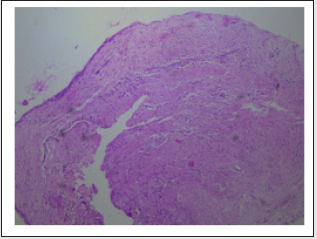
A 6-year-old female patient reported to the Department of Pedodontics and Preventive Dentistry, DY Patil School of Dentistry with a chief complaint of pain in the lower left back region of the mouth. On general examination, the patient was healthy without any significant past medical history. Intra oral examination revealed that the patient presented with a mixed dentition. The area of chief complaint had deep occlusal caries with loss of crown structure in relation with 74 and 75 (Figure 1). The primary molars were non vital and adjacent mucosa was apparently normal, with no signs of inflammation. An initial intra oral periapical radiograph was taken for radiological examination. which revealed a huge radiolucency with no signs of underlying premolar. Hence, a panoramic radiograph was advised (Figure 2) and it revealed the presence of a welldefined unilocular radiolucent cystic lesion with sclerotic border enveloping the crown of mandibular left second premolar. The first premolar was displaced medially while the second premolar was apically displaced close to the lower border of the mandible. After the clinical and radiological examination, a provisional diagnosis of the dentigerous cyst was made. Surgical enucleation of the cyst was chosen as the treatment of choice. The surgical intervention was carried out under general anaesthesia. Blood investigations (PT, PTT, INR) and cone beam computed tomography (CBCT) was done prior to the procedure. Both the primary mandibular molars were extracted followed by opening of the mucoperiosteal flap to disclose the cystic cavity. After the flap was opened, the cavity was identified and 3ml of cystic fluid was aspirated. The cystic lining enclosed both the premolars and hence were removed along with the soft tissue. The flap was then sutured to close the wound primarily. The specimen was fixed in 10% formalin and sent for a histopathological examination. The histopathologic examination confirmed the diagnostic hypothesis of a dentigerous cyst (Figure 3). The patient was followed up regularly for a month and was advised to maintain good oral hygiene. When the lesion was completely healed, prosthetic rehabilitation was done using fixed functional band and loop space maintainer (Figure 4).
Discussion
Dentigerous cysts are reported to be of two types – Developmental and inflammatory. The developmental type is most common and appears to be due to accumulation of fluid between the reduced enamel epithelium and enamel organ. In rare cases, the dentigerous cyst develops as a result of the intrafollicular spread of periapical inflammation from an overlying primary tooth. (Murakami et al 1995) [4]. Accordingly, in the present case, the presence of overlying nonvital necrotic primary mandibular first and second molars increase the possibility of being an inflammatory type of the dentigerous cyst. The nature of the causative tooth, size of the lesion and location influences the type of treatment required for the dentigerous cyst which includes enucleation with primary closure or marsupialization. Marsupialization of the cyst is the treatment of choice which gives a chance to the unerupted tooth to erupt in large cysts [3]. However, in the present case, the cystic sac was surrounded by the unerupted premolar and was firmly attached to it; hence, enucleation of the cyst along with the extraction of premolar was carried out [5]. The histologic examination of the specimen showed cystic lining composed of reduced enamel epithelium which was 2-3 cell layers thick and proliferative at some places. The outer connective tissue stroma showed inflammatory infiltrate. The aspirated cystic fluid was pink in colour and thick consistency. Correlating clinically, the features were suggestive of dentigerous cyst. Owing to the age of the patient and growth phase, it was decided to rehabilitate the patient with a suitable prosthesis. Various options were considered and finally based on the comfort and acceptance of the patient a fixed functional band and loop was fabricated and cemented. This would restore the occlusal function of lost primary teeth and will also maintain the space till the time patient develops permanent dentition and there is bone development for further fixed prosthesis [6]. For the fabrication of the appliance, a conventional band and loop was constructed. The acrylic teeth were placed in the edentulous area of the cast and stabilized with modelling wax. The occlusion was checked with the cast of the opposing arch and adjusted. Cold cure acrylic was used to attach the poetic to the loop. The completed appliance was then finished and polished. Trial fit was done in patient’s mouth and checked for soft tissue irritation or occlusal interferences and adjusted accordingly. The final cementation of the appliance was done using glass ionomer luting cement [7]. The patient was evaluated one week post cementation of the appliance and no complications were reported.
Follow up
The patient was followed up for 6 months with no reports of fracture of the appliance or food lodgment. Clinical and radiographic examination did not reveal any signs of recurrence of the cystic lesion.
Conclusion
Dentigerous cysts are rare in primary dentition and asymptomatic, usually diagnosed during routine radiographs. The sequelae of an untreated or undiagnosed cyst could be harmful to the patient’s future dental development. Thus, regular check-ups by the patient and close observation on the part of treating doctor are essential. This results in elimination of pathology and maintenance of dentition with minimum surgical intervention.
Financial Support
Nil.
Conflict of Interests
There is no conflict of interests.
References
- Ghoms (2016) Textbook of Oral Medicine 3rd
- Arjona-Amo, Manuel (2015) Conservative management of dentigerous cysts in children. Journal of clinical and experimental dentistry 7(5): 671-674.
- Demiriz, Levent (2015) Dentigerous cyst in a young child. European journal of dentistry 9(4): 599-602.
- Clauser C, Zuccati G, Barone R (1994) Simplified surgical orthodontic treatment of a dentigerous cyst. J Clin Orthod 28: 103-106.
- Vinothini V, Sanguida A, Selvabalaji A, Prathima GS, Kavitha M (2019) Functional Band and Loop Space Maintainers in Children. Case Rep Dent 2019: 4312049.
- S Gandhi, DL Franklin (2008) Presentation of a radicular cyst associated with a primary molar. European Archives of Paediatric Dentistry 9(1): 56–59.
- Morawala A, Shirol D, Chunawala Y, Kanchan N, Kale M (2017) Bismuth subnitrate iodoform parafin paste used in the management of inflammatory follicular cyst - Report of two cases. J Indian Soc Pedod Prev Dent 35(3): 269-274.
Editorial Manager:
Email:
pediatricdentistry@lupinepublishers.com

Top Editors
-

Mark E Smith
Bio chemistry
University of Texas Medical Branch, USA -

Lawrence A Presley
Department of Criminal Justice
Liberty University, USA -

Thomas W Miller
Department of Psychiatry
University of Kentucky, USA -

Gjumrakch Aliev
Department of Medicine
Gally International Biomedical Research & Consulting LLC, USA -

Christopher Bryant
Department of Urbanisation and Agricultural
Montreal university, USA -

Robert William Frare
Oral & Maxillofacial Pathology
New York University, USA -

Rudolph Modesto Navari
Gastroenterology and Hepatology
University of Alabama, UK -

Andrew Hague
Department of Medicine
Universities of Bradford, UK -

George Gregory Buttigieg
Maltese College of Obstetrics and Gynaecology, Europe -

Chen-Hsiung Yeh
Oncology
Circulogene Theranostics, England -
.png)
Emilio Bucio-Carrillo
Radiation Chemistry
National University of Mexico, USA -
.jpg)
Casey J Grenier
Analytical Chemistry
Wentworth Institute of Technology, USA -
Hany Atalah
Minimally Invasive Surgery
Mercer University school of Medicine, USA -

Abu-Hussein Muhamad
Pediatric Dentistry
University of Athens , Greece

The annual scholar awards from Lupine Publishers honor a selected number Read More...


