Lupine Publishers Group
Lupine Publishers
Menu
ISSN: 2637-6636
Research Article(ISSN: 2637-6636) 
Effect of Surface Treatment and Type of Resin Cement on Marginal Fit of Zirconia Crowns Volume 4 - Issue 2
Ali Sayed*, Yasser F Gomaa, and Sherif Adel Mohsen
- Department of Dental Biomaterials, Assiut University, Egypt
Received: April 08, 2020; Published: June 16, 2020
*Corresponding author: Ali Sayed, Department of Dental Biomaterials, Assiut University, Egypt
DOI: 10.32474/IPDOAJ.2020.04.000182
Abstract
This in vitro study was conducted to evaluate effect of surface treatment and two resin cements on marginal integrity of zirconia crowns.
Materials and Methods: Forty zirconia crowns (Amann Girrbach AG,Austri) were fabricated and divided into2 groups according to type of resin cement, group 1 Rely x ultimate clicker (3M Deutschland ,Germany ) and group 2 Rely x unicem (3M ESPE,USA) , each group was then subdivided into 4 groups according to type of surface treatment , no surface treatment, sandblasting, sandblasting with silane, sand blasting with MDP . After cementation each specimen was photographed using USB Digital microscope with a built-in camera (Scope Capture Digital Microscope, Guangdong, and China) for measuring and evaluation of gap width.
Results: group 2 showed higher significant mean of marginal gap than group 2.
Conclusion: group 1 Rely x ultimate clicker resin cement showed less marginal gap than group 2 Rely x unicem resin cement.
Keywords: Surface treatment; resin cement; zirconia
Introduction
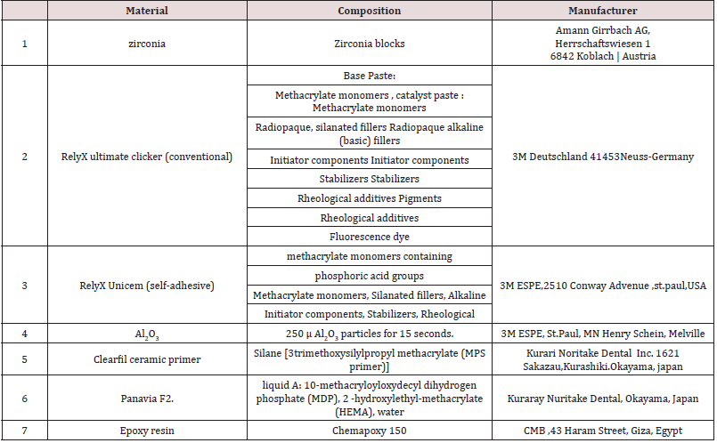
As esthetic restorative materials for crowns and fixed partial dentures (FPDs), all-ceramic systems can be used as alternatives to metal ceramic systems. During the last decade, zirconium dioxide (zirconia, ZrO) ceramics, which have superior mechanical properties [1], have been used increasingly for copings and frameworks of fixed restorations. An ideal all-ceramic dental material should exhibit excellent esthetic characteristics, like translucency, natural tooth color, outstanding light transmission and, at the same time, optimal mechanical properties, like flexural strength, fracture toughness and limited crack propagation at the functional and parafunctional load conditions, in order to ensure long-term service. To date, zirconia has been considered a suitable choice for dental restorations due to its good mechanical properties, toothcolored and natural appearance and low plaque accumulation [2- 4] . Zirconia (zirconium oxide) was introduced by Martin Heinrich Klaproth in 1789. This material is a non-cytotoxic metal oxide, is insoluble in water and has no potential of bacterial adhesion. In addition, it has radio-opacity properties and exhibits low corrosion [5]. There are 3 crystalline shapes of this material at different temperatures are as follows: cubic (c) tetragonal (t) monoclinic (m). Increasing the crystal volume, constrained by the surrounding ones, leads to a favorable compressive stress. This limits growth of cracks. Transformation toughening or “phase transformation toughening” (PTT) is the reported mechanism for exceptional flexural strength and fracture toughness of zirconia among all the other ceramics , hence Zirconia is considered as one of the most reliable ceramic materials [6,7]. Zirconia based fixed partial dentures (FPDs) have the potential to withstand physiological occlusal forces applied in the posterior region, and therefore provided interesting alternatives to metal-ceramic restorations. Although combined surface treatment and specific adhesives with a hydrophobic phosphate monomer such as methacryloyl oxydecyl dihydrogen phosphate (MDP), are currently considered reliable for bonding to zirconia ceramics, but further clinical and invitro studies are still needed to obtain long-term clinical information on zirconia-based restorations [8] (Figure 1).
Materials and Methods
Teeth selection, cleaning, storage and preparation
Atotal 40 extracted caries free human teeth were collected from clinics of faculty of Dentistry Al- Azhar University, Assiut branch, after taking permissions from patients for using their extracted teeth in research purposes. Caries free human upper first premolars extracted for orthodontic purposes from patient 18-20 years old were collected for standardization. Extracted teeth stored in 0.5% thymol not more than 2 months following esthetic protocol of faculty of dentistry Al- Azhar University, Assiut branch.
Specimens Preparation
A machined standard stainless steel was used with 5 mm height and 5 mm diameter with 10 degrees convergent axial walls, the dye was prepared with 50° shoulder finish line (1 mm thickness), forty polyvinyl siloxane impressions (silibest via m.Bonarroti ,cappanoli ,Italy).
Fabrication of zirconia crowns
Crowns made of a partially sintered zirconia ceramic material
by using CAD/CAM technology optical impression was made to the
epoxy specimen after being sprayed , using model scanning via :
desktop extra-oral scanner map 400 Amann girrbach)
a) Crowns were designed via (Exocad software ) with the
following parameters,
b) 0.05mml cement gap starting 1mml from the restoration
margins, Milling the designed restoration with (ceramill motion
25 axix machine manufactured by Amann girrbach).
Testing procedures
Each specimen was photographed using USB Digital microscope with a built-in camera (Scope Capture Digital Microscope, Guangdong and China) connected with an IBM compatible personal computer using a fixed magnification of 45X. A digital image analysis system (Image J 1.43U, National Institute of Health, USA) was used to measure and qualitatively evaluate the gap width. Within the Image J software, all limits, sizes, frames and measured parameters are expressed in pixels. Therefore, system calibration was done to convert the pixels into absolute real world units. Calibration was made by comparing an object of known size (a ruler in this study) with a scale generated by the Image J software. Specimens were held in place over their corresponding dies using a specially designed and fabricated holding device. Shots of the margins were taken for each specimen. Then morphometric measurements were done for each shot (3 equidistant landmarks along the cervical circumference for each surface of the specimen) (Mesial, buccal, distal, and lingual) [4]. Readings of each surface of specimen were taken, then the data obtained were collected, tabulated then statistically described in terms of mean and standard deviation (SD) (Table 1).
Results
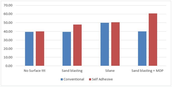
Rely x unicem showed the higher insignificant mean of marginal. According to following Table 2.
Discussion
Zirconia ceramics have superior strength, toughness, fatigue resistance and enhanced long-term durability than other ceramics Oilo et al. [9], Della Bona [10]. CAD/CAM system crowns demonstrated more accurate marginal fit than the traditional fabrication methods Mart´ınez et al. [11]. The treatment of zirconia ceramic surfaces with abrasive alumina enhances the surface roughness resulting in the increase of the effective contact surface between ceramic and resin cement which leads to the increase of the bond strength [12]. also improves micromechanical interlocking resulting to a strong and durable bond between resin/zirconia [13]. Silane coupling primers have been reported to increase the wettability of the surface but there is no chemical reaction occurs between the silane and surface of zirconia which may explain the low bond strength resulting in comparison to the other surface treatment methods [14]. In the present study maximum gap found in zirconia crowns cemented by self-adhesive resin cement after sand blasting with MDP surface treatment, this finding was in agreement with Yuksel et al. [15] which may be due to short setting time of conventional cement as opposed to that of the self- cure resin bonding system [16]. This finding disagreed with Kern et al. [17] which reported that selfadhesive resin cement (RelyX Unicem) produced better marginal continuity, and high adaptation to substrate.
References
- Christel P, Meunier A, Heller M, Torre JP, Peille CN (1989) Mechanical properties and short-term invivo evaluation of yttrium-oxide-partially-stabilized zirconia. J Biomed Mater Res 23: 45-61.
- Olsson KG, Fürst B, Andersson B, Carlsson GE (2003) A long-term retrospective and clinical follow-up study of In-Ceram Alumina FPDs. Int J Prosthodont 16:150-156.
- Bateli M, Kern M, Wolkewitz M, Strub JR, Att W (2013) A retrospective evaluation of teeth restored with zirconia ceramic posts: 10-year results. Clin Oral Investig 18: 4.
- Raigrodski AJ (2004) Contemporary materials and technologies for all ceramic fixed partial dentures: a review of the literature. J Prosthet Dent 92: 557-562.
- Dion I, Bordenave L, Levebre F (1994) Physicochemistry and cytotoxicity of ceramics. J Mater Sci Mater Med 5: 18-24.
- Scotti R, Kantorski KZ, Monaco C, Valandro LF, Ciocca L, et al. (2007) SEM evaluation of in situation early bacterial colonization on a Y-TZP ceramic: a pilot study. Int J Prosthodont 20: 419-22.
- Hannink RHJ, Kelly PM, Muddle BC (2000) Transformation toughening in zirconia containing ceramics. J Am Ceram Soc 83: 461-487.
- Futoshi Komine, Markus B Blatz, Hideo Matsumura (2010) Current status of zirconia-based fixed restorations. J Oral Sci 52(4): 531-539.
- Oilo M, Gjerdet N, Tvinnereim H (2008) The firing procedure influences properties of a zirconia core ceramic. Dent Mater 24(4): 471-475.
- Della Bona A, Pecho OE, Alessandretti R (2015) Zirconia as a dental biomaterial. Materials 8(8): 4978-4991.
- Martinez Rus F, Suarez MJ, Rivera B, Pradies G (2011) Evaluation of the absolute marginal discrepancy of zirconia-based ceramic copings. J Prosthet Dent 105: 108-114.
- Kern M (2009) Resin bonding to oxide ceramics for dental restorations. J Adhes Sci Technol 23: 1097-1111.
- Xie H, Tay FR, Zhang F, Lu Y, Shen S, et al. (2015) Coupling of 10-methacryloyloxydecyldihydrogenphosphate totetragonal zirconia: effect of pH reaction conditions oncoordinate bonding. Dent Mater 31: 218-225.
- Lei Zhu, Toru N, Shauzo K (2009) Effect of surface abrasion and silica coatingon tensile bond strength of resin cement to zirconia ceramics .INT Chin J Dent 9: 23-30.
- Yuksel E, Zaimoglu A (2011) Influence of marginal fit and cement types on microleakage of all- ceramic crown systems. Braz Oral Res 25(3): 261-266.
- Gu XH, Kern M (2003) Marginal discrepancies and leakage of all ceramic crowns: Influence of luting agents and ageing conditions.Int J Prosthodont 16: 109-116.
- Kern M (2009) Resin bonding to oxide ceramics for dental restorations. J Adhes Sci Technol 23: 1097-1111.
Editorial Manager:
Email:
pediatricdentistry@lupinepublishers.com

Top Editors
-

Mark E Smith
Bio chemistry
University of Texas Medical Branch, USA -

Lawrence A Presley
Department of Criminal Justice
Liberty University, USA -

Thomas W Miller
Department of Psychiatry
University of Kentucky, USA -

Gjumrakch Aliev
Department of Medicine
Gally International Biomedical Research & Consulting LLC, USA -

Christopher Bryant
Department of Urbanisation and Agricultural
Montreal university, USA -

Robert William Frare
Oral & Maxillofacial Pathology
New York University, USA -

Rudolph Modesto Navari
Gastroenterology and Hepatology
University of Alabama, UK -

Andrew Hague
Department of Medicine
Universities of Bradford, UK -

George Gregory Buttigieg
Maltese College of Obstetrics and Gynaecology, Europe -

Chen-Hsiung Yeh
Oncology
Circulogene Theranostics, England -
.png)
Emilio Bucio-Carrillo
Radiation Chemistry
National University of Mexico, USA -
.jpg)
Casey J Grenier
Analytical Chemistry
Wentworth Institute of Technology, USA -
Hany Atalah
Minimally Invasive Surgery
Mercer University school of Medicine, USA -

Abu-Hussein Muhamad
Pediatric Dentistry
University of Athens , Greece

The annual scholar awards from Lupine Publishers honor a selected number Read More...


