Lupine Publishers Group
Lupine Publishers
Menu
ISSN: 2637-6636
Research Article(ISSN: 2637-6636) 
Analysis of Marginal Bone Loss and Implant Success Rate After Implant Implantation Volume 7 - Issue 1
Hee Ja Na1 and Moon Sil Choi2*
1Department of Dental Hygiene, Honam University, Gwangju, Republic of Korea
2Department of Dental Hygiene, Songwon University, Gwangju, Republic of Korea
Received:October 21, 2021; Published: November 01, 2021
*Corresponding author: Moon Sil Choi, Department of Dental Hygiene, Songwon University, Republic of Korea
DOI: 10.32474/IPDOAJ.2021.07.000253
Abstract
Purpose: Patients with prosthesis-equipped post-implantation prosthesis performance were continuously observed, revealing the association of marginal bone loss with gender and age implant success and survival rates during a 1year study of clinical prosthesis placement; with results used as a reference for implant procedures and prognosis.
Methods: This study was conducted on 31 patients from January to February 2021 at Y Dental Hospital in Gwangju, with 20 men and 11 women equipped with prosthetics after implant procedure a year earlier. The subjects of the study were carried out with the G-power program, and 31 subjects were extracted. The distance from the shoulder of the implant to the attachment point of the dichotomous bone was measured at the center of the area of atrophy, checking both from the mesial region to the distal region using a radiation ruler taking photos with endocardial radiation as well as panoramic images. The subjects of the implant study were analyzed by age, gender, cause of release, duration of implant, implant length, implant width, implant assembly period, osteoplasty type, shield presence, match with corresponding teeth, implant survival symptom, implant success rate symptoms, and 1mm loss within a year. This study was conducted after approval.
Results: As a result of testing the contribution and statistical significance of individual independent variables to dependent variables, at a significance level of 0.05, the independent variable loses 1 mm at a ratio of (t=5.473, p=.00) in 5.473, p=.000). This loss significantly affects implant success rates.
Conclusions: Recognizing the importance of bone loss management within a year after implant procedure, dental staff and patients can achieve higher success and survival rates after implant procedure if they can be thoroughly managed within a year.
Keywords:Implant formula; marginal bone loss; success rate; survival rate; implant peripheral inflammation; Implant prosthesis
Introduction
The Margin bone of the implant is loaded around the implant over time and is involved in implant survival [1]. Factors affecting implant survival include peri-implant mucositis, the inflammation of local soft tissue without osteosis near the implant. This, along with local inflammatory lesions around the area of the implant affect implant viability [2]. In 2008, the 6th European Dental Workshop reported that 80 percent of implant subjects had mucosal inflammation and 28 between56 percent of those affected had implant peripheral inflammation [3,4]. The subjects measured the distance from the shoulder of the implant to the attachment point of the dichotomous bone using a radiation ruler. Photographs of the dental root were taken by endothelial radiation as well as panorama images taken within a year. The subjects of the implant study were analyzed by age, gender, cause of release, duration of implant, implant length, implant width, implant formulation period, osteoplasty type, shield presence, agreement, implant survival symptoms, implant success rate symptoms, and 1mm loss within a year. Implant Success Rate: The assessment for implant success considered liquidity, pain, retardation, radiolucent pathology, and the nonexistence of implant peripheral inflammation. Along with this, using the criteria of Zarb and Albrektson, subjects were studied for the non-presence of progressive bone absorption in accordance with Zarb and Albrektson’s criteria (less than 1mm within a year of implant assembly and not more than 0.2mm). Implant survival rates were assessed by Buser et al. [5,6]. The first assessment was based on no agitation during each implant clinical examination, no pain or subjective abnormalities, no inflammation around the implant, and no continuous radiation penetration around the implant [7]. There is a lack of research on the significance between the survival rate of implants and the loss of limbic bone in implant success rate, and the cause of limbic bone loss has been unresolved and contested so far.
Materials and Methods
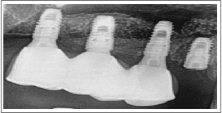
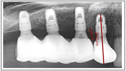
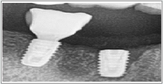
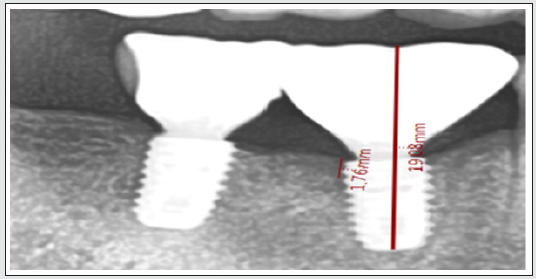
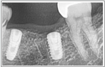
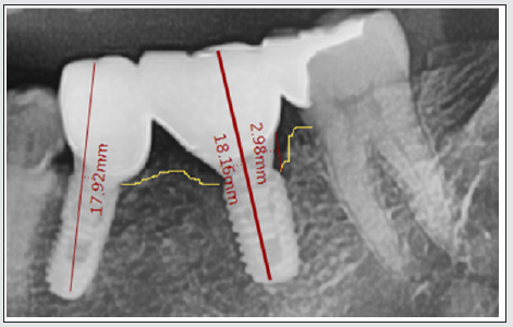
From January to February 2021, Y Dental Hospital in Gwangju conducted this study on 31 patients with 20 men and 11 women after installing prosthetics after implant procedures a year earlier. Thirty-one people were extracted from the study by conducting the G-power program. The distance from the shoulder of the implant to the attachment point of the dendritic bone was measured at the center of the area of atrophy, checking both from the mesial region to the distal region using a radiation ruler in the photo of the endocardial end of the mouth and a panorama image taken within a year. After implantation, the skeletal aspects of the bone (DP-80-P/PM2002 EC proline, plameca, Helsinki, Finland) were evaluated on radiographs (Figures 1-6). Subjects of the implant study were classified by age, gender, cause of release, reason for extraction, extraction period, implant length, implant width, implantation period, osteoplasty type, shield presence, match with corresponding teeth, implant landscape, implant prosthesis installation period, bone transplant type, shield film, , match with corresponding teeth , implant survival symptoms, implant success rate symptoms, along with implant non-survival rate symptoms and implant non-performance symptoms according to marginal bone loss of 1 mm within a year. All patients live in Gwangju. This study was conducted after deliberation by the Bio-Science Ethics Committee of Honam University (IRB) (NO 1041223-202011-HR- 24). Age, gender, cause of release, duration of release, length of implant, implant width, implantation period, implant prosthesis installation period was surveyed, with a 1 point on the likert 5-point scale meaning ‘very negative’, and 5 points on the scale meaning ‘very positive’. Implant survival rates were assessed by Buser et al. First, there would be no agitation during each implant clinical examination, second, no pain or subjective abnormalities, third, no inflammation around the implant, fourth, no continuous radiation penetration around the implant [7]. In the case of implant success rate symptoms, the questionnaire was set for those who had less than 1 mm (1 mm within a year of implant erosion and not more than 0.2 mm). Results were according to whether there were no issues with fluidity, pain, or perceptual abnormalities, as well as having any radiological permeability, pathology, or other implant periphery issues. There is a lack of research on the significance between the survival rate of implants and the loss of limbic bone in implant success rate, and the cause of limbic bone loss has been unresolved and contested so far.
Data analysis
SPSS 18.0 program was used for the collected data. General characteristics 13 used descriptive statistical analysis. In this study, in general characteristics and corresponding samples t-test, in correlation, the confidence interval is *Noticeable at 0.05 correction (both sides) **, Significant at correction coefficient 0.01 level (both sides), in ANOVA for regression models. In the middle and multiple regression analysis, a. Dependent variable: Implant successful were confidence interval.
Results
General characteristics
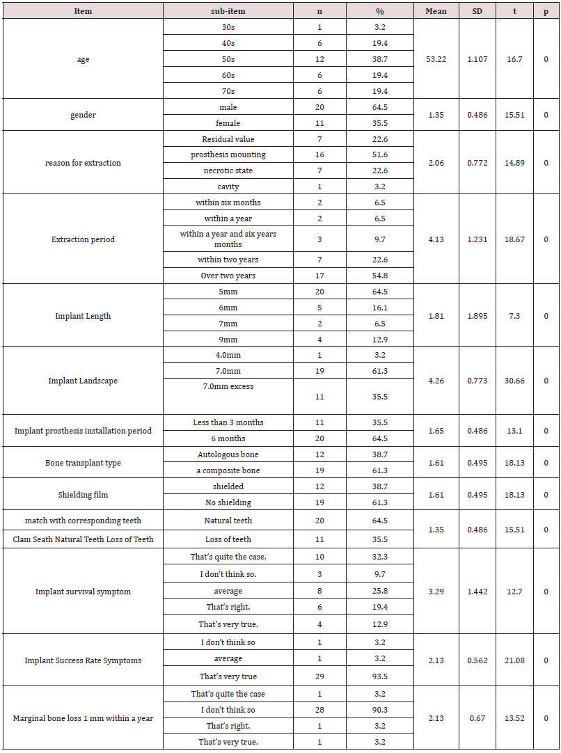
According to the general characteristics, one person was in their 30s (3.2%); next, (19.4%) were in their 40’s, twelve people were in their 50s (38.7%); six people were in their 60s (19.4%), and six people were in their 70s (19.4%). The mean and standard deviation is 53.2 (1.107), t=16.70, p=. 00. There were 20 men and 11 women for a ratio of 64.5 percent male, 35.5 percent female; 1.35 (.486) mean and standard deviation, and t = 15.51 and p = 0.00 respectively. The cause of release is 7 residual values (22.6%) and 16 prosthetics (51.6%) and 7 necrotic states (22.6%) and 1 cavity (3.2%) with the mean and standard deviation of 2.06 (2.772), t=14.89), and p=.00. The average and standard deviation are 4.13 (1.231), t=18.67 and p=.00 with 2 persons (6.5%) within 6 months, 3 persons (9.7%), 7 persons (22.6%) within 2 years, and 17 persons (54.8%) within 2 years. Implant length for 20 (64.5%) was normal. Implant length for 5 (16.1%) was normal. Two people had implant length of 7mm for a ratio of 6.5%. Four people had an implant length of 9mm for a ratio of (12.9%). The mean and standard deviation is 1.81 (1.895); t=7.30, p=.00 has a width of 4.0mm (3.2%) and 19 (61.3%). The average and standard deviations are 4.26 (.773), t=30.66, and p=.00 with 11 persons (35.5%) exceeding 7.0 mm. 11 people (35.5 percent) and 20 people (64.5 percent) are in 6 months. The mean and standard deviation are 1.65 (.486), t=13.10, p=.00. The type of osteophyte is 12 autogenous bones (38.7%) and 19 synthesis bones (61.3%), with an average and standard deviation of 1.61 (.495), t=.00. The average and standard deviation are 1.61 (.495), t=18.13 and p=.00. match with corresponding teeth. The mean and standard deviation are 1.35 (.486), t=15.51, p=.00 with 20 natural teeth (64.5%) and 11 lost teeth (35.5%).
The average and standard deviation are 3.29 (1.442), t=12.70, p=, and 10 (32.3%) who are not, and 8 (25.8%) who are normal, and 6 (19.4%) who are “very” (12.9%) who are 3.29 (1.442), t=12.70, and p=.00. The average and standard deviation are 2.13(.562), t=21.08, and p=.00 with one “not” (3.2%) and 29 “very” (93.5%) respectively. Within a year, 1 mm (3.2%) of 1 mm loss, 28 (1.2%) of 1.2 mm (90.3%), 1 (3.2%) of 1.4 mm, and 1 (1.2%) of 1.5 mm. The mean and standard deviation are 2.13 (670), t=13.52, p=.00 (Table 1). The corresponding sample t-test shows that the mean of Gender and cause of development is -.710 standard deviation is .864, t=-4.574 and the probability of significance is .000, which is significant. A significant difference was shown in 05. Age and age of release mean is-The 806 standard deviation is 1.424 and t=-3.153 and the significance probability is .000, which is the significance level A shows a significant difference in 05. This is a 1.516 standard deviation where 1.85, t=4.677, and the significance probability is .000, which is significant. A significant difference was shown in 05. Age and implantation period, the mean of 1.677 standard deviation was 1.166 with t=8.011 and the significance probability is .000, which is significant. A significant difference was shown in 05. Gender and implant success is -.774 The standard deviation is.805 and t=5.358 with a significance probability .000. It was shown that there is a significant difference in 05 (Table 2). In (Table 3), the bone grafts correlation coefficient is .467** and the shielding film and bone grafts correlation coefficient is .358* and Implant Length and bone grafts negative correlation coefficient is -.476** and the negative correlation coefficient of Implant Success Rate and antagonist teeth is similar. This is 457**.
*Noticeable at 0.05 correlation (both sides).
** Significant at correlation coefficient 0.01 level (both sides).
The correlation coefficient between Implant Length and Implant assembly period is –457** and the negative correlation coefficient between antagonist teeth and Implant Success Rate is -.457** and the correlation coefficient between antagonist teeth and bone grafts is similar. This is 357**. The correlation coefficient between bone grafts and anti-agonist teeth diagrams-.518** and the negative correlation coefficient of shielding film and antagonist teeth - .656**, all significantly vary at a significance level of 0.01. The shielding film and bone grafts correlation coefficients are .358*, Implant Length and bone grafts are negative correlation coefficients –393*, antagonist teeth and Implant Success Rate correlation coefficients are .369*, and the antagonist teeth and Implant Length are similar. All were significantly shown at the significance level of 0.05. Statistical significance test results for the success rate of 1mm loss and implantation within 1 year of the cartilage show that f statistics are 18.161, and the probability of significance is similar. At a significant level of 0.05 with 00, 1 mm loss within a year of the limbic bone significantly accounts for the implant success rate. The total level of satisfaction is 75% (56% according to the coefficient of correction) which describes the independent variables included in the model (Table 4). As a result of testing the contribution and statistical significance of individual independent variables, the independent variables that significantly affect the implant success rate at the significance level of 0.05 are lost 1 mm (t=5.473, p=).000) is affecting implant success rate (Table 5).
a: Dependent variable: Implant successful.
b: Predictor: (constant), implantation period, 1mm loss within 1.
a: Dependent variable: Implant successful.
Discussion
Much controversy exists in the field of implant advanced research regarding healing and tooth function for eating. An important issue regards why the amount of bone loss that occurs during the first year after implant surgery is greater than the amount of bone loss that occurs every year thereafter. Causes of rapid disfigurement include surgical damage, overload of glyphs, peripheral inflammation of the implant area, presence of micro-space, biologic width, and the critical module of the implant. Surgical damage is caused by heat generated by the tissue formation, engine speed, and applied force during implant surgery, which affects the loss of the initial implant limb by deformation. In advanced research, surgical damage is caused by heat generated by the colossal geometry of the periodontal tissue, engine speed, and force applied during implant procedures, which affects the initial implant limb bone loss [8-10]. Von Re cum et al. reported that in photoelasticity and stereoscopic finite element analysis, stress is concentrated at the site where the two objects first meet, and studies using photo elastic blocks similar to implant and bone tissue show a large amount of U, V-shaped stress patterns., t=13.52, p=.00 (Table 1). Several factors that affect the loss of implant margin bone include age, gender, length and breadth of implant, eating and smoking. Existing studies have shown that age and gender are not associated with loss of margin bone, and regarding the width of [11-13]. implants, large implants claim less loss of margin bone than standard types, but a recent paper has shown no significance between these.
It has been reported that longer implant length is more favorable in maintaining the margin bone than shorter ones [14-17]. In this study, the response sample t-test showed a mean of -.710 standard deviation, t=-4.574 and a corresponding significance probability. A significant difference was shown in 05. Age and duration of release, where the mean age is 806 standard deviation is 1.424 and t=-3.153 and the significance probability is .000, which gives a significance level showing A significant difference in 05. For gender and implant length, the mean is 1.516 standard deviation is 1.85, t=4.677, and the significance probability is .000, which is significant. A significant difference was shown in 05. For age and implant assembly, the mean is 1.677 standard deviation is 1.166 and t=8.011 with a significant probability of p= .00. A significant difference was shown in 05. The mean for gender and implant success is -.774. The standard deviation is.805 and t=5.358 with a significance probability of p= .00. It was shown that there is a significant difference in 05 (Table 2). In (Table 3), the correlation coefficient between bone grafting and tooth extraction period is .467**, the correlation coefficient between shielded film and bone grafting is .864**, and the negative correlation coefficient between implant length and tooth extraction period is -.476** and the negative correlation coefficient of implant success rate to assembly period is similar. This is 457**. Implant length and duration negative correlation coefficient is –457** and match and age negative correlation coefficient is -.457** and the coefficient of correlation between the agreement and the duration of the pitch negative is similar.
This is 357**. Correlation coefficient of negative with corrugated vegetables-.518** and negative correlation coefficient of shield and contrast - .656**, all significantly vary at a significance level of 0.01. The shield and foot length correlation coefficient are .358*, implant length and corrugated material show a negative correlation coefficient –393*, concordance and implant success rate are .369*, concordance and implant length are .369*, and both autologous and implant length negative correlation are significant at -393. If the implant is buried and the implant is equipped with overlying denture or fixed prostheses, the missing margin bone of the implant is 1.64mm and 0.05-0.13mm per year after the implant is buried, and the partial denture is supported by the implant every year. Factors, i.e., bone mesh, density of sponges, thickness of cortical bone, also are affected by surgical methods and the form of implants [18,19]. Therefore, even if the bone marrow is poor, the appropriate implant can be selected, and the surgical method can be improved to increase initial stability [20]. In this work we have discussed so far, we have analyzed success rates based on factors such as implant length, implant width bone type, shield presence, and analysis status for the match, and found no significant difference from implant success rate. In this study, the statistical significance test of 1mm loss and implant success rate within 1 year of the limb bone shows that the f statistic is 18.161, p=.00, which significantly describes the implant success rate of the limb bone within 1 year at the significance level of 05.
The total level of satisfaction is 75% (56% according to the coefficient of correction), which describes the independent variables included in the model. Table 4 and the test for the contribution and statistical significance of individual independent variables to dependent variables, and the independent variables that significantly affect the implant success rate at the significant level of 0.05 show loss of 1 mm within a year (t=5.473, p=).000) which has been shown to affect implant success rates. In other words, the analysis results, affecting the success rate of 1mm loss and implant within a year of the limb bone, have also been shown in this study. This study suggests that findings of 1mm increase implant success rate and survival rate within a year after prosthetic installation after implant procedure. Recognizing the importance of bone loss management within a year after implant procedure, dental staff and patients will thoroughly manage bone loss management within a year to maintain bone adhesion after implant procedure, prevent bone loss of implant, and improve [21] success rates and survival. The limits of this study are that the trace of the disappearance of the limb bone was studied within one year, and the future study is to examine the change of the limb bone loss by tracking it for more than two years.
Conclusion
This study was conducted on 31 patients from January to February 2021 at Y Dental Hospital in Gwangju, with 20 men and 11 women after installing prosthetics after implant procedures a year earlier. The subjects of the study were conducted with the G-power program and 31 people were studied. The distance from the shoulder of the implant to the dendritic attachment point was measured at the area of atrophy, checking both from the mesial region to the distal region using a radiation ruler in the photos taken at the time of prosthesis mounting and at the time of implant prosthesis installation.
a) According to the general characteristics, one person was in their 30s (3.2%); next, (19.4%) were in their 40’s, twelve people were in their 50s (38.7%); six people were in their 60s (19.4%), and six people were in their 70s (19.4%). There were 20 men and 11 women for a ratio of 64.5 percent male, 35.5 percent female.
b) As a result of the response sample t-test, the cause of gender and release, period and age of release, length of gender and implant, age and implantation period, and p= gender and implant success with a significance level of 000; results showed a significant difference in 05.
c) Bone graft marginal and extension period correlation coefficient is .467**, shield and bone graft marginal correlation coefficient .864**, implant length and extraction period have a negative correlation coefficient of -.476** and the implant success rate and implant placement period negative correlation coefficient are similar. This is 457**. For the Implant length and Extraction period. The negative correlation coefficient is –457** and the negative correlation coefficient match with corresponding teeth and age is -.457** and the correlation coefficient between match with corresponding teeth and the extraction period diagram is similar. This is 357**. The negative correlation coefficient for bone graft marginal and clamp is also -.518** and negative correlation coefficient of shield and clamp- .656**, all vary significantly at a significance level of 0.01. In addition, shield and extraction correlations are .358*, implant length and bone graft marginals show negative correlations –393*, match and implant success rates are .369*, match and implant length are .369*, and negative correlations of both autologous and implant length are significant at -393*.
d) Statistical significance test results for the success rate of 1mm loss and implantation within 1 year of the cartilage show that f statistics are 18.161, and the probability of significance is similar. At a significant level of 0.05 with 00, 1 mm loss within a year of the limbic bone significantly accounts for the implant success rate. The total level of satisfaction is 75% (56% according to the coefficient of correction) which describes the independent variables included in the model.
e) As a result of testing the contribution and statistical significance of individual independent variables, the independent variables that significantly affect the implant success rate at the significance level of 0.05 are lost, 1 mm (t=5.473, p=).000) shows an effect on the implant success rate.
Conflict of Interest
No potential conflict of interest relevant to this article was reported.
Acknowledgement
This study was supported by research fund from Songwon University [A2021-51].
References
- Alba Carrasco-García, Lizett Castellanos Cosano, José Ramon Corcuera Flores, Antonio Rodríguez Pérezrres Lagares, Guillermo Machuca Portillo (2019) Influence of marginal bone loss on peri-implantitis: systematic review of literature. J Clin Exp Dent 11(11): e1045-e1071.
- Søren Jepsen, Tord Berglundh, Robert Genco (2015) Primary prevention of peri-implantitis: managing peri-implant mucositis. J Clin Periodontol 42(16): 152-157.
- Berglundh T, Lindhe J, Marinello C, Ericsson I, Liljenberget B (1992) Soft tissue reaction to de novo plague formation on implants and teeth. An experimental study in the dog. Clin Oral Implants Res 3(1): 1-8.
- Iindhe J, Meyle J (2008) Peri-implant diseases: consensus Report of the Sixth European Workshop on Periodontology. J Clin Periodontol 35(8): 282-285.
- Albrektsson T, Branemark PL, Hansson HA, Lindstrom J (1981) Osseointerated titanium implants. Requirements for ensuring a long –lasting, direct bone-to-implant anchorage in mam. Acta Orthop Scand 52(2): 155-170.
- Angelis De F, Papi P, Mencio F, Rosella D, Carlo Di S, et al. (2017) Implant survival and success rates in patients with risk factors: results from a long-term retrospective study with a 10 to 18 years follow-up. Eur Rev Med Pharmacol Sci 21(3): 433-437.
- Buser D, Mericske-Stern R, Bernard JP, Behneke A, Behneke N, Hirt HP (1997) Long-term evaluation of non-submerged ITI implants. Part 1: 8-year life table analysis of a prospective multi-center study with 2359 implants. Clin Oral Implants Res 8(3): 161-172.
- Kang BW, Lee SM (2012) Awareness of periodontal diseases and implant management among implant wearers. J Korean Soc Dent Hyg 12(4): 759-770.
- Serino G, Strom C (2009) Peri-implantitis in partially edentulous patients: association with inadequate plaque control. Clin Oral Implants Res 20(2): 169-175.
- VonRecum A (1986) Handbook of biomaterial evaluation. New York: Macmillan Publishing Co 6: 202-205.
- Bryant SR, Zarb GA (2003) Crestal bone loss proximal to oral implants in older and younger adults. J Prosthet Dent 89(6): 589-597.
- M Sommer, J Zimmermann, L Grize, S Stübinger (2020) Marginal bone loss one year after implantation: a systematic review of different loading protocols. Int J Oral Maxillofac Surg 49(1): 121-134.
- Fartash B, Eliasson S, Arvidson K (1995) Mandibular single crystal sapphire implants: change in crestal bone levels over three years. Clin Oral Implants Res 6(3): 181-188.
- Andresson B, Odman P, Carlsson GE (1995) A study of 184 consecutive patients refered for single-tooth replacement. Clin Oral Implants Res 6(4): 232-237.
- Ivanoff CJ, Sennerby C, Johnsson B, Rangert B, Lekholm U (1997) Influence of implants diameters on the integration of screw implants: An experimental study in rabbits. Int J Oral Maxillofac Implants 26(2): 141-148.
- Tawil G, Mawla M, Gottlow J (2002) Clinical and radiographic evaluation of the 5 mm diameter regular platform Branemark fixture: 2 to 5 years follow up. Clin Implant Dent Relat Res 4(1): 16-26.
- Pylant, T, Triplet G, Key MC (1992) A retrospective evaluation of endoosseous titanium implants in the partially edentulous patient. Int J Oral Maxillofac Implants 7(2): 195-202.
- Huwiler MA, Pjetursson BE, Bosshardt DD, Salvi GE, Lang NP (2007) Resonance frequency analysis in relation to jawbone characteristics and during early healing of implant installation. Clin Oral Implants Res 18(3): 275-280.
- Ignacio Sanz-Sánchez, Ignacio Sanz-Martín, Elena Figuero, Mariano Sanz (2015) Clinical efficacy of immediate implant loading protocols compared to conventional loading depending on the type of the restoration: a systematic review. Clin Oral Implants Res 26(8): 964-982.
- Rabel A, Kohler SG, Schmidt-Westhausen AM (2007) Clinical study on the primary stability of two dental implant systems with resonance frequency analysis. Clin Oral Investig 11(3): 257-265.
- Cheng-En Sung, Cheng-Yang Chiang, Hsien-Chung Chiu, Yi-Shing Shieh, Fu-Gong Lin, et al. (2018) Periodontal status of tooth adjacent to implant with peri-implantitis. J Dent 70: 104-109.
Editorial Manager:
Email:
pediatricdentistry@lupinepublishers.com

Top Editors
-

Mark E Smith
Bio chemistry
University of Texas Medical Branch, USA -

Lawrence A Presley
Department of Criminal Justice
Liberty University, USA -

Thomas W Miller
Department of Psychiatry
University of Kentucky, USA -

Gjumrakch Aliev
Department of Medicine
Gally International Biomedical Research & Consulting LLC, USA -

Christopher Bryant
Department of Urbanisation and Agricultural
Montreal university, USA -

Robert William Frare
Oral & Maxillofacial Pathology
New York University, USA -

Rudolph Modesto Navari
Gastroenterology and Hepatology
University of Alabama, UK -

Andrew Hague
Department of Medicine
Universities of Bradford, UK -

George Gregory Buttigieg
Maltese College of Obstetrics and Gynaecology, Europe -

Chen-Hsiung Yeh
Oncology
Circulogene Theranostics, England -
.png)
Emilio Bucio-Carrillo
Radiation Chemistry
National University of Mexico, USA -
.jpg)
Casey J Grenier
Analytical Chemistry
Wentworth Institute of Technology, USA -
Hany Atalah
Minimally Invasive Surgery
Mercer University school of Medicine, USA -

Abu-Hussein Muhamad
Pediatric Dentistry
University of Athens , Greece

The annual scholar awards from Lupine Publishers honor a selected number Read More...


