Lupine Publishers Group
Lupine Publishers
Menu
ISSN: 2637-6636
Case Report(ISSN: 2637-6636) 
A Rare Case of Multiple Radix Entomolaris in the Primary Dentition of a 6 Year Old Girl: A Case Report Volume 8 - Issue 1
Shreya Rani1, Soni Patel1, Supreeya Patel2, Nishi Singh1, Arunima Ghose1 and Nilotpol Kashyap3*
- 1PG Student, Department of Pedodontics and Preventive Dentistry, Vananchal Dental College and Hospital, India
- 2Reader, Department of Pedodontics and Preventive Dentistry, Vananchal Dental College and Hospital, India
- 3Professor, Department of Pedodontics and Preventive Dentistry, Vananchal Dental College and Hospital, India
Received: August 03, 2022; Published: August 10, 2023
*Corresponding author: Nilotpol Kashyap, Professor, Department of Pedodontics and Preventive Dentistry, Vananchal Dental College and Hospital, India
DOI: 10.32474/IPDOAJ.2023.08.000296
Abstract
Radix entomolareis or supernumerary root is a rare anomaly of root. Radix entomoralis is more commonly seen in permanent dentition and rarely in deciduous dentition. The presence of an extra root can be a problem for the clinicians if the extra root is not radiographically evaluated as it may be left untreated during endodontic therapy or may cause injury to the developing tooth bud in cases of extraction.
Keywords: Radix entomolaris; supernumerary tooth; endodontic therapy
Introduction
Complete knowledge of variation in the primary dentition morphology is important in delivering better diagnosis and treatment for the young patients. But, in the literature of odontology the morphology, the pathologic characteristics and the evolution of the deciduous teeth is less explained compared to that of permanent dentition. ‘Radix entomolaris’ (RE) is a developmental anomaly of the root which can affect treatment planning as well as prognosis of any tooth [1]. It is also known as ‘extra distolingual root’ or a ‘distolingual root’ or an ‘extra third root’ [2]. Root in RE can be a short conical or a mature root of its usual length [3]. Tratman (1938) was the first scholar to discover the “racial” significance of three-rooted mandibular first molars, a trait that he found in high frequency in Asiatic populations. He also has reported that three rooted mandibular molars are rare with a frequency of < 1% in the deciduous dentition and common in the permanent dentition in the year 1938 [4]. Lack of knowledge of RE may lead to the failure in endodontic treatment of the tooth. Radiographic interpretation plays a major role in the successful endodontic treatment of such teeth [5].
Case Report
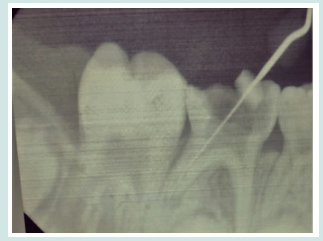
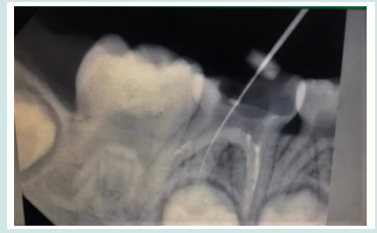
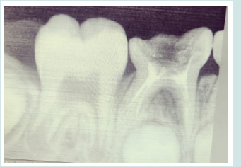
A girl child of 6 years had come to the department of pedodontic and preventive dentistry at the dental institution with a chief complaint of pain in the in the lower left back tooth region of jaw since past one week. On clinical examination she was having deep occlusal caries with respect to tooth number 85. Radiographically, the caries had progressed to the pulp chamber. The presence of third root/ additional root was also revealed. The diagnosis of chronic irreversible pulpitis with 85 was made (Figure 1). It was also found through radiograph that extra root was present with respect to 84 and 75 which were healthy. The tooth was isolated and access opening was done under local anesthetia and all the canals were located. Those were mesiobuccal, mesiolimgual, distobuccal and distolingual with the working length of 15mm in all the four canals (Figure 2). Cleaning and shaping of all the canals were done. Followed by obturation with metapex (Figure 3). The cavity was then sealed with permanent restorative material followed by stainless steel crown cementation.
Discussion
From the clinical point of view it is very important for a clinician to know about the anatomical variation present in teeth. The presence of three rooted permanent mandibular first molar and deciduous second molar has been reported widely in literature [6-8]. In our case report the presence of three rooted deciduous second molar were seen bilaterally i.e. 75 and 85. Supernumerary root has a predilection for the mangolian population with an incidence rate of 15.2% [9]. The etiology of supernumerary roots remains a mistery. It has been seen that if during the development of root, the epithelial sheath of Hertwig is folded or disrupted, it can lead to the formation of supernumerary root [10]. Hence, when performing pulpectomy in primary teeth, it is necessary for the clinician to be aware of the possibilities of an anomalous root.
If the extra canal is left unobturated in may lead to reinfection and treatment failure.
Conclusion
A clinician should know about the unknown variation of root morphology in order to perform a successful pulpectomy. Failure to obdurate an additional root canal in an extra root may result in failure of a pulpectomy or RCT in a primary or a permanent tooth respectively. Before proceeding with extraction or endodontic treatment a clinician should take two peri-apical radiograph at different angle to confirm the presence of an extra root in order to avoid trauma to developing tooth buds during extraction as well as for the successful endodontic treatments [11].
References
- Carabelli G (1844) Systematisches Handbuch Der Zahnheikunde. 2nd Vienna, Braumuller and Seidel pp. 114.
- De Moor RJ, Deroose CA, Calberson FL (2004) The radix entomolaris in mandibular first molars: an endodontic challenge. Int Endod J 37: 789-799.
- Calberson FL, De Moor RJ, Deroose CA (2007) The radix entomolaris and paramolaris: clinical approach in endodontics. J Endod 33: 58-63.
- Tratman EK (1938) Three rooted lower molars in man and their racial distribution. Br Dent J 64: 264-274.
- Nagaven NB, Umashankara KV (2012) Radix entomolaris and paramolaris in children: A review of the literature. J Indian Soc Pedod Prev Dent 30: 94-102.
- Badger GR (1982) Three rooted mandibular first primary molar. Oral Surg Oral Med Oral Pathol 53: 547.
- Mokhtari S, Mokhtari S (2013) Primary mandibular first molar with three reports. J Pak Oral Dent 33: 74-75.
- Mayhall JT (1981) Three-rooted deciduous mandibular second molars. J Can Dent Assoc 47: 319-321.
- Ferraz J, Pecora J (1992) Three rooted mandibular molars in patients of Mongolian, Caucasian and Negro origin. Braz Dent J 3: 113-117.
- Yadav S, Mishra P, Marwah N, Goenka P (2017) Bilateral multirooted first primary molar: A rare case report. J Indian Acad Oral Med Radiol 29: 63-66.
- Rani R (2019) Unilateral radix entomolaris in primary first molar: A rare entity. IJADS 5(3): 297-298.
Editorial Manager:
Email:
pediatricdentistry@lupinepublishers.com

Top Editors
-

Mark E Smith
Bio chemistry
University of Texas Medical Branch, USA -

Lawrence A Presley
Department of Criminal Justice
Liberty University, USA -

Thomas W Miller
Department of Psychiatry
University of Kentucky, USA -

Gjumrakch Aliev
Department of Medicine
Gally International Biomedical Research & Consulting LLC, USA -

Christopher Bryant
Department of Urbanisation and Agricultural
Montreal university, USA -

Robert William Frare
Oral & Maxillofacial Pathology
New York University, USA -

Rudolph Modesto Navari
Gastroenterology and Hepatology
University of Alabama, UK -

Andrew Hague
Department of Medicine
Universities of Bradford, UK -

George Gregory Buttigieg
Maltese College of Obstetrics and Gynaecology, Europe -

Chen-Hsiung Yeh
Oncology
Circulogene Theranostics, England -
.png)
Emilio Bucio-Carrillo
Radiation Chemistry
National University of Mexico, USA -
.jpg)
Casey J Grenier
Analytical Chemistry
Wentworth Institute of Technology, USA -
Hany Atalah
Minimally Invasive Surgery
Mercer University school of Medicine, USA -

Abu-Hussein Muhamad
Pediatric Dentistry
University of Athens , Greece

The annual scholar awards from Lupine Publishers honor a selected number Read More...


