Lupine Publishers Group
Lupine Publishers
Menu
ISSN: 2637-6636
Research Article(ISSN: 2637-6636) 
980 nm Diode Laser: A Good Choice for the Treatment of Pyogenic Granuloma Volume 7 - Issue 3
Merita Bardhoshi1*, Kevin Ndreu2, Ira Bollo3 and Enea Haxhiraj3
- 1Department of Oro-Maxillofacial, Faculty of Dental Medicine, Albania, Tirana
- 3Department of Public Health, Medicine Faculty Tirana, Albania
- 3Part time lecturer, Faculty of Dental Medicine, Tirana, Albania
Received: March 15, 2022; Published: April 01, 2022
*Corresponding author: Merita Bardhoshi, Department of Oro-Maxillofacial, Faculty of Dental Medicine, Albania, Tirana
DOI: 10.32474/IPDOAJ.2022.07.000265
Abstract
Pyogenic granuloma is a benign non/neo plastic mococutanous lesion . It is a reactional response to constant minor trauma and can be related to hormonal changes. Several methods can be applied to remove PG like surgery electrocauterisation and laser treatment. We report our experience for the treatment of two cases with pyogenic granuloma with diode laser 980 nm. Patients reported no pain or swelling after the treatment. The wounds were completely healed without scarring, providing good aesthetic results. Diode laser 980 nm is a good modality for the management of pyogenic granuloma.
Introduction
Introduction
Pyogenic granuloma is a benign non/neo plastic mucocutanous lesion . In the mouth PG is manifested as a sessile or pedunculated, resilient, erythematous, exophytic papule or nodule with a smooth or lobulated surface that bleed easily. 1 2 3 the size of PG varies from 2 mm to 2 cm in diameter. Occasionally the lesions may reach a diameter of up 5 cm. PG affect the gingiva, but may also occur on the lip, tongue, oral mucosa and palate. PG usually looks in colour from red/pink to purple [1-5]. Younger lesions are more likely to be red because of the high number of blood Vessels. Older lesions become pink. The exact cause of PG is unknown, Trauma is usually a precipitating factor. Several methods can be applied to remove PG like surgery electrocauterisation and laser treatment. There are several report about treatment of pulsed day laser , although it was only successfully employed in removing very small granuloma .In the past few years the continuous – wave carbon dioxide laser has proved to be an effective treatment option. In this study we report an experience in the treatment of PG with diode laser 980 nm [6-9].
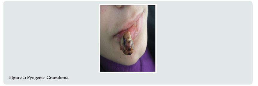
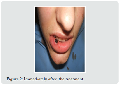
Two patients with PG were examinate in this study. They were treated with the 980 nm diode laser at the University of Tirana Dental School . An initial clinical examination consisting of the medical and dental history and a thorough extra and intra oral examination was performed . A complementary blood test , complete blood count and erythrocyte sedimentation rate test made it possible to exclude infectious diseases. The patients have no systemic complaints. The data collected was evaluated and a clinical diagnosis for the type of lesion was established Figure 1. Patients were given written and verbal information on the nature of laser treatment, and he signed informed consent forms were obtained prior to the treatment . The 980 nm diode laser was applied using the following parameter - continuous wave, 300 micrometer optical fiber and 3, 5 w power. The treatment was conducted under infiltration anesthesia . The treatment area was cooled by the application of ice for two to five minutes post operatively. All wounds were left open to heal by granulation and secondary epithelization, therefore no sutures were required (Figure 2). After the treatment analgesic medication was prescribed to be taken as required ,but no antibiotics were prescribed [11]. The follow-up visits were scheduled at ten days, one month, six months and one year after surgery. All of the lesions were photographically documented at all stages of treatment and healing.
Results
Post operatively there was no bleeding or pain. The power setting of 3, 5 w in continuous mode and focused contact handpiece appeared to lead to some superficial tissue necrosis associated with delayed wound healing. The wounds were completely healed after 3 to 4 weeks without scar formation and pigmentary changes (Figure 3). The cosmetic effect of laser therapy was evident. No recurrence was observed in the patients who were followed up one year after the surgery [12-15].
Discussion
Aesthetic treatment PG consists of the removal of the lesion. Current treatment modalities include chemical cauterization and surgical excision. However, these methods do not exclude the risk of complications such as scarring or pigmentary changes . These complications may lead to aesthetic problems for patients. Who accept the surgical removal of PG only with great difficulty. The conventional lip shape procedure is often painful , results insignificant bleeding and poses cosmetic concerns. Laser treatment of the lip does not compromise the importance of the lip , its discrete anatomic borders such as the vermilion border or its functionality. Various laser devices have been successfully used to treat PG including the 585 and 595 pulsed dye laser , the 1064 nm Nd. YAG and dioxide carbon laser. The pulsed dye laser is safe it can be used for lesions.
Conclusion
Treatment with 980 nm diode laser is a viable treatment option. The 980 nm diode laser beam is well absorbed by haemoglobin . The excision was well performed and suturing after surgery was not necessary because of good coagulation. The surgical period was significantly reduced. From the goods results obtained it was concluded that the application of 980 nm diode laser in the treatment of PG appears to be of beneficial effect.
References
- Jose Y, Antonio-Jesus E, Leonardo B, Cosme G (2009) Treatment of oral mucocele–scalpel versus CO2 Med Oral Patol Cir Bucal 1(9): 469-474.
- Ata J, Carrillo C, Bonet C, Balaguer J, Penarrocha M, et al. (2010) Oral mucocele: rewiew of the literature J Clin Exp Dent p. 18-21.
- Baurmash HD (2003) Mucoceles and ranulas. J Oral Maxillofacial Surgery 61: 369-378.
- Baurmash H (2002) The etiology of superficial oral mucoceles. J Oral Maxillofac Surg 60: 237-238.
- Jinbu Y, Kusama M, Itoh H, Matsumoto K, Wang J, Noguchi T (2003) Mucocele of the glands of Blandin-Nuhn clinical and histopathologic analysis of 26 cases. Oral Surg oral Med Oral Pathol Endod 95: 467-470.
- Bhaskar SN, Bolden TE, Weinmann JP (1956) Pathogenesis of mucoceles. J Dent Res 35: 863-874.
- Silva A Jr, Nikitakis NG, Balciunas BA, Meiller TF (2004) Superficial mucocele of the labial mucosa: a case report and review of the literature. Gen Dent 52: 424-427.
- Huang IY, Chen CM, Kao YH, Worthington P (2007) Treatment of mucocele of the lower lip with carbon dioxide laser. J Oral Maxillofac Surg 65: 855-858.
- Yamasoba T, Tayama N, Syoji M, Fukuta M (1990) Clinicostatistical study of lower lip mucoceles. Head Neck 12: 316-320.
- Romanos G, Nentwig G (1999) Diode laser 980 nm in Oral and Maxillofacial Surgical procedures Clinical observations Based on Clinical Applications. Journal of Clinical Laser Medicine and Surgery 17(5): 193-197.
- Romanos G (2001) Der Laser in der ChirurgieEinsteiger–Handbuch p. 52-53.
- Pick RM, Colvard MD (1962) Current status of Lasers in soft tissue dental surgery. J Periodontol 63: 589-602.
- Vitale M, Caprioglio C (2010) Lasers in dentistry. Practical textbook pp. 243-253.
- Gregnanin P, Cunha V, Hiramatsu L, Correa L (2010) Treatment of Mucocele of the lower lip with diode laser in pediatric patients: Presentation of 2 Clinical cases. Pediatric Dentistry 32(7): 539-541.
- Neckel C, Neustadt B (2012) Der Erbium: YAG und Diodenlaser in der aesthetisch–kosmetischen Zahnmedizin Jahrbuch Laser zahnmedizin p. 85-89.
Editorial Manager:
Email:
pediatricdentistry@lupinepublishers.com

Top Editors
-

Mark E Smith
Bio chemistry
University of Texas Medical Branch, USA -

Lawrence A Presley
Department of Criminal Justice
Liberty University, USA -

Thomas W Miller
Department of Psychiatry
University of Kentucky, USA -

Gjumrakch Aliev
Department of Medicine
Gally International Biomedical Research & Consulting LLC, USA -

Christopher Bryant
Department of Urbanisation and Agricultural
Montreal university, USA -

Robert William Frare
Oral & Maxillofacial Pathology
New York University, USA -

Rudolph Modesto Navari
Gastroenterology and Hepatology
University of Alabama, UK -

Andrew Hague
Department of Medicine
Universities of Bradford, UK -

George Gregory Buttigieg
Maltese College of Obstetrics and Gynaecology, Europe -

Chen-Hsiung Yeh
Oncology
Circulogene Theranostics, England -
.png)
Emilio Bucio-Carrillo
Radiation Chemistry
National University of Mexico, USA -
.jpg)
Casey J Grenier
Analytical Chemistry
Wentworth Institute of Technology, USA -
Hany Atalah
Minimally Invasive Surgery
Mercer University school of Medicine, USA -

Abu-Hussein Muhamad
Pediatric Dentistry
University of Athens , Greece

The annual scholar awards from Lupine Publishers honor a selected number Read More...


