Lupine Publishers Group
Lupine Publishers
Menu
ISSN: 2637-6628
Case Report(ISSN: 2637-6628) 
Swiss Cheese Pattern a Harbinger of Dementia or an Incidental Finding in an Unusual Case? Volume 3 - Issue 4
Faheem Arshad*
- Department of Neurology, National Institute of Mental Health and Neurosciences (NIMHANS), India
Received: December 20, 2019; Published: January 09, 2020
Corresponding author: Faheem Arshad, Department of Neurology, National Institute of Mental Health and Neurosciences (NIMHANS), Bengaluru, India
DOI: 10.32474/OJNBD.2020.03.000170
Abstract
Dilated Virchow Robin (VR) spaces are pial- line fluid filled structures which surround the walls of small penetrating vessels. In a severe form they develop a swiss cheese pattern or a cribriform pattern in straitum which may predispose to cognitive impairment. We report a patient with change in personality associated with diffuse atrophy, hypometabolism, microbleeds and swiss cheese striatum which is rare.
Keywords: Swiss cheese pattern; Dementia; Virchow robin space
Abbreviations: VR: Virchow Robin; MRI: Magnetic Resonance Imaging; FLAIR: Fluid Attenuated Inversion Recovery; CSF: Cerebrospinal Fluid
Introduction
Virchow Robin (VR) spaces are pial-lined, fluid filled,
structures which surround the walls of small penetrating arterioles
and venules as they course from subarachnoid space to brain
parenchyma. These often appear in basal ganglia and centrum
semi vale and are reflected in Magnetic Resonance Imaging(MRI)
brain as hypointense in T1-weighted images, hyperintense on
T2-weighted and hypointense on Fluid Attenuated Inversion
Recovery(FLAIR) images, thus distinguished from pathological
white matter lesions by persistent is intensity to Cerebrospinal
Fluid(CSF) on all sequences, lack of enhancement and sharply
defined margins. Dilated VR spaces can appear on neuroimaging
as single enlarged cavity(up to 2cm in diameter) or may appear as
hundreds of bilateral 1-2 mm foci in the basal ganglia, subcortical
white matter and sub insular area lateral to lentiform nucleus, a
pattern sometimes referred to as etat crible or cribriform or Swiss
cheese striatum.
Though a common finding in elderly population, studies have
shown that enlarged VR spaces are a marker of small vessel disease
and associated with incident dementia and depression. Multiple
mechanisms for giant VR spaces have been described which include
mechanical trauma due to CSF pulsation, fluid exudation due to
abnormalities of the vessel wall abnormality and ischemic injury
to perivascular tissue causing ex vacuo effect. The precise function
of VR spaces is not completely understood. They are believed to
serve as a lymphatic of brain also known as the glymphatic system
whereby CSF exchanges with the interstitial fluid within the brain
parenchyma, including clearing the interstitial solutes such as betaamyloid.
Epidemiology
Dilated Virchow robin spaces were described Durant-Fardel in 1843. In a study of healthy participants using high resolution images prevalence was 1.6%. In radiological studies involving patients VR spaces were found in 3% patients under 20 years of age. In addition, studies have shown higher rate of cognitive decline with dilated VR spaces, which is intriguing, and its elucidation may improve the complex understanding of role of vascular alterations and higher risk of cognitive decline [1]. Furthermore, dilated VR spaces have different topographically patterns in microangiopathies like cerebral amyloid angiopathy where they are mostly seen in centrum semiovale and hypertensive angiopathy where they are predominantly seen in basal ganglia [2]. To date, longitudinal data with regard to significance of dilated VR spaces in healthy older adults is scarce. However small number of studies have shown association with development of new onset dementia. Prevalence is more in vascular dementia compared to Alzheimer’s dementia and healthy controls.
Clinical presentation
Increased basal ganglia or centrum semiovale perivascular spaces have been associated with worse nonverbal reasoning and visuospatial cognitive abilities. Various clinical presentations have been reported which include parkinsonism, hemisensory symptoms and in addition depending on the size of the VR space causing mass effect. Dilated VR spaces have been associated with vascular dementia and also have been correlated with reduced cognitive function [3]. Thus for a clinician differential diagnosis of dilated spaces should always be considered in view of multiple mimics like multiple lacunar infarctions, cryptococcosis, multiple sclerosis, mucopolysaccharidosis, cystic neoplasms and arachnoid cysts. Knowledge of their signal changes in neuroimaging may help in differentiating these lesions from dilated VR spaces.
Clinical case
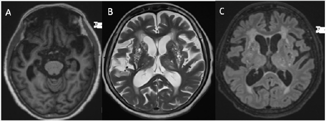
Figure 1: (A) T1-weighted MRI showed bilateral frontal and anterior temporal lobe atrophy; (B, C) T2/FLAIR hyperintensities showing confluent foci in the bilateral deep and periventricular white matter, and prominent perivascular spaces in bilateral basal ganglia suggestive of a swiss cheese pattern.

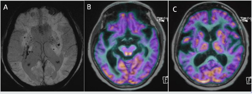
Figure 2: (A) SWI sequence showing multiple foci of blooming in bilateral basal ganglia, thalami, cerebral hemispheres, brainstem and left cerebellar hemisphere. (B) Positron emission tomography (PET) MRI brain showing hypometabolism in bilateral parietal lobes, medial and anterior temporal lobes and orbitofrontal cortex. (C) PET MRI brain showing hypometabolism in dilated VR spaces in the striatum.

60 year old female, presented with the 5 years history of change in personality associated with behavioural disturbances in the form of getting angry for trivial issues and screaming at family members, trying to pick things in front of her, telling that insects are crawling on her clothes. Since last 2 years caregivers report history of fluctuating restlessness, wandering aimlessly, muttering to self, disinhibited behaviour and apathy. Since last 6 months she had lost concern for family members, started becoming slow in her activities especially walking and became completely dependent for her daily activities of living. There was also history of incontinence without any concern. There was no history of food faddism, utilization behaviour, myoclonic jerks, seizure, recurrent falls, bulbar symptoms, weight loss, change in bowel habits. Patient was diagnosed case of hypertension since 5 years and was on medications. She was not cooperative for mental status examination. Her physical examination revealed mild bradykinesia, brisk reflexes, mildly wide based gait with reduced clearance and primitive reflexes were present. Blood investigation were all normal. MRI brain T1-weighted image (Figure 1A) showed bilateral frontal and anterior temporal lobe atrophy, confluent foci of T2/FLAIR hyperintensities (Figure1B and C) in the bilateral deep and periventricular white matter and T2 weighted image showed prominent perivascular spaces in bilateral basal ganglia suggestive of a swiss cheese pattern. In addition, there were multiple foci of blooming on Susceptible Weighted Images (SWI) (Figure 2A) are seen in the bilateral basal ganglia, thalami, cerebral hemispheres, brainstem and left cerebellar hemisphere. Positron Emission Tomography (PET) MRI brain (Figure 2B) showed hypometabolism in bilateral parietal lobes, medial and anterior temporal lobes and orbitofrontal cortex. PET MRI brain (Figure 2C) showed hypometabolism in dilated VR spaces in the striatum. Final diagnosis of mixed dementia associated with swiss cheese brain syndrome was considered and patient was managed as per standard guidelines for dementia. In addition to Vascular dementia, more commonly mixed dementia includes Alzheimer’s dementia, however patient had predominant features suggestive of Frontotemporal dementia which less commonly associated with microbleeds and dilated VR spaces as seen in this patient. Whether it is a combination of multiple dementias contributed by enlarged VR spaces will remain unanswered.
Conclusion
Dilated VR spaces may be an either an incidental finding on MRI without any pertinent manifestations. However, evidence suggests that these may be predisposing risk factor for cognitive impairment. Whether estimating the burden of perivascular spaces may help in predicting development of cognitive impairment and type of dementia, remains undetermined and may require further research.
References
- Zhu YC, Dufouil C, Soumare A, Mazoyer B, Chabriat H, et al. (2010) High degree of dilated virchow-robin spaces on mri is associated with increased risk of dementia. J Alzheimers Dis22(2): 663-672.
- Charidimou A, Boulouis G, Pasi M, Auriel E, van Etten ES, et al. (2017) MRI-visible perivascular spaces in cerebral amyloid angiopathy and hypertensive arteriopathy. Neurology88(12):1157-1164.
- Burnett MS, Witte RJ, Ahlskog JE (2014) Swiss cheese striatum: Clinical implications. JAMA neurology71(6):735-741.

Top Editors
-

Mark E Smith
Bio chemistry
University of Texas Medical Branch, USA -

Lawrence A Presley
Department of Criminal Justice
Liberty University, USA -

Thomas W Miller
Department of Psychiatry
University of Kentucky, USA -

Gjumrakch Aliev
Department of Medicine
Gally International Biomedical Research & Consulting LLC, USA -

Christopher Bryant
Department of Urbanisation and Agricultural
Montreal university, USA -

Robert William Frare
Oral & Maxillofacial Pathology
New York University, USA -

Rudolph Modesto Navari
Gastroenterology and Hepatology
University of Alabama, UK -

Andrew Hague
Department of Medicine
Universities of Bradford, UK -

George Gregory Buttigieg
Maltese College of Obstetrics and Gynaecology, Europe -

Chen-Hsiung Yeh
Oncology
Circulogene Theranostics, England -
.png)
Emilio Bucio-Carrillo
Radiation Chemistry
National University of Mexico, USA -
.jpg)
Casey J Grenier
Analytical Chemistry
Wentworth Institute of Technology, USA -
Hany Atalah
Minimally Invasive Surgery
Mercer University school of Medicine, USA -

Abu-Hussein Muhamad
Pediatric Dentistry
University of Athens , Greece

The annual scholar awards from Lupine Publishers honor a selected number Read More...


