Lupine Publishers Group
Lupine Publishers
Menu
ISSN: 2637-4722
Case Report(ISSN: 2637-4722) 
Greig Syndrome: A Rare Disease - Case Report Volume 2 - Issue 1
Anwar A Mithwani1*, Adnan A Mithwani2, Muhammad Ziad Shama3, Assem Ahmad Kadrey3, Abdullah Alomar Almeshrif3 and Amerullah Malik4
- 1Consultant Pediatrician/Neonatologist, Maternity and Children Hospital, Al Kharj, Saudi Arabia
- 2Rockyview General Hospital, Internal Medicine, Calgary AB, Canada
- 3Neonatologist, Maternity and Children Hospital Al Kharj, Saudi Arabia
- 4Consultant Neonatologist, Maternity and Children Hospital, Al Kharj, Saudi Arabia
Received:January 21, 2019 Published: January 25, 2019
Corresponding author: Anwar A Mithwani, Consultant Pediatrician/Neonatologist, Maternity and Children Hospital, Al Kharj, Saudi Arabia
DOI: 10.32474/PAPN.2019.02.000129
Abstract
Grieg cephalopolysyndactyly syndrome (GCPS) is a rare congenital genetic disorder present at birth, characterized through physical abnormalities, primarily affecting the development of the limbs, head, and face (craniofacial malformations). We report a case of a full term, newborn female, large for gestational age with 4.2 kg. Symptoms included transient tachypnea of the newborn and craniofacial dysmorphism. The mother was G5P4 with gestational diabetes, and a reported case of polyhydramnios but did not have any previously affected babies. The craniofacial dysmorphism anomalies consisted of macrocephaly (Head circumference of 39 cm with a standard deviation of 2+ for her age), frontal bossing with a broad forehead, a groove between the frontal bone, widely spaced eyes, bilateral polysyndactyly, a thumb and an extra digit fused bilaterally in the toes. However, imaging such as CXR & skeletal survey were normal. Her tachypnea partially improved and only occurred intermittently during feeds, maintained her vital signs well and remained at room air, feeding on demand. Consults with neurology, ENT and ophthalmology specialists in hospital did not add any new findings. She was then discharged from the hospital with referral to genetics to assess the child and counsel the family as an outpatient.
Background
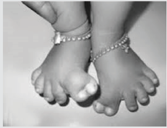
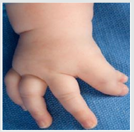
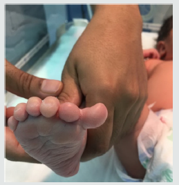
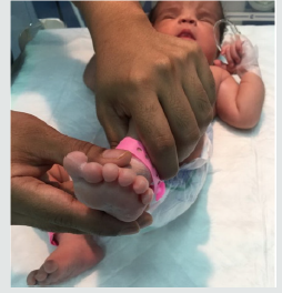
Typical Grieg cephalopolysyndactyly syndrome (GCPS) is characterized by a preaxial polydactyly or a mixed pre- and/or postaxial polydactyly, true wide spaced eyes, and macrocephaly. Individuals with mild GCPS may have subtle craniofacial findings. The mild end of the GCPS spectrum is a continuum with preaxial polysyndactyly type IV and crossed polydactyly (preaxial polydactyly of the feet and postaxial polydactyly of the hands plus syndactyly of fingers 3-4 and toes 1-3). Individuals with severe GCPS may have seizures, hydrocephalus and intellectual disability. This condition israre and its prevalence is unknown. Grieg syndrome has a wide range of varieties depending on the Mutations in the GLI3 gene related to chromosome 7, and the presence of continuous intermittent tachypnea did not report as a primary fetcher. Certain cranial sutures may close prematurely (craniosynostosis). Such irregular closure of the sutures may cause the head to appear shaped abnormally (scaphocephaly, trigonocephaly, or plagiocephaly). Rarely, less than 10% of affected individuals may have more serious medical problems including seizures, delayed development, and intellectual disability, build-up of fluid inside the skull (hydrocephalus), and abnormalities affecting the nerve fibers (corpus callosum) that connect the two cerebral hemispheres of the brain, may be present (Figures 1 & 2).
Case Presentation
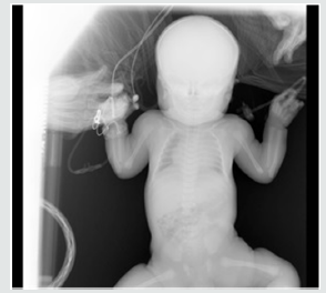
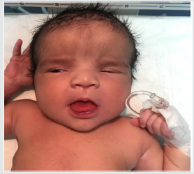
A full term, newborn female, large for gestational age weighing 4.2kg. Her symptoms included transient tachypnea of the newborn and craniofacial dysmorphism. The mother was a G5P4 with gestational diabetes and polyhydramnios with a history of shoulder dystocia in her previous baby, which was delivered by caesarean section. The Apgar score was 7 at 1 minute and 9 at 5 minutes. The craniofacial dysmorphism anomalies consisted of macrocephaly (Head circumference of 39 cm with a standard deviation of 2+ for her age), frontal bossing with a broad forehead, a groove between the frontal bone, widely spaced eyes, bilateral polysyndactyly, extra thumb, and an extra digit fused bilaterally in the toes. The parents did not have a family history of any reported inherited diseases. At birth, the baby was distressed, tachypneic with subcostal & intercostal retractions. Therefore, she was managed in Neonatal intensive care unit in an incubator with suitable heat, meticulous cardiorespiratory monitoring. She was kept NPO, provided with fluid resuscitation, supplemental oxygen. Neonatal sepsis was ruled out though sepsis work-up. Later nasogastric tube feeding was initiated and 5 days later was transitioned to oral feeding gradually as tolerated. However, she continued to have intermittent tachypnea which appeared to improve gradually but was still evident during & after her feeds. This continued until her discharge, which was on Day-37 (Figures 3-6).
Investigations
Imaging such as skeletal survey, Abdominal ultrasound, transfontanelle ultrasonography and a CT brain were done which were all normal except for macrocephaly without any hydrocephalus. Blood work included CBC, extended electrolyte panel and sepsis work-up including blood cultures were all negative. Echocardiography was also done to check for any valvular abnormalities but did not show any anomalies. Consultation with specialists in neurology, ophthalmology and ENT did not add any other findings. Although, genetic counseling was recommended but there were no facilities available for this and therefore, mutation analysis could not be performed.
Treatment
Managed in the neonatal intensive care unit in an incubator with suitable heat and meticulous cardiorespiratory monitoring. She was kept NPO, provided with fluid resuscitation, supplemental oxygen. Surgical consultation was done for elective repair of polydactyly with follow-up with the outpatient department.
Outcome and Follow-Up
The baby was treated in the NICU for 30 days for intermittent tachypnea especially during & after feeds. She was then shifted to PICU according to our admission and discharge policy, managed there for 8 days and was then discharged on breast feeding with proper education of the parents, and referral for Outpatient Department appointment follow-up for further surveillance. This was done to monitor for any changes in the occipitofrontal circumference (OFC) and continuous evaluation and treatment as needed for developing hydrocephalus or any other CNS abnormalities showing signs of increased intracranial pressure, developmental delay, loss of milestones, and/or seizures. On followup with neurology, the growth, and rest of the neurological workup remained normal.
Discussion
Grieg cephalopolysyndactyly syndrome (GCPS) is a rare
congenital genetic disorder present at birth, characterized through
physical abnormalities, primarily affecting the development of the
limbs, head and face (craniofacial malformations). The features
of this syndrome are highly variable, ranging from mild to severe.
Patients with this condition typically have one or more extra
fingers or toes (polydactyly), or an abnormally wide thumb, or
a big toe (hallux). The skin between the fingers and toes may be
fused (cutaneous or osseous syndactyly). In some individuals, the
syndrome may include permanently flexed fingers (camptodactyly),
dislocation of the hip, protrusion of a portion of the large intestine
through an abnormal opening in the muscular wall that lines the
lower abdominal cavity (inguinal hernia). This disorder is also
characterized by an abnormally large head size (macrocephaly), and
a high, prominent or protruding forehead (frontal bossing), high
anterior hairline; a broad nasal bridge; and/or widely spaced eyes
(ocular hypertelorism). In some cases, the fibrous joints (sutures)
between certain bones in the skull may be abnormally wide and
may close unusually late in development; but, in rare individuals,
certain cranial sutures may close prematurely (craniosynostosis).
Such irregular closure of the sutures may cause the head to
appear unusually shaped (scaphocephaly, trigonocephaly, or
plagiocephaly). Rarely, less than 10% of affected individuals may
have more serious medical problems including seizures, delayed
development, and intellectual disability, build-up of fluid inside the
skull (hydrocephalus), and abnormalities affecting the nerve fibers
(corpus callosum) that connect the two cerebral hemispheres of the
brain may be present.
We have to consider these related disorders:
a. Acrocallosal syndrome (ACLS): A rare genetic disorder
typically inherited in an autosomal recessive pattern.
b. Oro-facial-digital syndrome: A rare genetic disorder
includes split tongue, splits in the jaw, midline cleft lip.
c. Pfeiffer syndrome (acrocephalosyndactyly type V) is
generally accepted to be the same as Noack syndrome inherited
in an autosomal dominant pattern.
Mutations in the GLI3 gene related to chromosome 7 can
cause Grieg cephalopolysyndactyly syndrome In most cases. This
gene provides instructions for making a protein that controls gene
expression, which plays a role in the normal shaping or patterning
of many organs and tissues before birth, so it is inherited in an
autosomal dominant pattern from either parent, or in rare instances
of this disorder are sporadic, and occur in people with no history of
the condition in their family result of a new mutation (gene change)
in the affected individual. The risk of passing the abnormal gene
from an affected parent to an offspring is 50% for each pregnancy
regardless of the gender of the child. In most individuals with the
severe form of the disorder, it is caused by a deletion of the entire
GLI3 gene. The larger the deletion encompassing GLI3 gene is
(greater than 300 kb), the more likely the individual will show
these uncommon symptoms.
Learning Points
a) The diagnosis of GCPS is based on clinical findings and
family history.
b) An Enlarged skull (macrocephaly) in ultrasound imaging
during a pregnancy have to be considered.
c) The diagnosis at birth is based upon clinical evaluation,
identification of characteristic physical findings. (Head
circumference greater than a 97th centile, distance between the
pupils).
d) Procedures including X-rays and transfontanyl ultrasound
and CT scanning reveals the extent of bone fusion in several
occurrences of osseous syndactyly.
e) In future significant development delay or intellectual
disability should have genetic testing through sequence
analysis to detect possible changes in GLI3 gene.
f) Treatment may require the efforts of a team of such as
pediatricians, orthopedists, plastic surgeons, physical and
occupational therapists, and/or other health care professionals.
References

Top Editors
-

Mark E Smith
Bio chemistry
University of Texas Medical Branch, USA -

Lawrence A Presley
Department of Criminal Justice
Liberty University, USA -

Thomas W Miller
Department of Psychiatry
University of Kentucky, USA -

Gjumrakch Aliev
Department of Medicine
Gally International Biomedical Research & Consulting LLC, USA -

Christopher Bryant
Department of Urbanisation and Agricultural
Montreal university, USA -

Robert William Frare
Oral & Maxillofacial Pathology
New York University, USA -

Rudolph Modesto Navari
Gastroenterology and Hepatology
University of Alabama, UK -

Andrew Hague
Department of Medicine
Universities of Bradford, UK -

George Gregory Buttigieg
Maltese College of Obstetrics and Gynaecology, Europe -

Chen-Hsiung Yeh
Oncology
Circulogene Theranostics, England -
.png)
Emilio Bucio-Carrillo
Radiation Chemistry
National University of Mexico, USA -
.jpg)
Casey J Grenier
Analytical Chemistry
Wentworth Institute of Technology, USA -
Hany Atalah
Minimally Invasive Surgery
Mercer University school of Medicine, USA -

Abu-Hussein Muhamad
Pediatric Dentistry
University of Athens , Greece

The annual scholar awards from Lupine Publishers honor a selected number Read More...


