Lupine Publishers Group
Lupine Publishers
Menu
ISSN: 2637-4722
Short Communication(ISSN: 2637-4722) 
Detecting Ehlers-Danlos Syndrome Early in Life Is an Urgent Priority Volume 2 - Issue 3
Hamonet C1*, Ducret L2 and Bahloul H3
- 1Department of medicine, University Paris-East-Creteil [UPEC] PRM, France
- 2Department of Center of diagnosis ELLAsante, Paris
- 3Department of Integrative medicine system, France
Received: August 08, 2019 Published: August 19, 2019
Corresponding author: Hamonet C, Department of medicine, University Paris-East-Creteil [UPEC] PRM, France
DOI: 10.32474/PAPN.2019.02.000139
Abstract
Ehlers-Danlos syndrome is a frequent hereditary disease that affects all the connective tissue and is transmitted to all the children of an affected person. A diagnosis is possible, from birth, or shortly thereafter, when observing a clubfoot, hip dislocation, intestinal intussusception, acute umbilical or inguinal parietal hernia, hemorrhages [cutaneous, oral, gastric, intestinal, nasal], persistent constipation, regurgitation and vomiting during bottle-feeding, or false roads. These symptoms are often the cause of false allegations of abuse with children withdrawal and wrongful parents or false accusations of Munchausen disease “by delegation” in a parent, the mother most often.
Keywords: Ehlers-Danlos; hereditary disease; early diagnosis; ecchymosis; fractures; children abuse; hypemobility
Introduction
Ehlers-Danlos syndrome is a frequent [1] hereditary disease that affects all the connective tissue and is transmitted to all the children of an affected person. It’s ancient [2,3] description by Tschernogobow, Moscow, 1881 and Ehlers, Copenhagen, 1900 has undergone an important clinical and pathophysiological update for the last 20 years. A reliable diagnosis, made on clinical grounds alone, is now possible in the absence of a biological marker. It is characterized by tissue fragility, including bones, diffuse and generalized dysproprioception. Most doctors remain unaware of this condition, exposing children to iatrogenic accidents and parents to false accusations of child abuse [4]. This goes to show the importance of knowing how to carry out this diagnosis from the beginning of the child’s life.
Reminder of Diagnostic Criteria
Confusion can be observed in the descriptions available to
doctors and patients: new communication technologies allow
everyone to have access to all information and to express, without
control, all kinds of opinions. This situation is particularly
devastating in Ehlers-Danlos disease, which has not been taught
to doctors, and is expressed by mainly subjective signs, of which
anxiety and imaginative creativity are part. Clinicians [5-9] have
progressively regrouped, on large series of patients, clinical
criteria which, by their singular association, allow the diagnosis:
intense fatigue, diffuse pain, disruption of motor control by
dysproprioception and dystonia, skin fragility, joint hypermobility,
hemorrhage, hyperacusis, gastric reflux, autonomic dysfunction.
These clinical signs’ expression is variable evolving on a permanent
background with episodes of crisis triggered by activity, trauma,
hormonal factors (pregnancies, periods, menopause), climate
change. Paucisymptomatic forms are identified by the presence in
the family of a more complete clinical picture. The evolution, in the
long term is very variable but an accentuation in adulthood is often
observed.
Which signs should trigger an Ehlers-Danlos Syndrome
diagnosis in a newborn or a very young child?
A diagnosis is possible, from birth, or shortly thereafter, when
observing a clubfoot, hip dislocation, intestinal intussusception,
acute umbilical or inguinal parietal hernia, hemorrhages (cutaneous,
oral, gastric, intestinal, nasal), persistent constipation, regurgitation
and vomiting during bottle-feeding, or false roads, intolerance to
milk, very flexible joints, dislocated seats (shoulders, hips, finger
knees) or joint crunches, particularly thin skin, irritable and easy to stretch, injury long or difficult to treat. Sometimes, frequent crying,
evokes abdominal pain. Occurrence of “spontaneous fracture”
for minimal trauma, epiphyseal or diaphyseal, costal but also
cranial should suggest, before any other diagnosis, Ehlers-Danlos
syndrome, because of its significant incidence which is estimated
at 2% [1]. The cause may be low vitamin D level present in this
pathology and possibly related to insufficiency of its manufacture
by a pathological skin. Cerebral lesions can also be observed due
to the lack of brain protection by pathological meninges and the
shock waves’ violence transmitted by overly lax tissues, as we have
shown, with Professor Daniel Fredy with diffusion tensor MRI [10]
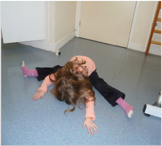
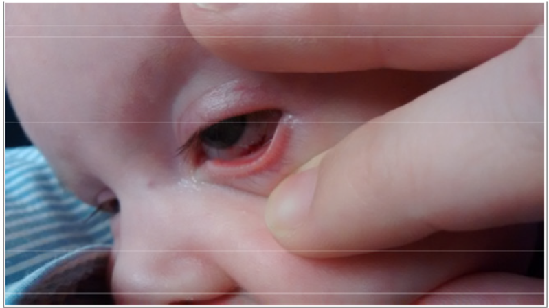
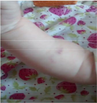
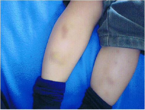
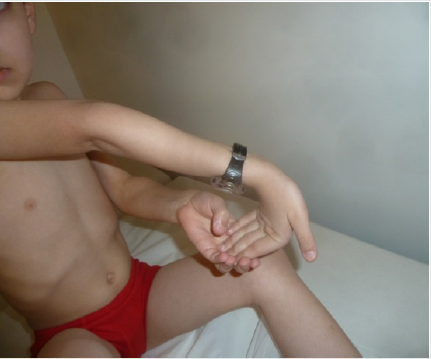
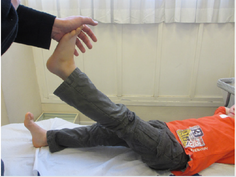
(Figures 1-7).
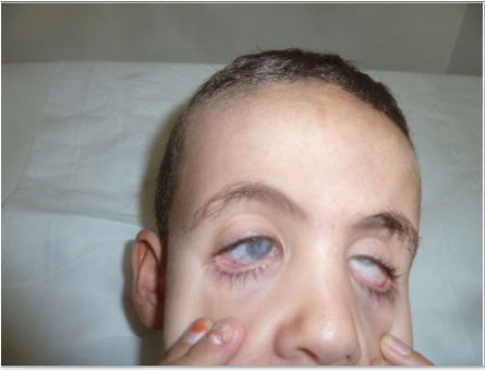
Figure 3: 12 years old boy with Ehlers-Danlos syndrome. Totally blind because of Sclerotic Necrosis of both eyes after Trauma and infection when he was Two years old
Fractures, hematoma and cerebral lesions are often the cause of false allegations of abuse with children withdrawal and wrongful parents’ convictions with psycho-emotional consequence. Serious adverse impact is also medical because of concerned children’s lack of treatment. We must associate the false accusations of Munchausen disease “by delegation” in a parent, the mother most often [11].
Conclusion
Ehlers-Danlos is most likely, in the history of medicine, the disease that despite (or because of?) Its frequency estimated at 2% [1] remains the most ignored and is the cause of the greatest number of misdiagnosis. This is what Rodney Grahame [11] says when he writes: “It is incomprehensible that most doctors, specialized or not, do not detect it, do not know how to diagnose it, or, of course, how to treat it. Many, even, deny its existence, often causing the patient and his family dramatic and unnecessary suffering.” The decisive diagnostic argument is brought by the demonstration of clinical signs of certainty in one of the parents, since this disease is systematically transmitted to all children. They must be searched consistently whenever one or more of the guidance signs are discovered, especially if parental abuse is suspected.
References
- Tinkle B, Castori M, Berglund B, Cohen H, Grahame R, et al. (2017) Hypermobile Ehlers-Danlos Syndrome: Clinical description and natural history. Am J Med Genet Part C Semin Med Genet 175(1): 48–69.
- Tschernogobow N A (1892) über einen Fall von Cutis Lockering laxa first meeting of Moscow Dermatologic and venerologic society. Monatshefte für paraktsche Dermatologie, Hamburg, 14: 76.
- Ehlers E. Cutis laxa, Neigung zu Haememorragien in der Haut, lockering mehrerer Articulationen, Dermatologissche Zeitschrift.
- Holick MF, Hossein-Nezhad, Tabatabaei F (2017) multiple fractures in infants who have Ehlers-Danlos/hypermobility syndrome and or vitamine D deficieciency. A case series of 72 infans whose parents were accused of child abuse and neglect. DermatoEndicrinology 9(17).
- Grahame R, Bird HA, Child A (2000) The revised (Brighton 1998) criteria for thr diagnosis of benign joint hypermobilty syndrome (BJHS). J Rheumatology 27: 1777-1779.
- Hamonet C, Ravaud PH (2012) (all Ehlers-Danlos about 664 cases). Statistical analysis of clinical signs from 644 patients with a Beighton scale > 4/9. First international Symposium on the Ehlers-Danlos syndrome. September 8-9 Ghent.
- Chopra P, Tinkle B, Hamonet C, Brock I, Gompel A, et al. (2017) Pain management in the Ehlers–Danlos syndromes. American Journal of Medical Genetics, Part C Seminars in Medical Genetics 175(1).
- Marino Lamari N (2017) Systemic Manifestations of Ehlers-Danlos Syndrome Hypermobility Type. MOJ Cell Science & Report 4(2): 9–13.
- Hamonet C, Frédy D, Lefèvre J H, Bourgeois Gironde S, Zeitoun J D (2016) Brain injury unmasking Ehlers-Danlos syndromes after trauma: The fiber print. Orphanet Journal of Rare Diseases 11: 45
- Hamonet C, Jouvencel M, Manicourt D, Holick M (2019) Ehlers-Danlos et fausses accusations de maltraitances. Gaz. Pal. 5 mars, n° GPL343y8, p. 19
- Hamonet C, Ehlers-Danlos (2018) The disease forgetten by medicine, l'Harmattan.

Top Editors
-

Mark E Smith
Bio chemistry
University of Texas Medical Branch, USA -

Lawrence A Presley
Department of Criminal Justice
Liberty University, USA -

Thomas W Miller
Department of Psychiatry
University of Kentucky, USA -

Gjumrakch Aliev
Department of Medicine
Gally International Biomedical Research & Consulting LLC, USA -

Christopher Bryant
Department of Urbanisation and Agricultural
Montreal university, USA -

Robert William Frare
Oral & Maxillofacial Pathology
New York University, USA -

Rudolph Modesto Navari
Gastroenterology and Hepatology
University of Alabama, UK -

Andrew Hague
Department of Medicine
Universities of Bradford, UK -

George Gregory Buttigieg
Maltese College of Obstetrics and Gynaecology, Europe -

Chen-Hsiung Yeh
Oncology
Circulogene Theranostics, England -
.png)
Emilio Bucio-Carrillo
Radiation Chemistry
National University of Mexico, USA -
.jpg)
Casey J Grenier
Analytical Chemistry
Wentworth Institute of Technology, USA -
Hany Atalah
Minimally Invasive Surgery
Mercer University school of Medicine, USA -

Abu-Hussein Muhamad
Pediatric Dentistry
University of Athens , Greece

The annual scholar awards from Lupine Publishers honor a selected number Read More...


