Lupine Publishers Group
Lupine Publishers
Menu
ISSN: 2637-6628
Case Report(ISSN: 2637-6628) 
Successful Surgical Treatment of Arnold-Chiari Malformation Combined with a Pontocerebellar Cyst and Unilateral Hearing Loss Volume 4 - Issue 1
Shamil Safin1,2, Ilmira Gilemkhanova1,3, Khristina Derevyanko1,4 and S Muruga Subramaniam1,5,6*
- 1Department of Neurosurgery and medical rehabilitation with an ICPE (Institute of Continuing Professional Education) course, Bashkir State Medical University, Russia
- 2Department of Neurosurgery, GG Kuvatov Republican Clinical Hospital, Russia
- 3Department of Neurosurgery, Republican Children’s Clinical Hospital, Russia
- 4Department of Neurology, Clinic of Bashkir State Medical University, Russia
- 5Departments of Surgery, Division of Neurotrauma, Malaysia
- 6Melaka Manipal Medical College, Manipal University, Malaysia
Received: May 15, 2019; Published: June 04, 2020
Corresponding author: S Muruga Subramaniam, Department of Neurosurgery and medical rehabilitation with an ICPE (Institute of Continuing Professional Education) course, Bashkir State Medical University, UFA, Russia
DOI: 10.32474/OJNBD.2020.04.000178
Abstract
Background: Cochlear dysfunction, is considered one of the difficult topical and important areas in modern otorhinolaryngological practice. Diagnosis and treatment of cochlear dysfunction is even more complicated in combined pathologies. There have been reported only few cases of a combined with Arnold-Chiari malformation with hearing impairment.
Case description: 34 years old male, was admitted in Republican Clinical Hospital G.G. Kuvatov under neurosurgical care. Patients main complaints was hearing loss associated with “tinnitus” on the left sided, morning headaches, dizziness, and unstable gait. The patient noted a gradual loss of hearing on his left ear over the years. He was diagnosed to have Arnold chiari malformation type 1 with combined hearing loss.
Conclusion: Good planned surgical strategies and operative techniques the hearing loss was restored, and his neurological symptoms was relieved.
Keywords: Arnold-Chiari malformation; Pontocerebellar cyst; Hearing impairment
Abbreviations and Acronyms: ACM: Arnold Chiari Malformation; MRI: Magnetic Resonance Imaging
Introduction
Arnold Chiari malformation (ACM) is a group of congenital malformations of the hindbrain development that affect structural relationships of the cerebellum, brain stem, supracervical spinal cord and base of skull [1]. Diagnosing ACM is usually with certain difficulties, since not all cases of cerebellar tonsils descent below the great occipital foramen with clinical manifestations. The clinical signs of ACM themselves are very polymorphic. M.A. Valiulin (1996), reported that the initial manifestations of type I ACM usually with cervical-occipital and facial pains, hearing impairments, dizziness, balance disorders, ataxia, facial hypesthesia, dysphagia, dysphonia, diplopia, nystagmus, arm pains and weakness in the limbs. In patients with type I ACM combined with myelosyringosis, the first symptoms of the disease appear later stage [2].
Some journals reported cases of hearing impairment in patients with type I ACM in its onset [3,4]. K.S. Paul et al. (1983) reported tinnitus in 9.9% of patients among 71 patients with type I ACM examined [5]. Studies done by R.E. Rydell, J.L. Pulec (1971) with 130 patients with Arnold Chiari type I, discovered bilateral progressive hearing impairment in 22.3% of patients [6]. We are reporting a clinical case of Arnold-Chiari malformation complicated by a pontocerebellar cyst and left-sided hearing loss.
Case Description
34 years old, male was admitted for treatment at Republican
Clinical Hospital G.G. Kuvatov with chief complaint of hearing
loss, “tinnitus” on the left sided, morning headache, dizziness,
and unstable gait. The patient noted a gradual loss of hearing in
the left ear over the years. No family history with similar illness.
Neurological examination noted convergent nystagmus, moderate
truncal ataxia, clinically with a combination of cerebellar ataxia.
Neuropsychological examination no cognitive impairment.
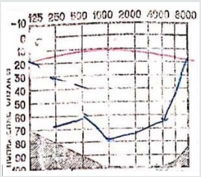
Patient underwent an audiometric test and noted a reduction
on the left ear with 65dB, and right side with normal readings.
We came up to a diagnosis of stage 4 left sided hearing loss with
bordering deafness.
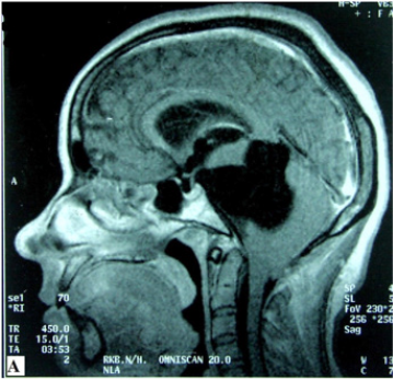
Magnetic resonance imaging (MRI) of the brain noted arnold
chiari malformation (ACM) with 10 mm descent of cerebellum
tonsils, with a dorsal compression at the level of the cervicalmedullary
junction, and a cyst at the left cerebellopontine angle
(Figure 1,2).
Diagnosis of Arnold-Chiari malformation with left-sided
pontocerebellar cyst was made. After a careful evaluation and
discussion with patient, surgical option of treatment was planned
after discussing with patient. Surgical option mainly to relieve the
nerve compression especially the 8th nerve by pontocerebellar cyst.
The patient underwent resection of cerebellum tonsils, with an
opening of cistern connecting the pontocerebellar cerebrospinal
fluid cavity and airtight fascioduraplasty.
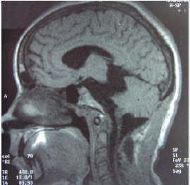
Postoperative MRI of the brain reveals regression of the left
cerebellopontine angle cyst (Figure 3,4). Dorsal compression is
relieved at the level of the cervical-medullary junction. During
the recovery period patient feels improvement in his left ear.
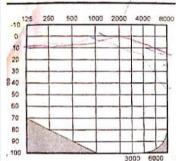
Audiometric test noted to have 30 dB, and diagnosis of stage 1 leftsided
hearing loss.
Discussion
The major discussion of ACM and intervention remain
debatable. In 1999 Milhorat TH et al. published data on prospective
analysis of 346 patients with Arnold-Chiari malformation of which
36% suffered from hearing loss.
They identified 4 possible mechanisms of damage of the eighth
pair of cranial nerves:
a) Nerve fibers tension as a result of brain stem descent.
b) Nuclei compression of the eighth pair of cranial nerves by
cerebellum tonsils.
c) Ischemic damage to the nuclei due to compression of the
cerebellar artery.
d) Cochlear damage, transmission of increased pressure of
cerebrospinal fluid through the abnormal cochlea.
The data of other local and international authors are reduced
to the fact that hearing impairment is associated with stretching
of auditory nerves and compression of the brain stem in the region
of the great occipital foramen [7,8]. G. Bertrand (1973) proposed a
hypothesis of ischemic damage to the cochlear nuclei of the brain
stem when the posterior inferior cerebellar artery is damaged and
moves down along with the cerebellum tonsils and is subject to
compression at the level of the great occipital foramen [9].
Good recovery of hearing after the surgical treatment to relieve
the compression of the nerve fibers by the pontocerebellar cyst
resulting from cerebrospinal fluid disturbances associated with the
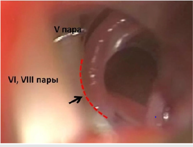
Arnold-Chiari malformation (Figure 5).
Abbreviations and Acronyms:
a. ACM – Arnold Chiari malformation
b. MRI – Magnetic Resonance Imaging
Conclusion
A good selection of patients and studying the comorbid, progression of the disease requires an additional diagnostics tool. After a clinical examination and neuroimaging, a diagnosis of ACM and a pontocerebellar cyst combined with unilateral hearing loss was established. After successful surgical treatment, the patient’s hearing was restored, which is confirmed by audiometric data. Thus, an assessment of the otolaryngological status makes clearer the possible objective of malfunctions of the peripheral and central parts of the sensory systems of the brain, mainly located in the area of interest of the pathological process, which is important when deciding on the tactics of surgical treatment.
References
- Bertrand G (1973) Dynamic factors in the evaluation of syringomyelia and syringobulbia. Clin Neurosurg20:322-333.
- Kumar A, Patni A, Charbel F (2002) The Chiari I Malformation and the Neurotologist. OtolNeurotol23(5):727-735.
- Milhorat TM, Nishikawa M, Kula RW, Yosef D Dlugacz (2010) Mechanisms of cerebellar tonsil herniation in patients with Chiari malformations as a guide to clinical management. Acta Neurochir (Wien)152(7):1117-1127.
- Milhorat H, Chou W, Elizabeth M, Kotzen M, Marcy CSpeer (1998) Chiaril Malformation: Description of Syndrome, Clinical Manifestation, and Inheritance Patterns in 364 Symptomatic Patients.Sep Neurosurgery; 43(3): 674-674.
- Milhorat TM, Nishikawa M, Kula RW, Yosef D Dlugacz (2010) Mechanisms of cerebellar tonsil herniation in patients with Chiari malformations as a guide to clinical management. Acta Neurochir (Wien)152(7):1117-1127.
- Paul KS, Lye RH, Strang FA, Dutton J (1983) Arnold-Chiari malformation. Review of 71 cases. J Neurosurg58(2):183-187.
- Rydell RE, Pulec JL (1971) Arnold-Chiari Malformation: Neuro-otologic Symptoms. Arch Otolaryngol 94(1): 8-12.
- Sperling NM, Franco RA, Milhorat TH (2001) Otologic Manifestations of Chiari I Malformation.Otology & Neurotology 22(5): 678-681.
- ValiulinMA (1996) Syringomyelia and Chiari malformation: initial clinical manifestations and results of surgical treatment: Abstract. MA Valiulin-SPb PP:23.

Top Editors
-

Mark E Smith
Bio chemistry
University of Texas Medical Branch, USA -

Lawrence A Presley
Department of Criminal Justice
Liberty University, USA -

Thomas W Miller
Department of Psychiatry
University of Kentucky, USA -

Gjumrakch Aliev
Department of Medicine
Gally International Biomedical Research & Consulting LLC, USA -

Christopher Bryant
Department of Urbanisation and Agricultural
Montreal university, USA -

Robert William Frare
Oral & Maxillofacial Pathology
New York University, USA -

Rudolph Modesto Navari
Gastroenterology and Hepatology
University of Alabama, UK -

Andrew Hague
Department of Medicine
Universities of Bradford, UK -

George Gregory Buttigieg
Maltese College of Obstetrics and Gynaecology, Europe -

Chen-Hsiung Yeh
Oncology
Circulogene Theranostics, England -
.png)
Emilio Bucio-Carrillo
Radiation Chemistry
National University of Mexico, USA -
.jpg)
Casey J Grenier
Analytical Chemistry
Wentworth Institute of Technology, USA -
Hany Atalah
Minimally Invasive Surgery
Mercer University school of Medicine, USA -

Abu-Hussein Muhamad
Pediatric Dentistry
University of Athens , Greece

The annual scholar awards from Lupine Publishers honor a selected number Read More...


