Lupine Publishers Group
Lupine Publishers
Menu
ISSN: 2637-4692
Case Report(ISSN: 2637-4692) 
Telescopic Hybrid Denture Prosthesis with Anterior Metal Ceramic Crowns Volume 4 - Issue 3
Sushmita VP1*, Vinaya Bhat2 and Chethan Hegde2
- 1Department of Prosthodontics, Sri Ramakrishna Dental College and Hospital, India
- 2Department of Prosthodontics, AB Shetty Memorial Institute of Dental Sciences, India
Received: February 05, 2020; Published: February 13, 2020
Corresponding author: Sushmita VP, Senior Lecturer, Department of Prosthodontics, Sri Ramakrishna Dental College and Hospital, Coimbatore, India
DOI: 10.32474/MADOHC.2020.04.000187
Introduction
While restoring patients with poor oral hygiene, it is mandatory to consider the self-cleansing ability of the prosthesis to reduce the risk of progression of the disease. The use of telescopic retainers continues to allow treatment options for prostheses that facilitate access for cleaning by the patient and/or dentist and retain questionable teeth longer. The concept of removable telescopic partial denture resulted from clinical experiments with the tooth supported complete denture [1]. It is also considered as a removable periodontal prosthesis, as it provides cross arch splinting of the periodontally compromised teeth and helps in equally distributing the forces along the long axis of the abutment [2-6]. In addition, a telescopic prosthesis can be designed to be cemented with light cement, which can satisfy a patient’s need for a fixed prosthesis, but also allow for removal by a dentist to carryout prophylactic procedure to maintain hygiene. In this case report, a patient was successfully rehabilitated with a removable telescopic crown retained hybrid denture with metal ceramic crown on the remaining anterior teeth.
Case Report
Patient aged 45 years reported to the Department of Prosthodontics, AB Shetty Memorial Institute of Dental Sciences, Mangalore, with the chief complaint of multiple missing teeth and a desire to replace them. On examination there was loss of vertical dimension with collapse of occlusion. The remaining teeth 13, 21, 22, 23, 31, 32, 33, 34, 35, 42 and 43 (Figure 1) were found to be periodontally compromised and with deep carious lesions. After periodontal and radiographic examination 21, 31, 32, 42 were found to have poor prognosis and it was decided to extract these teeth, other teeth were endodontically treated and used for retention of the prosthesis.
Diagnostic impressions were made, and casts were fabricated. Tentative jaw relation was recorded and facebow transfer was done using artex rotofix facebow. The study casts were mounted in an arcon semi adjustable articulator. Mounted study casts were analyzed. Endodontic therapy was carried out for the remaining salvaged teeth. A telescopic hybrid prosthesis with porcelain fused to metal crown for the anterior teeth was planned to rehabilitate the patient.
Fabrication of primary coping
a) A tentative jaw relation was recorded, and the casts were
mounted. It was observed that the patient had class II jaw
relation, with 7mm of horizontal overlap of anterior teeth.
b) After endodontic treatment was completed, the teeth
were prepared to receive the primary copings. Clearance was
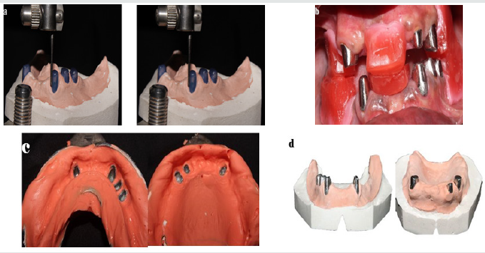
checked by using the tentative jaw relation record (Figure 2a-
2d).
c) Wax patterns for primary coping were fabricated and
surveyed to ensure parallelism. Casting was done using base
metal alloy. Then the copings were trimmed and checked for the
fit in the patient’s mouth, following which a pickup impression
was made (Figure 3) and casts were poured.
d) Final milling was done using two degree tapered milling
bur and they were cemented using resin cement U200 from 3M
(Figure 4).
Figure 2: a) Surveying of primary copings; b) Tryin of primary coping; c) Pickup impression of the primary copings, d) Cast made from the pickup impression
Establishing anterior occlusal plane
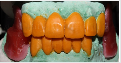
a) Anterior teeth were aesthetically modified, and missing
teeth were fabricated with wax (Figure 5).
b) An index was made from this wax pattern using heavy
body putty material. Later it was duplicated using tooth colored
composite resin (protemp) to form a template. Template was
trimmed, finished and polished. It was then tried in the mouth
to determine the anterior plane of the maxillary teeth according
to aesthetics and phonetics.
c) After establishing the plane of the maxillary anterior
teeth, the position of the lower anterior was also decided in the
similar manner based on phonetics, overjet and overbite (Figure
6). Anterior guidance was determined based on phonetics and
aesthetics.
Fabrication of metal framework
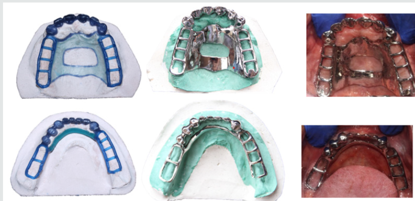
a) A wax pattern for anterior metal ceramic bridge was
fabricated using the template and cut back was done (Figure 7).
b) Major connector and minor connectors were designed,
and casting was done.
c) The metal framework was tried in the patient’s mouth for
fit and aesthetics.
Completion of the prosthesis
a) Ceramic layering was done for both maxillary and
mandibular anterior teeth. An aesthetic try in procedure was
carried out (Figure 8a & 8b)
b) Definitive jaw relations were recorded, and the casts were
mounted with the help of facebow transfer and centric relation
record.
c) Posterior teeth were arranged and try in was carried out
to ensure balanced occlusion in the mouth.
d) Acrylization of the posterior teeth was carried out taking
precautions not to damage the completed ceramic crowns.
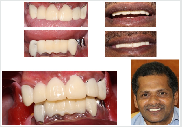
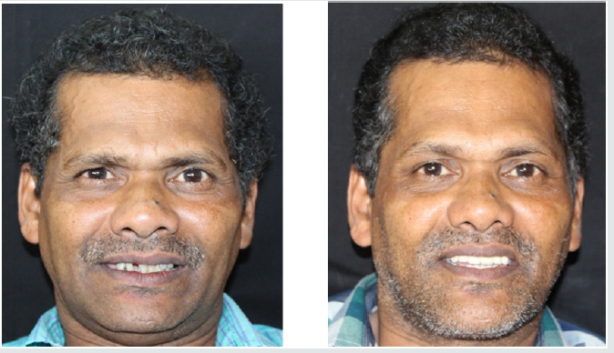
e) The acrylised prosthesis was trimmed, polished and
inserted in the mouth (Figure 9). Checked for occlusal contacts
in centric and eccentric movements.
f) Patient was kept on a standard recall regimen
Discussion
Preventive prosthodontics emphasizes the importance of
any procedure that can delay or eliminate future Prosthodontic
problems. Miller in 1958 stated that the maxilla and mandible
were designed to house the teeth and not to support artificial
denture [1]. In order to prevent alveolar ridge resorption and to
maintain the ridge height the tooth was retained and dentures
were constructed over it, hence the denture had good support
During 1960s the concept of telescopic crown retained overdenture
came into existence wherein the teeth were prepared and primary
coping were given with parallel or tapered walls which will receive
a secondary crown. The precisely made primary coping and
secondary crown will have friction fit which will aid in retention of
the prosthesis.
The weaker the tooth smaller will be the abutment preparation
with more taper and the primary coping. While longer will be the
primary coping with parallel walls in periodontally sound tooth.
The longer and less tapered walls will provide better retention
[1,2]. Telescopic crown retained removable dentures are indicated
when there is presence of one third or less complement of alveolar
bone, with unfavorable crown root ratio, loss of attached gingiva,
remaining teeth poorly situated/tilted and with presence of
adequate vertical dimension.
The advantage of this prosthesis includes, retention of natural
teeth, maintaining proprioception, distribution of occlusal force
to the alveolar bone equally and reduced bone resorption. It
provides good support to the prosthesis and aids in retention of
the prosthesis. It also acts as a periodontal splint for the remaining
compromised teeth [3]. However, tedious laboratory process,
increased cost, bulkiness of the prosthesis, loss of retention in long
run limits the use of this kind of prosthesis. The major disadvantage
is it is impossible to reline this kind of prosthesis [4-8].
In the present patient, the metal ceramic crowns that
were given for anterior teeth help in reducing the bulk of the
conventional telescopic removable partial denture. Apart from
the above, the main advantage of using hybrid prosthesis is that,
whenever the prosthesis is subjected to horizontal occlusal forces,
there is dislodgement of the denture from the primary coping
on the abutment. This reduces the load transfer to the abutment
thereby increasing its life expectancy. Pezzoli et al. [7] evaluated the
biomechanics of load transfer in telescopic denture and found out
that the occlusal load is distributed uniformly to the abutments and
edentulous areas [9].
Conclusion
A patient was successfully rehabilitated using telescopic metal ceramic crown for the remaining anterior natural teeth to support and retain a posterior removable partial denture was discussed. However, successful long-term treatment outcome would be accompanied by a routine periodontal and prosthodontic maintenance procedures.
References
- Juliana PA, Henking (1982) Overdentures: Brewer AA, Morrow RM (Eds.), Journal of Dentistry, Mosby,Elsevier, USA, pp. 190.
- Breitman JB, Nakamura S, Freedman AL, Yalisove IL (2012) Telescopic retainers: an old or new solution? A second chance to have normal dental function. J Prosthodont 21(1):79-83.
- Bhat VS, Prasad KD, Malli P (2015)Periodontal prosthesis-review. NITTE5(1):97-102.
- Weaver JD (1989) Telescopic copings in restorative dentistry. J Prosthet Dent 61(4):429-433.
- Langer A (1981) Tooth-supported telescope restorations. J Prosthet Dent 45(5):515-520.
- Hou GL, Tsai CC, Weisgold AS (1997) Periodontal and prosthetic therapy in severely advanced periodontitis by the use of the crown sleeve coping telescope denture. A longitudinal case report. Aust Dent J42(3):169-174.
- Pezzoli M, Rossetto M, Calderale PM (1986) Evaluation of load transmission by distal-extension removable partial dentures by using reflection photoelasticity. J Prosthet Dent56(3):329-337.
- Goswami R, Mahajan P, Siwach A, Gupta A (2013) Telescopic overdenture: Perio-prostho concern for advanced periodontitis. Contemp Clin Dent4(3):402-405.
- Ragini B, Chandrasekar A, Praveen M, Aarti S, Gautham, et al. (2013) Telescopic overdenture. Journal of Orofacial Research3(1):57-62.

Top Editors
-

Mark E Smith
Bio chemistry
University of Texas Medical Branch, USA -

Lawrence A Presley
Department of Criminal Justice
Liberty University, USA -

Thomas W Miller
Department of Psychiatry
University of Kentucky, USA -

Gjumrakch Aliev
Department of Medicine
Gally International Biomedical Research & Consulting LLC, USA -

Christopher Bryant
Department of Urbanisation and Agricultural
Montreal university, USA -

Robert William Frare
Oral & Maxillofacial Pathology
New York University, USA -

Rudolph Modesto Navari
Gastroenterology and Hepatology
University of Alabama, UK -

Andrew Hague
Department of Medicine
Universities of Bradford, UK -

George Gregory Buttigieg
Maltese College of Obstetrics and Gynaecology, Europe -

Chen-Hsiung Yeh
Oncology
Circulogene Theranostics, England -
.png)
Emilio Bucio-Carrillo
Radiation Chemistry
National University of Mexico, USA -
.jpg)
Casey J Grenier
Analytical Chemistry
Wentworth Institute of Technology, USA -
Hany Atalah
Minimally Invasive Surgery
Mercer University school of Medicine, USA -

Abu-Hussein Muhamad
Pediatric Dentistry
University of Athens , Greece

The annual scholar awards from Lupine Publishers honor a selected number Read More...


