Lupine Publishers Group
Lupine Publishers
Menu
ISSN: 2637-4692
Case Report(ISSN: 2637-4692) 
Orthodontic Management of Bilateral Ectopic Maxillary Canines in Young Patient Using Non-Extraction Technique – A Case Report Volume 5 - Issue 4
Santoshni Samal1*, Ratna Renu Baliarsingh1, Prayas Ray1 and Althwaf Shahjahan2
- 1Department of Pedodontics and Preventive Dentistry, SCB Dental College and Hospital, India
- 2PGT, SCB Dental College and Hospital, India
Received: September 09, 2022; Published: September 26, 2022
Corresponding author: Santoshni Samal, Post PG doctor, Department of Pedodontics and Preventive Dentistry, SCB Dental College and Hospital, Cuttack, Odisha, India
DOI: 10.32474/MADOHC.2022.05.000218
Abstract
Ectopic buccally erupted maxillary canines are one of the most frequently encountered conditions in orthodontic practice with different etiology like lack of space, early loss of a primary canine, ankylosis, neoplastic formation, root dilacerations, and an abnormal lateral root position concerning an erupting canine. Diagnosis is crucial for proper intervention. Management can be done by extraction or non-extraction technique. This case report describes a 14 year old boy who reported the same problem and was managed by a noninvasive method, HYRAX followed by fixed orthodontics treatment.
Introduction
An ectopic maxillary canine is one of the most common orthodontics problems in dentistry with a prevalence of 1-2% [1]. There are numerous systemic and local etiology-like disturbances associated with the follicle of the unerupted tooth that may influence the direction of eruption and contribute to the displacement of the maxillary canine [2], lack of space, early loss of a primary canine, ankylosis, neoplastic formation, root dilacerations, and an abnormal lateral root position concerning an erupting canine [3,4]. Diagnosis can be based on clinical examination where canines are buccally aligned or out of the arch with moderate to severe crowding. Radiographs and cast analysis are useful to show the arch length discrepancy. Management of ectopic canine is complex using extraction methods in young patients so noninvasive methods can also be an option for growing children. This case report showed the management of ectopic maxillary canine using HYRAX as a nonextraction technique followed by fixed orthodontics.
Case Report
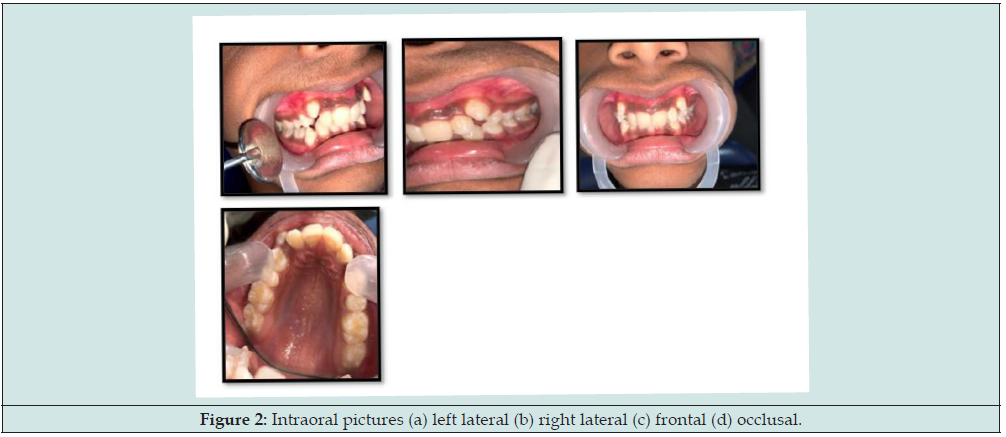
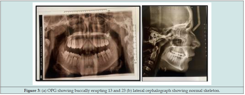
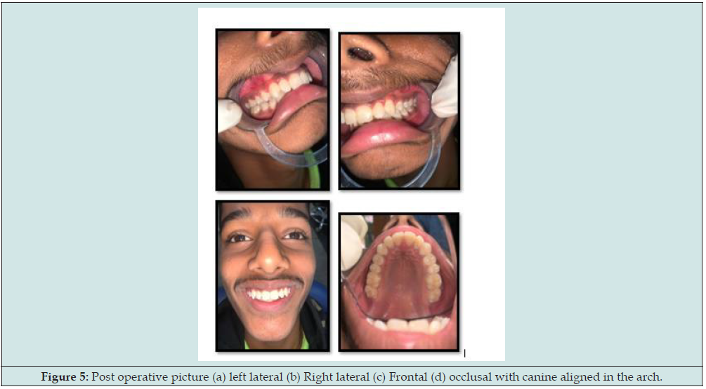
A 14 years old boy reported to the Department with an unaesthetic appearance and proclined corner teeth (Figure 1a). Medical history was not relevant. The patient had no dental history. Family history was not significant. On Extraoral examination, the patient had a symmetrical face, on smiling Bilateral proclined maxillary canines were present (Figures 1a-1c), and mandibular teeth were normal. On intraoral examination, Bilateral ectopic maxillary canines were present which were buccally displaced. Class, I molar relationship on Both sides was present (Figures 2a- 2d). Maxillary arch showed moderate crowding with Overjet- 1 mm, and overbite- 2mm. Cast analysis showed a length discrepancy of 11 mm in the maxillary arch. A treatment plan was made using the Non extraction method of correction with Rapid maxillary expansion (RME) HYRAX placement for 3 months which was Activated – 2 turns/day for 2 weeks followed by fixed orthodontics treatment and followed by the Retention phase (Figures 3a & 3b). After 1 year, the patient’s ectopic canines were aligned in the arch with normal overjet and overbite.
Figure 3: (a) OPG showing buccally erupting 13 and 23 (b) lateral cephalograph showing normal skeleton.
Discussion
Ectopic or buccally erupted maxillary canine can be due to individual variations in eruption patterns and timing. Early diagnosis can be done for proper intervention by panoramic radiographs and cast analysis. The amount of space available and the amount of space required can be calculated by space analysis with a full set of orthodontics dental casts. There are four treatment options for impacted teeth: observation, intervention, relocation, and extraction [5]. Untreated impacted canines may determine arch length discrepancies, loss of vitality of adjacent teeth, follicular cysts, canine ankylosis, infections, and pain (Figures 4a- 4d). As a consequence of this, orthodontic treatment is strongly recommended [6,7]. The orthodontic management of these difficult cases requires the use of specific orthodontic mechanics, which could allow a perfect three-dimensional correction [8]. Opening of palatal sutures can be done in young children using a HYRAX expander (Figures 5a-5d). HYRAX helps in the opening of mid-palatal sutures and gains some space in the arch which allows movement of ectopic canines in the arch. After the 1-year patient had aligned canines with normal overjet and overbite [9].
Figure 4: Intraoperative pictures (a) left lateral (b) Right lateral (c) frontal (d) occlusal with HYRAX placed.
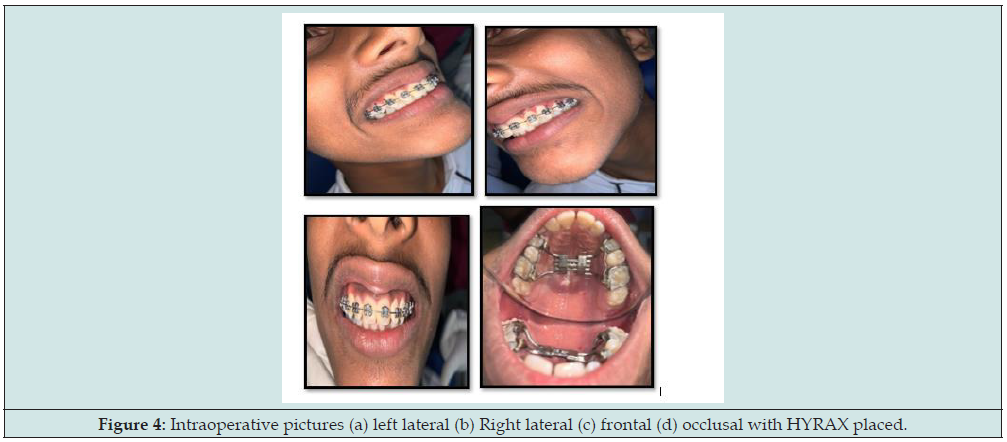
Figure 5: Post operative picture (a) left lateral (b) Right lateral (c) Frontal (d) occlusal with canine aligned in the arch.
Conclusion
Impaction of a maxillary canine is a frequent occurrence and requires a multi-disciplinary approach for proper management. Early recognition and implementation of interceptive treatment are mandatory. In growing children noninvasive method can be chosen after an adequate diagnosis and treatment plan. so, it is concluded that HYRAX with orthodontic treatment can be considered an effective therapy for bilateral impacted canine correction.
References
- Fleming P, Scott P, Heidari N, Dibiase A (2009) Influence of radiographic position of ectopic canines on the duration of orthodontic treatment. Angle Orthod 79(3): 442-446.
- Fearne J, Lee RT (1988) Favorable spontaneous eruption of severely displaced maxillary canines with associated follicular disturbance. Br J Orthod 15(2): 93-98.
- Ngan P, Hornbrook R, Weaver B (2005) Early timely management of ectopically erupting maxillary canines. Semin Orthod 11: 152-163.
- Bishara SE (1992) Impacted maxillary canines: A review. Am J Orthod 101(2): 159-171.
- Becker A (2012) Orthodontic treatment of impacted teeth. 3rd
- Watted N, Proff P, Reiser V, Shlomi B, Abu-Hussein M, et al. (2015): CBCT; In Clinical Orthodontic Practice: Journal of Dental and Medical Sciences 14(2): 102-115.
- Watted N, Abu-Hussein M, Awadi O, Borbély P (2015) Titanium Button with Chain by Watted for Orthodontic Traction of Impacted Maxillary Canines Journal of Dental and Medical Sciences 14(2): 116-127.
- Jacobs SG (1999) Localization of the unerupted maxillary canine: How to and when to. Am J Orthod Dentofacial Orthop. 115(3): 314-322.
- Haas AJ (1970) Palatal expansion - Just the beginning of dentofacial orthopedics. Am J Orthod 57(3): 219-255.

Top Editors
-

Mark E Smith
Bio chemistry
University of Texas Medical Branch, USA -

Lawrence A Presley
Department of Criminal Justice
Liberty University, USA -

Thomas W Miller
Department of Psychiatry
University of Kentucky, USA -

Gjumrakch Aliev
Department of Medicine
Gally International Biomedical Research & Consulting LLC, USA -

Christopher Bryant
Department of Urbanisation and Agricultural
Montreal university, USA -

Robert William Frare
Oral & Maxillofacial Pathology
New York University, USA -

Rudolph Modesto Navari
Gastroenterology and Hepatology
University of Alabama, UK -

Andrew Hague
Department of Medicine
Universities of Bradford, UK -

George Gregory Buttigieg
Maltese College of Obstetrics and Gynaecology, Europe -

Chen-Hsiung Yeh
Oncology
Circulogene Theranostics, England -
.png)
Emilio Bucio-Carrillo
Radiation Chemistry
National University of Mexico, USA -
.jpg)
Casey J Grenier
Analytical Chemistry
Wentworth Institute of Technology, USA -
Hany Atalah
Minimally Invasive Surgery
Mercer University school of Medicine, USA -

Abu-Hussein Muhamad
Pediatric Dentistry
University of Athens , Greece

The annual scholar awards from Lupine Publishers honor a selected number Read More...


