Lupine Publishers Group
Lupine Publishers
Menu
ISSN: 2637-4692
Review Article(ISSN: 2637-4692) 
Correction of a Latero Nasal Cancrum Oris Defect Using the Webster Advancement Flap Technique; a Hypothesis Volume 4 - Issue 5
Zilefac Brian Ngokwe1*, Cheboh Cho Fon1, Ntep Ntep David Bienvenue1, Elage Epie Macbrain1, Nokam Kamdem Stephane1, Kouamou Tchiekou Audrey1 and Bengondo Messanga Charles1
- 1Department of Oral surgery, Maxillofacial surgery and Periodontology, Faculty of Medicine and Biomedical Sciences; University of Yaoundé I, Cameroon
Received:June 22, 2021 Published:August 10, 2021
Corresponding author: Zilefac Brian Ngokwe, Department of Oral surgery, Maxillofacial surgery and Periodontology, Faculty of Medicine and Biomedical Sciences; University of Yaoundé I, Cameroon
DOI: 10.32474/MADOHC.2021.04.000200
Abstract
Background: The destructive facial effects of cancrum oris remain a major problem for surgeons and the patients themselves. Although known since antiquity, the epidemiology, pathophysiology, and etiology of the disease remain a subject of debate. Further research is needed to identify more exactly the causative agents. Only in the twentieth century were effective drugs (sulfonamides and penicillin) against noma developed, as well as adequate surgical treatment for the sequelae of noma. These modes of treatment remain inaccessible for the many present-day victims of noma because of their extreme poverty. The only truly effective approach to the problem of noma throughout the world is prevention, namely, combating the extreme poverty with measures that lead to economic progress. Here, we explore the potential use of a flap technique as a solution to the extensive facial tissue destruction that occurs in Noma.
Objective: To propose the Webster advancement flap technique as a possible solution to latero nasal NOMA management. Results: The potential benefits and results of using a flap technique could be the little chances of graft rejection, the good blood supply associated with flaps and the esthetically conserved nature of the procedure. Thus, it could become an alternative to the often radical and inesthetic solution of skin grafts.
Conclusion: Surgeons often have to make tough choices in the selection of an appropriate technique when faced with the management of NOMA. Thus, we propose a conservative and esthetic method of going around this problem.
Keywords: Noma; Webster Flap
Introduction
Necrotising ulcerative stomatitis is an orofacial gangrene that mainly occurs in young children [1]. Untreated, it is generally lethal within a few weeks. Patients who survive noma frequently suffer severe sequelae such as facial disfigurement, trismus, oral incontinence and speech problems [2]. It is caused by a combination of malnutrition, debilitation by diseases like measles, and intraoral infections [2]. The global incidence of noma is 140 000cases, as estimated by the WHO, of which the majority lives in the savannas directly South from the Sahara Desert [2]. There are various classifications based on the location and the severity. The WHO classification based on the severity is as follows [3]:
A. STAGE I: Acute necrotizing gingivitis stage
B. STAGE II: Oedema stage
C. STAGE III: Gangrenous stage
D. STAGE IV: Scarring stage
E. STAGE V: Sequelae stage
And based on localization: [4]
Simple clinical forms
a) Cheek perforations
b) Commissural destruction
c) Superior commissurolabial mutilation
d) Inferior commissurolabial mutilation
e) Superomedian labial amputation
f) Inferomedian labial amputation
More extensive forms
a. Jugomasseteric mutilation
b. Labiomental amputation
c. Labionasal mutilation
d. Labiomaxilloseptocolumellar amputation.
Complex forms
a. Lateral hemifacial lesion
b. Labiopalatal amputation
c. Labiogeniomandibular mutilation
These classifications are the most widely used for both the diagnosis and the prognosis of NOMA cases.
Background
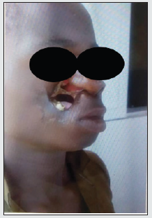
The destructive effects of cancrum oris or NOMA remain debilitating and can be psychologically damaging for the individuals affected. We go by the hypothesis that various techniques can be used for the reconstructive surgery of these patients. Surgical rehabilitation of noma sequelae is directed at the functional and cosmetic improvement of the face. The basic principle is to release the trismus or ankylosis and to replace lost tissue by transferring local tissue flaps and, when the loss is substantial, from other parts of the body [5]. One of the flap techniques is the Webster advancement flap technique which uses the adjacent jaw tissues for the recovery of lost tissue. Here, we discuss a case of NOMA and propose the Webster advancement flap technique as a solution.
Objective
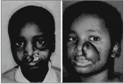
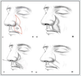
To propose the Webster advancement flap technique as a possible solution to latero nasal NOMA management (Figures 1 & 2).
Discussion
The treatment plan of NOMA depends on the severity of the facial destruction and include multiple surgical techniques such as tissue grafts, facial flaps and esthetic artificial prosthesis [3]. The social impact of surgical rehabilitation is rewarding with better chances for education, employment and marriage after surgery [1]. Tissue grafts remain the most extensively used reconstruction techniques due to the ease and availability of tissue from other parts of the body [3]. These result in good functional recuperations of the patients [3]. However, the esthetics aspect of these techniques is often very unsatisfactory as tissues taken from other parts of the body are not always similar to those of the facial region [6] (Figure 3) where massive defect of the palate, lips, cheek, and nose in a 10-year-old boy with mandibular constriction was reconstructed with a latissimus dorsi musculocutaneous free flap vascularized by the facial artery. Part of the flap was used to line the oral cavity. This can have psychologically damaging effects for the self-esteem of the concerned children and thus compromise their quality of life.
Figure 3: (Left) Massive defect of the palate, lips, cheek, and nose in a 10-year-old boy with mandibular constriction. (Right) Postoperative result after latissimus dorsi musculocutaneous free flap vascularized by the facial artery. Part of the flap was used to line the oral cavity [4].

With the advancement in esthetic surgery techniques and medicine in general, better post-surgical renditions are expected from surgeons by patients with an emphasis on the esthetic aspect of the surgeries [7]. We therefore propose the Webster advancement flap which is a conservative surgical procedure which relies on the use of adjacent jaw tissues to repair the loss of tissue. This method is based on the principle that the best form of reconstruction comes from tissue that most closely resembles the tissue being replaced-in this case, the cheek [4]. Because of the lack of excess skin in the areas between the lip and nose, many reconstructions for this area use medial jaw advancement [4]. To correct the maxillary bone defect, a scapula transplant can be used as it proves to be resourceful in the restoration of the maxilla [8]. The nasolabial fold provides color- and texture-matched tissue to the upper and lower lips. An excellent blood supply based on the facial arteries and a natural-appearing scar at the donor site reinforces this flap as a useful adjunct [4]. Webster advancement flap is a useful technique for defects of the upper lip that require musculocutaneous advancement from adjacent cheek tissue. Simple advancement would cause bunching at the perialar folds. This excision is essentially an elliptical excision with the upper part shifted laterally to avoid the nostril. The perialar skin may be preserved as a caudally based flap to reconstruct columellar or nostril floor defects [4].
Conflict Of Interest
All the authors do not have any possible conflicts of interest.
Conclusion
There are many difficulties faced by surgeons for the reconstruction of NOMA tissue defects and this is reflected in the complexity of the decision-making on what therapies to use for the tissue reconstruction. The decision-making must also take into account the esthetics of the facial region as many patients lay much emphasis on this aspect of the surgery. We thus propose a more efficient and esthetic reconstruction procedure which can greatly help provide better outcomes.
References
- Srour ML, Marck K, Baratti Mayer D (2017) Noma: Overview of a Neglected Disease and Human Rights Violation. Am J Trop Med Hyg 96(2): 268-274.
- Marck KW (2003) Cancrum oris and noma: some etymological and historical remarks. Br J Plast Surg 56(6): 524-529.
- (2017) World Health Organization. Regional Office for Africa. Poster for early detection and management of noma. Brazzaville: WHO, Regional Office for Africa.
- Montandon D, Lehmann C, Chami N (1991) The Surgical Treatment of Noma. Plast Reconstr Surg 87(1): 76-86.
- Farley ES, Amirtharajah M, Winters RD, Taiwo AO, Oyemakinde MJ, Fotso A, et al (2020) Outcomes at 18 mo of 37 noma (cancrum oris) cases surgically treated at the Noma Children’s Hospital, Sokoto, Nigeria. Trans R Soc Trop Med Hyg 114(11): 812-819.
- Adeniyi S, Taiwo A, Ibikunle A, Braimah R, Gbotolorun O, Ogbeide M, et al. (2017) Pattern of tissue destruction among patients diagnosed with cancrum oris (Noma) at a Northwestern Nigerian Hospital, Sokoto. Saudi J Oral Sci 4(2): 101-105.
- Agbara R, Fometeb B, Omejec KU, Obiadazieb AC, Ajikeb SO (2018) Treatment of complications following orofacial gangrenous infection in a resource limited setting: Experiences and a proposed classification. Egypt J Ear Nose Throat Allied Sci 19(2): 58-63.
- Vinzenz K, Holle J, Würinger E (2008) Reconstruction of the Maxilla with Prefabricated Scapular Flaps in Noma Patients. Plast Reconstr Surg 121(6): 1964-1973.

Top Editors
-

Mark E Smith
Bio chemistry
University of Texas Medical Branch, USA -

Lawrence A Presley
Department of Criminal Justice
Liberty University, USA -

Thomas W Miller
Department of Psychiatry
University of Kentucky, USA -

Gjumrakch Aliev
Department of Medicine
Gally International Biomedical Research & Consulting LLC, USA -

Christopher Bryant
Department of Urbanisation and Agricultural
Montreal university, USA -

Robert William Frare
Oral & Maxillofacial Pathology
New York University, USA -

Rudolph Modesto Navari
Gastroenterology and Hepatology
University of Alabama, UK -

Andrew Hague
Department of Medicine
Universities of Bradford, UK -

George Gregory Buttigieg
Maltese College of Obstetrics and Gynaecology, Europe -

Chen-Hsiung Yeh
Oncology
Circulogene Theranostics, England -
.png)
Emilio Bucio-Carrillo
Radiation Chemistry
National University of Mexico, USA -
.jpg)
Casey J Grenier
Analytical Chemistry
Wentworth Institute of Technology, USA -
Hany Atalah
Minimally Invasive Surgery
Mercer University school of Medicine, USA -

Abu-Hussein Muhamad
Pediatric Dentistry
University of Athens , Greece

The annual scholar awards from Lupine Publishers honor a selected number Read More...


