Lupine Publishers Group
Lupine Publishers
Menu
ISSN: 2637-4692
Research Article(ISSN: 2637-4692) 
Computerised Occlusal Analysis Utilising T Scan™ and it’s Implications in Periodontology Volume 2 - Issue 3
Sreelakshmi CK, Kishore HC, Prabhuji MLV, Rudrakshi C, Prafulla Thumati and Kalaiarasi S
- Department of Periodontology, Krishnadevaraya College of Dental Sciences, India
Received: May 07, 2018; Published: May 29, 2018
Corresponding author: Prabhuji MLV, NO.57, Department of Periodontology, Krishnadevaraya College of Dental Sciences, Gayathri Devi Park Extension, Opposite to Stella Maris School, Bangalore: 560003
DOI: 10.32474/MADOHC.2018.02.000138
Abstract
Introduction: The periodontium has its own adaptive capacity to the forces exerted on the teeth, this varies in different persons and in the same persons at different times. In current practise, the occlusal factors acting on the tooth are examined by articulation paper marks, waxes, pressure indicator paste, etc. However, the drawbacks of these methods are that they do not detect simultaneous contact nor do they quantify time and force. This study was conducted to determine and to analyse the distribution of occlusal loading forces, occlusal contact variability and its correction in the management of periodontitis using T-Scan III™.
Methods: A total of ten chronic periodontitis patients were selected for the study. Scaling and root planing procedure and occlusal analysis using T-Scan III™ were performed on them. The amount of occlusal forces was found to be highly concentrated either on the right or the left side of the dental arches. Thus, the occlusal adjustments were carried out to minimize and dissipate the abnormal occlusal forces.
Result: T-Scan analysis was repeated to analyse for the distribution of occlusal forces which depicted a reduction in the amount of occlusal forces and these forces were distributed nearly equally on the dental arch.
Conclusion: Comparing and analysing the occlusal distribution of forces prior to and after the occlusal adjustments using T-Scan III™ system showed equilibrium in occlusal force distribution.
Keywords: Occlusion, Periodontium, Periodontitis, T-Scan system, Intercuspal Position
Introduction
The periodontium has its own adaptive capacity to the occlusal forces exerted on the tooth. The occlusal trauma is not to be considered as an etiological factor for periodontitis, its presence may exacerbate the disease severity. Occlusal adjustments in teeth have been recommended for premature contacts or interferences (Junqueira RB [1]). The effect of these occlusal forces on the periodontium is dictated by its magnitude, direction, duration and frequency (Newman et al [2]). An individual’s occlusal status is generally described by two major characteristics: a) intra-arch relationship i.e. the relationship of the teeth within each arch to a smoothly curving line of occlusion and b) inter-arch relationship i.e. the pattern of which occlusal contacts between the upper and lower teeth (Profitt [3]). The disharmonies in these occlusal relationships increase the noxious inputs to the neuromuscular system, as well as, stress induced clenching, causing increased muscle activity and spasm pain (Weinberg, [4]). Premature occlusal contacts are frequently detected in subjects with chronic periodontitis and are significantly correlated with its severity. The posterior teeth with high occlusal forces in patients with untreated chronic periodontitis may reflect occlusal trauma associated periodontal conditions, that could probably increase the risk of further periodontal break down (Zhou).
In current practice, the occlusal factors acting on the teeth are examined by articulation paper marks, waxes, pressure indicator paste, etc. are some of the tools available to assess the forces of the occlusion and also to evaluate the occlusal contacts. However, the drawbacks of these methods are that they do not detect simultaneous contact nor do they quantify time and force. There is no scientific correlation between the depth of the colour of the mark, its surface area, amount of force, or the contact timing sequence that results as the articulation paper marks is used. (Schelb et al [5]; Halperin et al. [6]). This method solely relies on the combination of dental articulating paper and patient feel.
Commonly used quantitative occlusal approaches include a computer aided video system, a photo occlusion method and the T– Scan II™ system (Millstein, [7]; Arcan and Zandman, [8]). Recently, Bio Research Associates, Inc and Tekscan Inc corporations have developed a commercially available system (T-Scan III™) that overcomes the known limitations of articulation paper marks (Kerstein, [9]). It quantifies and displays relative occlusal force information, so that the clinician can minimize repeated errors of incorrect occlusal contact selection. There are numerous upcoming computer based treatment modalities, but many clinicians have not overcome the traditional methods for examining the occlusion. This study was conducted to evaluate the distribution of occlusal loading forces and correction of any occlusal discrepancies using Tekscan (T-Scan III™).
Materials and Methods
Patient Selection
Approval for the study was provided by the Institutional Ethical Committee which is established in accordance with the Declaration of Helsinki. In this study, a total of ten systemically healthy patients with Chronic Generalized Periodontitis were selected from the Out Patient section of the Department of Periodontology, Krishnadevaraya College of Dental Sciences, Bangalore, India. The inclusion criteria were subjects between the age group of 30 to 39 years, systemically healthy, subjects with atleast 20 functioning teeth, subjects who are co-operative and are able to attend follow up. Patients with aggressive periodontitis, bleeding disorders, gross oral pathology, subjects who had received any medications such as non steroidal anti inflammatory drugs (NSAIDs) within the last six months, Pregnant and lactating mothers were excluded from the study. Prior to the occlusal analysis, full-mouth supra and subgingival scaling and root planing procedure were performed. Occlusal analysis was performed using T-Scan III™ System (Tekscan, Inc Boston, MA) available in the department of periodontology, Krishnadevaraya College of dental sciences and hospital.
Occlusal Analysis Using Tekscan (T-Scan III™)
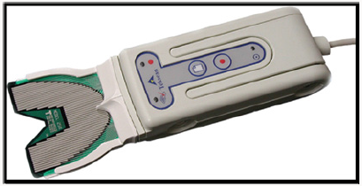
Subsequent to performing full mouth supra and subgingival scaling and root planing procedure, the subjects were seated in a comfortable position for the occlusal analysis using T-Scan III™ System (Figure 1). Before conducting the experiment, each subject practiced moving their mandible from the rest position to the intercuspal position (ICP) using a comfortable amount of biting pressure. After the subjects were familiar with the procedure, the operator put the sensor support of the T-Scan III™ System into their mouths and recorded the contact conditions when occluding. The sensor was set on the handle in advance and was positioned close to the maxillary occlusal plane. The subjects were then instructed to close their mouths in the ICP. The T-Scan III™ system was used to record the process as the mandible moved from the rest position to the ICP and then back to the rest position. Each subject repeated this procedure three times and the results were recorded. The T-Scan III™ system recorded the changes in occlusal contact at a time interval of 0.01s producing dynamic change data. The recorded data included contact locations and relative occlusive forces. The T-Scan III™ system measures the occlusal movement from the resting position to the ICP and the asymmetries of occlusal centres and force distribution (Liu et al., [10]). After the occlusal analysis using T-Scan III™ the occlusal distribution of forces were determined and corrected. A T-Scan analysis was performed after the correction of occlusal forces to analyse for the equal distribution of the occlusal forces.
Results
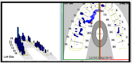
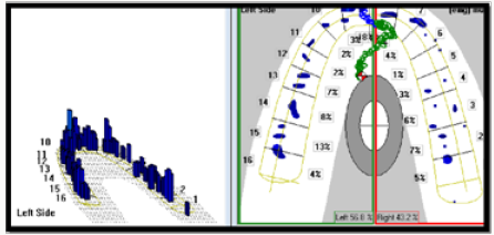
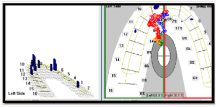
After a thorough scaling and root planing procedure, occlusal analysis using T-Scan III™ was performed and the amount of occlusal forces was noted in the 2D and 3D graph (Figures 2-3a). The amount of forces was found to be highly concentrated either on the right or on the left dental arch. Therefore, the occlusal adjustments were carried out to minimize and dissipate the occlusal forces on to right and left dental arches. After the occlusal adjustments, T-Scan analysis was repeated to analyse for the distribution of occlusal forces which depicted a reduction in the amount of occlusal forces and these forces were distributed nearly equally on inter and intra arches.
Discussion
Occlusal trauma threatens periodontal longevity and impedes successful therapy. Occlusal discrepancies especially balancing interferences are associated with periodontal breakdown during periodontal maintenance (Reinhardt and Killeen [1]) Teeth with occlusal discrepancies had significantly deeper probing depth and worse prognosis (Harrel and Nunn [11]). On treating them through selective grinding and or occlusal appliances during periodontal therapy is considered as an integral part of overall treatment of periodontal disease.
The evolution of T-Scan from 1st generation to 8th generation (1847-2015) has revolutionized the concept of occlusal analysis. The “T-Scan I” was invented 25 years ago, and since then, the entire system has undergone hardware, sensor, and software revisions, such that today’s “T-Scan III” system (version 7.0) is vastly improved over the earlier systems. (Reeta Jain et al., [12]).
The T-Scan III™ system utilizes a 0.1-mm-thick sensor made with a flexible material which minimizes the errors of mandibular deviation caused by excessively thick or hard sensors, on occluding. The parameters of this system include introduction time, occlusal center, track of occlusal center and percentage of occlusal force distribution, which can provide a reference for analyzing instant occlusal conditions. These parameters can also form a time track distribution of the points of maximum occlusion and occlusal force through contact of the teeth with the sensor. This information is beneficial for quantitative research in occlusal equilibrium (Liu et al., [10]). Early T-scan I™ and II™ system investigations demonstrated either mixed predictability of force reproduction, insufficient accuracy or a lack of force reproduction capability (Koos et al., [13]).
The T-Scan III™ system sensor comprises two polyester films as substrate, conductive ink as an interlayer and a vertical and horizontal woven wire grid with approximately 1500 sensor points. Since polyester films have high tear and strain resistance the thin polyester films in the T-Scan III™ system can endure occlusal force and change form during occlusal movement. The sensor is approximately 60μm thick, which does not hinder the subjects when conducting different types loops. After collecting the data, relevant software can be utilized to conduct quantitative analysis of the changes in occlusal contact points and force overtime and then compute a distribution of the occlusal forces. Compared to previous systems, the T-Scan III™ system has improved accuracy and repeatability. In measuring force distribution, the system can analyze the percentage of force distribution of one tooth, the anterior and posterior occlusal forces and the left and right occlusal forces. Regarding occlusal center, the system can record the track of occlusal center from the initial contact position to the final position (Throckmorton et al., [14]). Computerized occlusal analysis using T-Scan III™ guides the selection of tooth contacts for occlusal adjustment (Qadeer et al., [15]), also provides objective digital data for determining the quality of the orthodontic end-result, and observing the occlusal force distribution in natural dentition (Qadeer et al., [16]).
A number of researchers have indicated that the T-Scan III™ system measures the relative occlusal force of the subjects instead of the numerical values of the absolute occlusal forces (Hirano et al., [17]). However, research indicates that the T-Scan III™ system can be used as a measurement tool in clinical experiments and the sensitivity setting can be adjusted to measure the occlusal force at different ranges (Kerstein, [18]). Occlusal forces affect natural teeth to a different degree and they go through a primary intra socket and secondary intra alveolar movements (Dario, [19]; Garg, [20]; Kerstein, [21]; Sadamori et al., [22]). In this study, the amount of occlusal forces which was analyzed using T-Scan III™ system was corrected and re analysed again for the occlusal force distribution. The analysis presented a nearly equal distribution of forces and if these occlusal forces are unbalanced it could lead to temporomandibular disorders and masticatory muscle pain. Thus, the analysis of the distribution of occlusal loading forces using T-Scan III™ system provides an aid in treating and as well as managing the periodontal deterioration and preventing further periodontal breakdown [23].
Conclusion
T-Scan III™ system has shown to aid in correction of occlusal discrepancies and thus contributing in equal occlusal force distribution on either dental arches. Further, long term studies using T-Scan III™ system may be required to assess the role of occlusion and its correction along with Phase I Therapy in preventing periodontal breakdown.
Acknowledgement
The authors extend their gratitude to the subjects who participated in the study.
References
- Richard A Reinhardt, Amy C Killeen (2015) Do Mobility and Occlusal Trauma Impact Periodontal Longevity? Dent Clin North Am 59(4): 873- 883.
- Newman MG, Carranza FA, Takei HH, Klokkevold PR (2006) Clinical Periodontology. 10th edn. Missouri. Saunders pp.467.
- Profitt WR (1986) on the aetiology of malocclusion. British Journal of Orthodontics 13: 1-11.
- Weinberg LA (1983) The role of stress, occlusion, and condyle position in TMJ dysfunction-pain. Journal of Prosthetic Dentistry 49(4): 532-545.
- Schelb E, Kaiser DA, Bruki CE (1985) Thickness and marking characteristics of occlusal registration strips. Journal of Prosthetic Dentistry 54(1): 122-126.
- Halperin GC, Halperin AR, Noting BK (1982) Thickness strength and plastic deformation of occlusal registration strips. Journal of Prosthetic Dentistry 48(5): 575-578.
- Millstein PL (1984) A method to determne occlusal contact and non contact areas: Preliminary report. Journal of Prosthetic Dentistry 52(1): 106-110.
- Arcan M, Zandman F (1984) A method for in vivo quantitative occlusal strain and stress analysis. Journal of Biomechanics 17(2): 67-79.
- Kerstein RB (2004) Combining technologies: a computerized occlusal analysis system synchronized with a computerized electromyography system. Journal of Craniomandibular Practice 22(2): 96-109.
- Liu CW, Chang YM, Shen UF, Hong HH (2014) Using the T-scan III system to analyze occlusal function in mandibular reconstruction patients: A pilot study. Biomedical Journal 38(1):52-57.
- Nunn ME, Harrel SK (2001) The effect of occlusal discrepancies on periodontitis. I. Relationship of initial occlusal discrepancies to initial clinical parameters. Journal of Periodontology 72(4): 485-94.
- Reeta Jain, Ravudai Jabbal, Shweta Bindra, Swati Aggarwal (2015) T- Scan a digital pathway to occlusal perfection: a review Annals of Prosthodontics and Restorative Dentistry 1(1): 32-35.
- Koos B, Godt A, Schille C, Goz C (2010) Precision of an instrumentation method of analyzing occlusion and its resulting distribution of forces in the dental arch. Journal of Orofacial Orthopedics 71(6): 403-410.
- Throckmorton GS, Rasmussen J, Caloss R (2009) Calibration of T-scan sensors for recording bite forces in denture patients. Journal of Oral Rehabilitation 36(9): 636-643.
- Qadeer S, Kerstein R, Kim RJ, Huh JB, Shin SW (2012) Relationship between articulation paper mark size and percentage of force measured with computerized occlusal analysis. Journal of Advanced Prosthodontics 4(1): 7-12.
- Qadeer S, Yang L, Sarinnaphakorn L, Kerstein RB (2016) Comparison of closure occlusal force parameters in post-orthodontic and nonorthodontic subjects using T-Scan(R) III DMD occlusal analysis. Journal of Craniomandibular Practice 34(6): 395-401.
- Hirano S, Okuma K, Hayakawa I (2002) In vitro study on accuracy and repeatability of T-scan 2 system. Kokubyo Gakkai Zasshi 69: 194-201.
- Kerstein RB (2006) Delayed implant loading in cases with implants and natural teeth. Aesthetic Dentistry Fall 16-20.
- Dario LJ (1995) How occlusal forces change in implant patients: a clinical research report. Journal of American Dental Association 126(8): 1130-1132.
- Garg AK (2007) Analyzing dental occlusion for implants: Tekscan’s T-Scan III. Dental implant logy update 18(9): 66-70.
- Kerstein RB (2001) Non simultaneous contact in combined implant and natural teeth occlusal schemes. Practical Procedures in Aesthetic Dentistry 13(9): 751-755.
- Sadamori S, Kotani H, Abekura H, Hamada T (2007) Quantitative analysis of occlusal force balance in intercuspal position using the dental Presale system in patients with temporomandibulat disorders. International Chinese Journal of Dentistry 7: 43-47.
- Ash MM, Ramfjord SP (1982) Occlusion. 3rd edn. Philadelphia. WB Saunders Co

Top Editors
-

Mark E Smith
Bio chemistry
University of Texas Medical Branch, USA -

Lawrence A Presley
Department of Criminal Justice
Liberty University, USA -

Thomas W Miller
Department of Psychiatry
University of Kentucky, USA -

Gjumrakch Aliev
Department of Medicine
Gally International Biomedical Research & Consulting LLC, USA -

Christopher Bryant
Department of Urbanisation and Agricultural
Montreal university, USA -

Robert William Frare
Oral & Maxillofacial Pathology
New York University, USA -

Rudolph Modesto Navari
Gastroenterology and Hepatology
University of Alabama, UK -

Andrew Hague
Department of Medicine
Universities of Bradford, UK -

George Gregory Buttigieg
Maltese College of Obstetrics and Gynaecology, Europe -

Chen-Hsiung Yeh
Oncology
Circulogene Theranostics, England -
.png)
Emilio Bucio-Carrillo
Radiation Chemistry
National University of Mexico, USA -
.jpg)
Casey J Grenier
Analytical Chemistry
Wentworth Institute of Technology, USA -
Hany Atalah
Minimally Invasive Surgery
Mercer University school of Medicine, USA -

Abu-Hussein Muhamad
Pediatric Dentistry
University of Athens , Greece

The annual scholar awards from Lupine Publishers honor a selected number Read More...


