Lupine Publishers Group
Lupine Publishers
Menu
ISSN: 2637-4692
Case Report Article(ISSN: 2637-4692) 
Types and Dimensions of Molar Root Trunk Correlated with Molars Affected Class III Furcation Involvement in Taiwanese Volume 5 - Issue 1
Betül Taş Özyurtseven*
- Department of Oral and Maxillofacial Surgery, Faculty of Dentistry, Gaziantep University, Turkey
Received: October 20, 2021 Published: November 09, 2021
Corresponding author: Betül Taş Özyurtseven, Department of Oral and Maxillofacial Surgery, Faculty of Dentistry, Gaziantep University, Turkey
DOI: 10.32474/MADOHC.2021.05.000203
Abstract
Odontomas are developmental anomalies consisting of enamel, dentin, pulp and cementum. They are divided into two types as compound and complex odontomas depending on their clinical and histological features. Complex odontomas are irregular lesions that do not resemble teeth in appearance but consist of dental tissues and they are seen less common. The eruption of the complex odontomas are uncommon with only few cases stated in the literature. Our aim was to present a case of unusually large complex odontoma erupted into oral cavity located in the left posterior maxilla treated with surgical excision
Introduction
Odontomas are developmental anomalies known as
hamartomas consisting of enamel, dentin, pulp and cementum.
Varying amounts of odontogenic epithelium and mesenchymal
tissue are found during the development period. Odontomas are
divided into two as compound and complex odontomas. While the
compound odontoma consists of many small tooth-like structures,
the complex odontoma is an irregular lesion that does not resemble
teeth in appearance but consists of dental tissues. Compound
odontomas are often seen in the anterior maxilla, complex
odontomas are more common in the molar area in both jaws [1].
The etiology of odontomas remains unclear although local
trauma to primary dentition, epithelial cell rests of Malassez,
inflammatory processes or hereditary diseases (Hermann’s
syndrome, Gardner Syndrome) are considered as possible factors
[2]. Odontomas account for approximately 20% of all odontogenic
tumors of the jaws 20% [3] and noticed at the second and third
decade of life, usually during routine radiographic examination
[2]. Odontomas are seen radiolucent initially, while complex
dontomas gets irregular radiopaque image in later phase and
shows radiopacity with irregular tooth density and is surrounded
by a thin radiolucent halo. In its treatment, surgical excision for
the complete removal of the lesion is adequate. Eruption of the
odontoma is extremely rare for only 1.6% of the cases [4]. Exposure
of the odontoma to the oral cavity can cause inflammation and/or
infection as well as pain. In the present case, halitosis, recurrent
suppuration and expansion was seen. We present a giant erupted
complex odontoma located in the posterior maxilla.
Case Report
A 23-year-old female referred to Oral and Maxillofacial Surgery
Clinic, with an unusual tissue on the posterior maxilla. Patient had
no complaints but halitosis. She was unaware of the lesion until
a dentist noticed it. Extraoral examination showed no expansion
or asymmetry. There was no associated paresthesia. Patient was
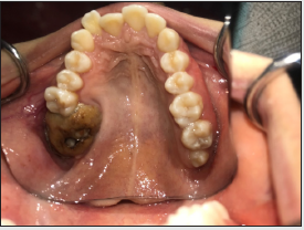
systemically healthy. Intraorally there was a calcified brownish
yellow mass on the posterior left maxilla. Second and third molars
were missing. Adjacent soft tissue was normal showing no signs of
infection and patient stated no pain (Figure 1). Cone beam computed
tomography (CBCT) images showed a solid radiopaque mass
surrounded by a radiolucent halo, displaced the maxillary sinus,
associated with an impacted molar tooth. Displacement had caused
a reduction in the volume of the left maxillary sinus and sinus floor
appeared as uninterrupted, but the sinus was blocked (Figure 2A-
2C). Bone walls around the mass was intact except the erupted part.
Dimensions of the mass were 28.58 mm in anteroposterior, 23.20
mm in transverse and 30.40 mm in superoinferior axis. Adjacent
first molar tooth had grade 2 mobility because of bone resorption
in the area. In the light of clinical and radiographic data, it was
decided to perform surgical removal with the initial diagnosis of
erupted complex odontoma. The differential diagnosis for the
lesion was osteoma, ossifying fibroma, ameloblastic fibroma and
fibro-osseous lesions [5]. Considering the size of the lesion, surgery
was planned under general anesthesia.
Surgical removal of the odontoma was performed through
intraoral approach. A mucoperiosteal flap was raised to reveal
the tumor (Figure 3A). Excision of tumor and fibrous capsule was performed along with removal of the impacted molar (Figure
3B, Figure C). Adjacent first molar was extracted due to lack of
bone support. Maxillary sinus examination showed intact bony
walls. The buccal fat pad was mobilized and advanced to cover
the remaining defect and buccal flap was scored in the horizontal
plane for primary closure of the soft tissue (Figure 3D-3F).
Histopathologic examination revealed haphazard deposition of
dental tissues confirming the preliminary diagnosis of complex
odontoma. Postoperative recovery was uneventful and clinical and
radiographic examination after 12 months showed no recurrence
(Figure 4).
Figure 1: Intraoral image showed the calcified brownish yellow mass with normal adjacent soft tissue.

Figure 3: Intraoperative exposure of the tumor via intraoral approach (A). Excision of the tumor along with fibrous capsule (B). Bone defect following the total removal of the lesion (C). Primary closure of the defect with buccal fat pad mobilization and buccal flap scoring (D and E). Macroscopic aspect of the lesion and fibrous capsule along with extracted first molar and impacted molar teeth (F).

Discussion
Odontomas are hamartomatous lesions placed under benign mixed odontogenic tumors in World Health Organization classification, published in 2017 [1,6]. Complex odontomas are less common than compound odontomas and for complex odontoma, prevalence favors females.1 Unlike our case complex odontomas are usually located in the mandible and a meta-analysis conducted on 3065 cases revealed that only 18.3% of maxillary odontomas were located at the posterior/molar area [7]. Eruption of the odontomas to the oral cavity is very rare and was stated as 1.6% of all cases [4]. And erupted odontomas are mostly related with impacted teeth [8]. Also, 50% of the complex odontomas were reported be associated with impacted teeth [9]. Preservation of the impacted teeth may be beneficial especially on cases with compound odontoma in the anterior region of the jaws.10 Unfortunately, in our case, there was not enough bone support after the removal of the odontoma and the location was unreachable for orthodontic movement of the tooth. Surgical excision of the odontoma is well studied and efficient way for treatment with no recurrence. Recurrence is only reported in case of early excision of the tumor in the non-calcified phase [7]. Local anesthesia is usually sufficient for surgical excision of the tumor, but general anesthesia is required especially in large and/or hard-to-reach lesions.
Conclusion
This case presents a scarce erupting complex odontoma. Eruption can cause symptoms in cases otherwise silent. Surgical excision of the tumor gives favorable results, without recurrence and usually helps to preserve adjacent tissues.
References
- Zhuoying C, Fengguo Y (2019) Huge erupted complex odontoma in maxilla. Oral Maxillofac Surg Cases 5(1): 100096.
- Satish V, Prabhadevi M, Sharma R (2011) Odontome: A Brief Overview. Int J Clin Pediatr Dent 4(3): 177-185.
- Olgac V, Koseoglu BG, Aksakalli N (2006) Odontogenic tumours in Istanbul: 527 cases. Br J Oral Maxillofac Surg 44(5): 386- 388.
- Amado Cuesta S, Gargallo Albiol J, Berini Aytés L, Gay Escoda C (2003) Review of 61 cases of odontoma. Presentation of an erupted complex odontoma. Med Oral 8(5): 366-373.
- Bueno NP, Bergamini ML, Elias FM, Braz-Silva PH, Ferraz EP (2020) Unusual giant complex odontoma: A case report. J Stomatol Oral Maxillofac Surg 121(5): 604-607.
- Adel EI-Naggar K, John Chan KC, Jennifer Grandis R, Takashi Takata PJS (2017) WHO classification of Head and Neck tumors. IARC Press: Lyon.
- Hidalgo-Sánchez O, Leco-Berrocal MI, Martínez-González JM (2008) Meta analysis of the epidemiology and clinical manifestations of odontomas. Med Oral Patol Oral Cir Bucal 13(11): 730- 734.
- Bagewadi SB, Kukreja R, Suma GN, Yadav B, Sharma H (2015) Unusually large erupted complex odontoma: A rare case report. Imaging Sci Dent 45(1): 49- 54.
- da Silva VS de A, Pedreira R do PG, Sperandio FF, Nogueira DA, de Carli ML, et al (2019) Odontomas are associated with impacted permanent teeth in orthodontic patients. J Clin Exp Dent 11(9): 790- 794.
- Isola G, Cicciù M, Fiorillo L, Matarese G (2017) Association between odontoma and impacted teeth. J Craniofac Surg 28(3): 755- 758.

Top Editors
-

Mark E Smith
Bio chemistry
University of Texas Medical Branch, USA -

Lawrence A Presley
Department of Criminal Justice
Liberty University, USA -

Thomas W Miller
Department of Psychiatry
University of Kentucky, USA -

Gjumrakch Aliev
Department of Medicine
Gally International Biomedical Research & Consulting LLC, USA -

Christopher Bryant
Department of Urbanisation and Agricultural
Montreal university, USA -

Robert William Frare
Oral & Maxillofacial Pathology
New York University, USA -

Rudolph Modesto Navari
Gastroenterology and Hepatology
University of Alabama, UK -

Andrew Hague
Department of Medicine
Universities of Bradford, UK -

George Gregory Buttigieg
Maltese College of Obstetrics and Gynaecology, Europe -

Chen-Hsiung Yeh
Oncology
Circulogene Theranostics, England -
.png)
Emilio Bucio-Carrillo
Radiation Chemistry
National University of Mexico, USA -
.jpg)
Casey J Grenier
Analytical Chemistry
Wentworth Institute of Technology, USA -
Hany Atalah
Minimally Invasive Surgery
Mercer University school of Medicine, USA -

Abu-Hussein Muhamad
Pediatric Dentistry
University of Athens , Greece

The annual scholar awards from Lupine Publishers honor a selected number Read More...


