Lupine Publishers Group
Lupine Publishers
Menu
ISSN: 2644-1403
Review Article(ISSN: 2644-1403) 
Painful Shoulder at the Hemiplegic Volume 1 - Issue 3
Saloua Khalfaoui* and El Mustapha El Abbassi
- Department of Physical Medicine and Rehabilitation of the Military Instruction Hospital, Morocco
Received:May 22, 2019; Published: May 29, 2019
Corresponding author: Saloua Khalfaoui, Department of Physical Medicine and Rehabilitation of the Military Instruction Hospital, Morocco
DOI: 10.32474/GJAPM.2019.01.000115
Abstract
The pain of the shoulder at the hemiplegic is one of the main complications arisen post-stroke. The causes are multiple and in most of the cases, they are associated with pre-existent or added risk factors; where from the interest of a premature diagnostic coverage with implementation of the means of prevention and curative treatment to favor the neurological recovery.
Keywords: Hemiplegia; Painful shoulder; Prevention and treatment
Introduction
Post stroke hemiplegia (CVA) is a common pathology occurring in France with an incidence of 140000 new cases / year, half of which has a motor disability. It is responsible for numerous complications contributing to the increase of the initial handicap [1]. Shoulder pain is one of the major complications reported in hemiplegics, a topic of daily physical and rehabilitation concern. The prevalence of this pain can be as high as 84% of cases [2] and its onset during the first year of the stroke affects 30% of hemiplegic patients. According to Bender [3], the origin of this shoulder pain is divided into four classes: articular, muscular. Neurological or complex regional pain syndrome type I.
Anatomical Reminder of the Glenohumeral Joint
This is one of the 5 joints of the shoulder joining the humeral head to the glenoid cavity of the scapula. It is synovial spheroid type providing 3 degrees of freedom (flexion/extension, abduction/ adduction, internal rotation/external rotation). It has two articular surfaces, the glenoid cavity and the humeral head. The glenoid cavity is oriented outwards and forwards. It is pisciform and almost flat. The humeral head has the shape of 1/3 of a sphere, directed upwards, inwards and backwards and seat of two reliefs the major and minor tubercle and two cervical anatomical and surgical. The glenoid bead is a fibrocartilage that fits around the glenoid cavity, is triangular in shape, and increases the concavity and surface. The means of union are of two types: passive and active.
Liabilities
The capsule: forms capsular folds in the lower part to allow the abduction movement. The synovium lines the inner side of the capsule. There are three groups of ligaments: the coraco-humeral ligament, which is composed of 2 upper and lower bundles, the upper and lower middle glenohumeral ligaments, which define 2 points of weakness superior and inferior and the transverse humeral ligament. Active: they are represented by the muscles of the rotator cuff (over and under thorny, small round), the tendon of the long head of the biceps and triceps and the deltoid. The management of these pains is of great importance because its persistence and accentuation interfere with rehabilitation and motor recovery of the upper limb as well as patient involvement [4,5].
Physiopathology of the hemiplegic shoulder
Once the neurological involvement is there, the periarticular muscles of the shoulder no longer play their role
of congruence. The latter is ensured by the capsulo-ligamentary elements from where the fragility at
hemiplegic.
Risk factors [3]
The occurrence of shoulder pain in the hemiplegic is related to the presence of certain risk factors such as:
I. Glenohumeral subluxation (between 17 and 80% of cases), considered as an indirect and insufficient factor [6].
II. The hemorrhagic cause of stroke with involvement of the premotor area.
III. Peripheral neuropathy by stretching the subscapular and circumflex nerve due to glenohumeral subluxation.
IV. Denervation of the median nerve.
V. Presence of a sensitivomotor deficit near the shoulder.
VI. Presence of a depressive syndrome.
VII. Appearance of spasticity of the upper hemiplegic limb.
VIII. Rehabilitation technique adopted (such as pulley therapy).
IX. Presence of metabolic disorders (dyslipidemia, diabetes, uricemia, dysthyroidism).
X. The management of pain must be comprehensive because the cause is often multifactorial.
Etiology of shoulder pain in hemiplegic patients
Pain of osteoarticular origin [7]
¬Retractile capsulitis or frozen shoulder with a prevalence between 43% and 77% in the hemiplegic [8], it corresponds to a retraction of the capsule which is part of the means of passive union of the glenohumeral joint to the origin of the limitation of the articular amplitudes in passive especially the flexion and the external rotation with a classic evolution in three phases (pain, stiffness and recovery). The sub-acromial conflict: during the ascent of the humeral head, the inextensible and rigid slide where the supraspinous tendon passes no longer allows it to slide normally, a source of degenerative lesions due to tendon suffering objectified by the Neer test. Tendinous lesions occurring between 35 and 55% of cases [7]. It is partial or total tendinopathy of the rotator cuff muscles due or aggravated by forced mobilizations, traction, subluxation and trauma by fall. The diagnosis is confirmed by magnetic resonance imaging.
Pain of muscular origin
¬Spasticity: speed-dependent hypertonia corresponding to the exaggeration of the myotatic muscle reflex following stretching. In the hemiplegic, it concerns the adductor muscles, internal rotators and pronators of the upper limb, responsible for a triple flexion attitude. Subluxation of the humeral head: this corresponds to the permanent movement down and in front of the humeral head with respect to the scapular glenoid, following the paralysis of the fixing muscles. It can cause pain in 50% of cases [8], but hemiplegic patients with subluxation do not necessarily have shoulder pain [1]. In other words, there is no correlation between the degree of dislocation and the intensity of pain [7]. The neuropathic pain by peripheral or central sensitization: Neuropathic pain is due to damage to the central or peripheral nervous system without tissue damage, in contrast to mechanical pain. The initial cause of shoulder pain is due to the sensitization phenomenon that results in the lowering of the pain threshold and therefore each stimulus can become painful. The regional complex syndrome type I: formerly called algoneurodystrophie or shoulder-hand syndrome. Its etiology and physiopathology remain unknown. Its incidence in the hemiplegic can go up to 48% and occurs mainly in the presence of the other etiologies of the painful shoulder [9]. The evaluation of the different pains must be regular by using for each type the corresponding scales and questionnaires (numerical scale, visual analogue scale, verbal scale, DN4 questionnaire ...), with impact on the quality of life and the activities of the daily life.
Evolution over time [7]: According to the studies, the time of onset of pain does not match the age of the stroke. The appearance and intrication of risk factors also play a role in the progressive mode of pain in hemiplegic patients [10,11].
Preventive management: by acting on controllable factors
Evolution dans le temps [7]: Selon les études, le moment d’apparition des douleurs ne correspond pas à l’ancienneté de l’AVC. L’apparition et l’intrication des facteurs de risque, joue aussi un rôle dans le mode évolutif des douleurs chez l’hémiplégique [10,11].
a) Preventive management: by acting on controllable factors
b) Replace barbiturates with other anti-epileptics.
c) Avoid traction on the hemiplegic limb.
d) Treat spasticity.
e) To fight against so-called decoaptation positions, source of intra and extra-articular lesions.
f) Circulatory massage and lymphatic drainage of the entire hemiplegic upper limb.
g) Handling is an important modality, consisting in giving much more interest to the hemiplegic shoulder:
h) When transferring, do not pull on the hemiplegic limb.
i) During reversals, insist on self-mobilization by the healthy side.
j) Start the dressing by the side reached.
k) Pulley therapy and the use of the gallows are no longer recommended [12].
l) Education of caregivers, caregivers and patients to prevent shoulder trauma.
m) This is the teaching of strategies to correctly position the affected member.
n) Do not mobilize the shoulder beyond 90° of anterior or lateral elevation unless the external bellows of the scapula.
o) Functional electrical stimulation resulting in muscle contraction and transcutaneous nerve stimulation with analgesic effect often meet. No application modality is defined in this support. No studies have shown a decrease in the incidence of pain by electrical stimulation, but it could prevent subluxation [1].
p) In the supine position: shoulder elevated from the healthy side, 60° abduction, 30° of anterior elevation, elbow flexed to 30°, hand semi-pronation on a foam support with fingers extended apart and thumb abducted.
q) In lateral decubitus: if positioned on the healthy side, the hemiplegic arm must be installed in abduction, on a pillow and if the patient is placed on the affected side, the hemiplegic arm must be well cleared forward and in ante pulsion.
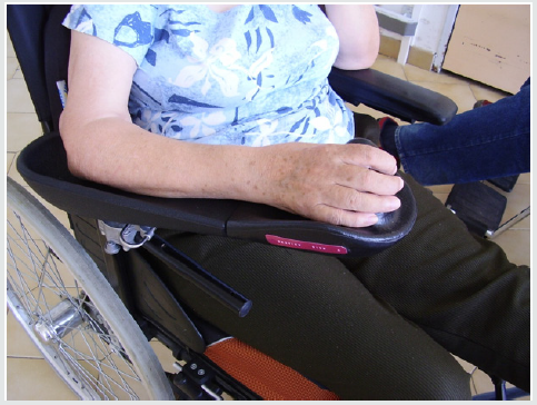
r) When seated in the wheelchair: the armrest must be adapted to the height of the arm with posterior stop to prevent slipping back of the elbow (risk of anterior subluxation of the humeral head), always withdrawing traction during transfers (Figure 1).
s) When standing, taping is used to keep and monitor correct alignment of the glenohumeral joint. It is recommended for the prevention of pain.
t) Curative management: is limited to symptomatic treatment and must take into account the different risk factors [5].
Physical means: to relieve pain by
Cryotherapy and heat: some authors suggest their use, others not because they increase the central pain.
The laser: can be used to reduce the intensity of the pain provided that it does not exceed more than 50 joules per treatment and 8 joules for each pain point [13].
a. Aromatherapy associated with acupuncture or acupressure is effective in decreasing the duration of pain treatment.
b. Bobath-type therapy that is more effective than cryotherapy in reducing pain.
c. The neuro-musculo-proprioceptive facilitation method is more effective than the Bobath type therapy.
d. The combination of relaxation techniques and EMG biofeedback is effective in hypertonic shoulder pain [14].
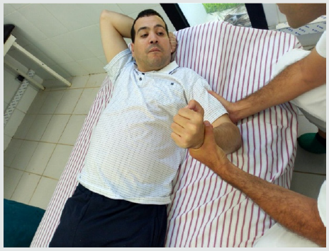
e. Do not forget to reduce spasticity before performing mobilizations on the shoulder (Figure 2).
Mirror therapy: is a method that has proven effective in relieving central pain after stroke and those associated with complex regional pain syndrome type I [7].
Medication Means Correspond to Classical Pharmacological Medical Interventions
i. Simple analgesics and nonsteroidal anti-inflammatory drugs are recommended as first-line therapy in the initial phase and in the absence of contraindications, but their efficacy is unproven.
ii. Oral or injectable corticosteroids are to be prescribed for a short duration without exceeding ten days.
iii. Block infiltrations such as suprascapular block and pectoral muscles are used to reduce pain and increase mobility.
iv. Botulinum toxin type A is used to fight against spasticity with effect from the first week and effectiveness for four months.
v. Tricyclic antidepressants and anticonvulsants are effective in central pain.
vi. Surgical means: in the absence of results on the reduction of the pain by the aforementioned means.
Their places become more and more limited in view of the good results obtained by the recent methods of rehabilitation. We cite:
1) Looping the long portion of the biceps around the coracoid process.
2) Implantation of a muscle microstimulator near the axillary nerve [15]: may be indicated if pain secondary to subluxation.
3) Psychology: in case of central pain lasting more than two months, psychotherapy, hypnosis and relaxation are proposed to relieve psychic suffering.
4) Les infiltrations par bloc comme le bloc supra-scapulaire et celui des muscles pectoraux sont utilisés pour diminuer la douleur et augmenter la mobilité.
5) La toxine botulinique type A est employée pour lutter contre la spasticité avec effet dès la première semaine et efficacité pendant quatre mois.
6) Les antidépresseurs tricycliques et les anti-convulsivants sont efficaces dans les douleurs centrales.
7) Surgical means: in the absence of results on the reduction of the pain by the aforementioned means. Their places become more and more limited in view of the good results obtained by the recent methods of rehabilitation.
We Cite
I. Looping the long portion of the biceps around the coracoid process.
II. Implantation of a muscle microstimulator near the axillary nerve [15] may be indicated if pain secondary to subluxation. Psychology: in case of central pain lasting more than two months, psychotherapy, hypnosis and relaxation are proposed to relieve psychic suffering.
Conclusion
The management of the painful shoulder in the hemiplegic must be multidisciplinary given the strong link existing with the heterogeneous factors that may appear and evolve throughout the initial pathology. The diagnosis remains difficult to establish because of the intrication of other complications of stroke, hence the importance of the establishment of preventive and curative means of fighting this pain to promote neurological recovery and improve the quality of life.
References
- Gliez B, Jacquin-Courtois S, Rode G (2012) l’épaule du patient hémiplégique La Lettre du Rhumatologue.
- Pignon L (2016) Épaule hémiplégique douloureuse : facteurs mécaniques. Kinesither Rev
- Bender L, McKenna K (2001) Hemiplegic shoulder pain : defining the problem and its management. Dis. Rehab 23: 698-705.
- Duncan PW, Zorowitz R, Bates B, Choi JY, Glasberg JJ, et al. (2005) Management of Adult Stroke Rehabilitation Care: a clinical practice guideline. Stroke 36(9): e100-e143.
- Brigittebouchot-Marchal (2006) Que faut-il faire de l’épaule de l’hémiplégique ? Kinesither Rev (61): 43-45.
- Dursun E, Dursun N, Ural C (2000) Glenohumeral joint subluxation and reflex sympathietic dystrophie in hemiplegics patients. Arch Phys Med Rehab 81: 944-946.
- Blennerhassett JM, Gyngell K, Crean R (2010) Reduced active control and passive range at the shoulder increase risk of shoulder pain during inpatient rehabilitation poststroke: an observational study. J Physiother 56(3): 195–196.
- Roosink M, Renzenbrink GJ, Buitenweg JR, Van dongen RTM, Geurts ACH (2011) Somatosensory symptoms and signs and conditioned pain modulation in chronic post-stroke shoulder pain. J Pain Off J Am Pain Soc 12(4): 476–85.
- Kocabas H, Levendoglu F, Ozerbil OM, Yuruten B (2007) Complex regional pain syndrome in stroke patients. Int J Rehabil Res 30: 33-38.
- Layadi K, Lahouel F, Rouai F, Talem Z, EL-habil F, et al. (2009) Les troubles orthopédiques chez les hémiplégiques par accidents vasculaires cérébraux : expérience du service de médecine physique du CHU Oran. Journal de Réadaptation Médicale : Pratique et Formation en Médecine Physique et de Réadaptation 29(3): 99–104.
- Lindgren I, Jonsson AC, Norrving B, Lindgren A (2007) Shoulder pain after stroke: a prospective population-based study. Stroke 38(2): 343– 348.
- Opsommer E, Knutti IA, Zwissig C, Eberlé G (2016) La prévention et le traitement de la douleur de l’épaule après un accident vasculaire cérébral (AVC): Recommandations pour la pratique clinique. Bureau d’Echange des Savoirs pour des pratiques exemplaires de soins. Lausanne (Suisse).
- Murie C (2010) Le laser en physiothérapie. PHT-2320 : Modalités électrothérapeutiques 2. Université de Montréal.
- Page T, Lockwood C (2003) Prevention and management of shoulder pain in the hemiplegic patient. JBI Reports 1(5): 149-165.
- Yu (2004) Intramuscular Neuromuscular Electric Stimulation for Poststroke Shoulder Pain: A Multicenter Randomized Clinical Trial». Archives of physical medicine and rehabilitation 85(5): 695-704.

Top Editors
-

Mark E Smith
Bio chemistry
University of Texas Medical Branch, USA -

Lawrence A Presley
Department of Criminal Justice
Liberty University, USA -

Thomas W Miller
Department of Psychiatry
University of Kentucky, USA -

Gjumrakch Aliev
Department of Medicine
Gally International Biomedical Research & Consulting LLC, USA -

Christopher Bryant
Department of Urbanisation and Agricultural
Montreal university, USA -

Robert William Frare
Oral & Maxillofacial Pathology
New York University, USA -

Rudolph Modesto Navari
Gastroenterology and Hepatology
University of Alabama, UK -

Andrew Hague
Department of Medicine
Universities of Bradford, UK -

George Gregory Buttigieg
Maltese College of Obstetrics and Gynaecology, Europe -

Chen-Hsiung Yeh
Oncology
Circulogene Theranostics, England -
.png)
Emilio Bucio-Carrillo
Radiation Chemistry
National University of Mexico, USA -
.jpg)
Casey J Grenier
Analytical Chemistry
Wentworth Institute of Technology, USA -
Hany Atalah
Minimally Invasive Surgery
Mercer University school of Medicine, USA -

Abu-Hussein Muhamad
Pediatric Dentistry
University of Athens , Greece

The annual scholar awards from Lupine Publishers honor a selected number Read More...


