Lupine Publishers Group
Lupine Publishers
Menu
ISSN: 2644-1403
Case Report(ISSN: 2644-1403) 
Mucoepidermoid Carcinoma; Clinical Case Report Volume 2 - Issue 5
Miranda Nava Gabriel1*, Gallo Frias Luis Gilberto2, Pimentel Diaz Alejandro Yami2 and Ginger Ramirez Diana Guadalupe3
- 1Neurologist, Major of Military Armed Forced of Mexico, USA
- 2Medical Doctor, Lamar University, Mexico
- 3Nutritionist, Master’s in Human Development, Mexico
Received: February 26, 2020; Published: March 10, 2020
Corresponding author: Miranda Nava Gabriel, Neurologist, Major of Military Armed Forced of Mexico, USA
DOI: 10.32474/GJAPM.2020.02.000150
Abstract
Mucoepidermoid carcinomas are rare and comprise 5% of head and neck malignancies and are seen in 2.6 cases for every 100,000 people. We report the case of a 49 year old blacksmith from Guadalajara, Mexico who presents to office and who is later diagnosed with a mucoepidermoid carcinoma located in the left side of the submandibular gland, tumor was treated with two surgery for removal of the mass with patient now being seen under periodic moments.
Background
Currently mucoepidermoid carcinomas are very rare they count for less than 5% of head and neck tumor and most of them occur in Young adults and women, nowadays it can be divided into three types one for mucin producing cells, another one for intermediate or clear cells and squamoid cells. Three grades are given to classify the tumor which are grade 1 for a tumor that does not usually metastasize and that is cured by an appropriate surgery, grade two is given to tumors that are in an in-between spectrum with grade 3 and that have a risk for developing disease progression and a certain mortality rate, and finally a grade 3 tumor is one with a major risk for presenting positive lymph nodes and disease progression and related mortality [1].
Case Report
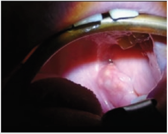
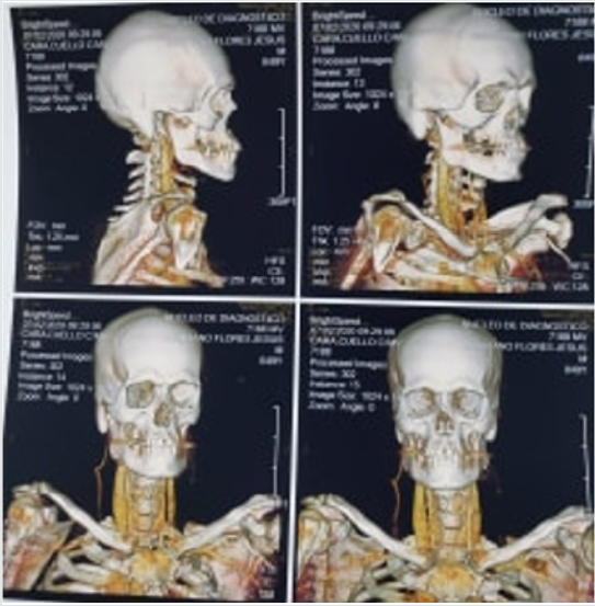
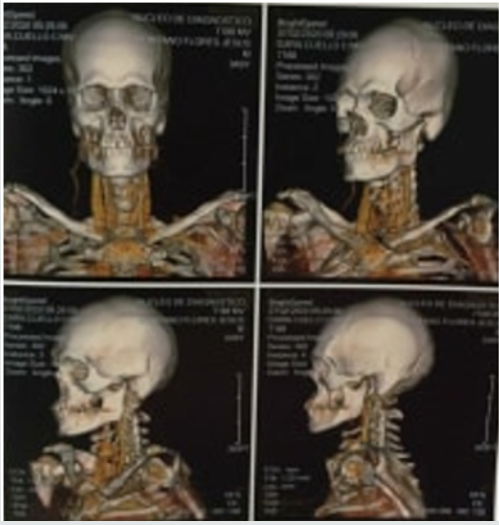
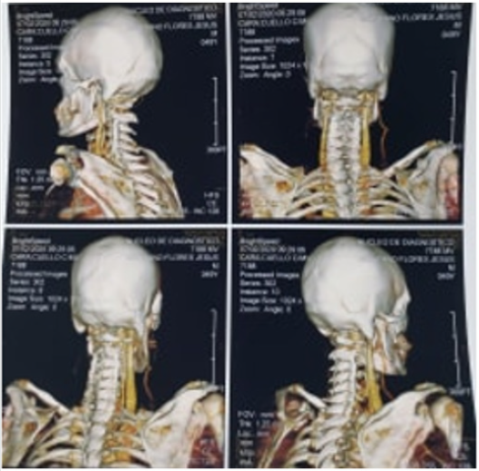
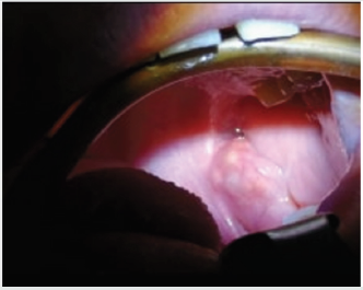
A 49 year old man presented to office in the San Martin Clinic located in Guadalajara, Mexico with a history of 6 month halitosis, as well as 2 year history of swelling in his lower left side of the retromolar region, the swelling had increase in size gradually, with medical and dental history being normal until now patient did not have any chronic illness or history of smoking, he drank occasionally and had no major problem in his personal background, he was performed an intraoral examination a solitary well defined oval shaped erythematous swelling on the left side of the retromolar area is seen with a size of about 2.0 x 1.5 cm with irregular borders, it was not painful to the touch and had a firm tenderness (Figure 1). A Computed Tomography Scans for Head and Neck with contrast material revealed a ganglion located in the submandibular gland a 1b stage pair of ganglions augmented in size located in suprahyoid and infrahyoid muscles with a size of 18 x 14 mm located anterior to the submandibular gland the other ganglions observed had a diameter of 10 mm or below (Figures 2-4).
Treatment
After evaluation patient was schedule to perform two surgeries for removal of the tumour which were carried out on the San Martin Clinic, surgical intervention included wide excision of the tumour located the submandibular gland and second surgery included wide excision of lymph nodes located in adjacent areas in the suprahyoid muscles and infrahyoid muscles reconstruction.
Pathology
A biopsy was taken during surgery and it was analyzed by a pathologist where I teas diagnosed as a low-grade stage 1 malignant mucoepidermoid carcinoma with dimension of 2 x 3 x 1 cm with granulomatous chronic peripheric inflammatory process (Figure 5) [2].
Discussion
These type of tumors are very rare they comprise only 5% of neoplasms and are seen in 0.4-2.6 for every 100,000 cases around the world, the mucoepidermoid tumor affects parotid and minor salivary glands in adults and is mostly seen in women and Young adults, most of the cases arise in the parotid gland with this case accounting for only 2-4% of the cases because it was seen in the submandibular gland, this patient is currently under treatment he was performed two surgeries for removal of ganglions located in neck and in the submandibular gland, highest prevalence for this type of tumor is around the fifth decade of life and they can be asymptomatic like in this case with the patient having few to no symptoms. It has a pluripotent cell origin and as we mention can be classified into three stages [3].
References
- Shuting B, Rashna C, Esther ABS (2012) Salivary Mucoepidermoid Carcinoma: A Multi-Institutional Review of 76 Patients. Head Neck Patho 7(2): 105-112.
- Samiksha JJ, Sushma D, Savitha A, Anirban C, Chaitanya B(2016) Mucoepidermoid carcinoma of the palate: A rare case report. J Indian Soc Periodontol 20(2): 203-206.
- Ramaraju D, Ramlal G, Harisha A, Srikanth G (2014) Mucoepidermoid Carcinoma. BMJ Case Rep

Top Editors
-

Mark E Smith
Bio chemistry
University of Texas Medical Branch, USA -

Lawrence A Presley
Department of Criminal Justice
Liberty University, USA -

Thomas W Miller
Department of Psychiatry
University of Kentucky, USA -

Gjumrakch Aliev
Department of Medicine
Gally International Biomedical Research & Consulting LLC, USA -

Christopher Bryant
Department of Urbanisation and Agricultural
Montreal university, USA -

Robert William Frare
Oral & Maxillofacial Pathology
New York University, USA -

Rudolph Modesto Navari
Gastroenterology and Hepatology
University of Alabama, UK -

Andrew Hague
Department of Medicine
Universities of Bradford, UK -

George Gregory Buttigieg
Maltese College of Obstetrics and Gynaecology, Europe -

Chen-Hsiung Yeh
Oncology
Circulogene Theranostics, England -
.png)
Emilio Bucio-Carrillo
Radiation Chemistry
National University of Mexico, USA -
.jpg)
Casey J Grenier
Analytical Chemistry
Wentworth Institute of Technology, USA -
Hany Atalah
Minimally Invasive Surgery
Mercer University school of Medicine, USA -

Abu-Hussein Muhamad
Pediatric Dentistry
University of Athens , Greece

The annual scholar awards from Lupine Publishers honor a selected number Read More...


