Lupine Publishers Group
Lupine Publishers
Menu
ISSN: 2644-1403
Research Article(ISSN: 2644-1403) 
Assessment and Correction of the Rachis Dysfunctions with Beninese Visually Impaired Subjects Volume 1 - Issue 5
Salifou Kora Zaki Yarou1*, Mouhamed Mansourou Lawani1, Toussaint Kpadonou2, Etienne Alagnidé2, Lafiou Yessoufou1 and Germain Houngbédji2
- 1Laboratory of Biomechanics and Performance (LaBioP) INJEPS-UAC, Africa
- 2Department of Rehabilitation and Fonctionnal Readjustment CNHU-HKM, Africa
Received: July 01, 2019; Published: July 11, 2019
Corresponding author: Salifou Kora Zaki Yarou, Laboratory of Biomechanics and performance INJEPS-UAC, Africa
DOI: 10.32474/GJAPM.2019.01.000123
Abstract
Background: Visual impairment, cause of a less stress on the physical abilities, is a source of loss of flexibility and muscle incompetence. Identify and correct spinal dysfunctions at the musculoskeletal level from visually impaired subjects. Methods: A prospective study with a descriptive and analytical carried on 53 visually impaired subjects including 38 “blinds” and 15 partially sighted. These patients were evaluated, using Schöber, Sorensen and Shirado-Ito tests, the flexibility of their trunks and the endurance of their abdominal and back muscles, before and after 15 sessions of 30 minutes of Iso stretching exercises. Results:1) The ratio trunk flexion ÷ trunk extension different from 2 (TF/TE≠2) during the first test, indicates, at insignificant level by sex, age and importance of the deficiency, the loss of flexibility (0.4≤P≤0.7) and/or extensibility (0.2≤P≤0.4) of the core muscles of all visually impaired subjects. 2) The average time ratio of flexor and extensor maintaining (F/E) during the first test not from 0.7 to 0.8 indicates a poor tone distribution between the two muscle groups of the trunk. 3) Notwithstanding the significant gains at 5% level recorded in endurance of the flexors and extensors muscles of the trunk from the boys B1 from the age of 15-19 years and 20-24 years, the balance of muscle tone (F/E=0.79) was obtained in the group of older subjects at the end of exercise sessions. Conclusion: The study reveals the rachis dysfunctions highlighted by a loss of flexibility and/or extensibility of the trunk and a tone imbalance between the abdominal muscles and the one of the back from all the visually impaired subjects. It has allowed to improve significantly in 15 sessions of Iso stretching exercises, the rachimetric normality of the participants.
Keywords: Visual Impairment; Dysfunction; Iso Sstretching; Rachimetric Normality
Introduction
A research on people suffering from visual disability which has the mobility of complex lumbo-pelvic femoral as major concern, meets a key demand [1]. Indeed, these people who live often in situations of social exclusion because of a deficiency, are usually forced to immobility or to be in reclined position and are afflicted by complications in cardiopulmonary and musculoskeletal level. Thus, long-term decubitus can lead for example to a mismatch stress with skeletal muscle changes that can worsen a pre-existing heart failure, a bronchial congestion or lower respiratory tract diseases [2,3]. As with any individual, it seems that the adapted physical activity is more useful for this category of persons characterized by a “hypodynamy”, a lack of self-confidence, a lack of initiative and involvement in physical activities [4,5], a joint restriction, an insufficient knowledge of the body patterns and a postural attitude affected [6-8]. Indeed, for proper operation of the vertebral spine, every anatomical system must respond and be organized as smoothly as possible with all the body. Unfortunately, with blindness the trunk wedged between the arms and the thighs hyper mobile [1] for sighted and relatively immobile for the visually impaired, will undergo without any doubt whatsoever many constraints, especially given that the psychic, so physical, retreat [9] of the latter will accentuate the lack of control of this region generally forgotten, unknown and sometimes painful. This study focus specifically to individuals suffering from visual impairment, therefore from a disability that may limit their autonomy and their ability to develop their functional capacities and motor skills. It tried to answer the lack of simple method that can allow the visually impaired to autonomously improve his muscular skeletal conditions, including the one of the trunks, by practicing iso stretching exercises reproducible at home.
Material and Methods
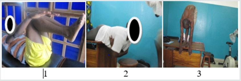
The equipment used in the study consists essentially of three exercise bikes Gymna Ergo-Fit 450, Polar branded, equipped with an electronic counter and that allowed the visually impaired subjects to warm up before doing the Isotretching exercises. There were also a Heart-rate monitor RS 100 type, branded Polar Electro Oy, Professorintie 5 FIN-90440 Kempele Finland, that was used to take the heart rate at rest and during warm-up ; a treatment table used during the Sorensen tests; gym mats providing security and comfort that were used during Isostretching exercises; a stopwatch Oregon scientific 500-Lap Memory which was used to determine the maintaining time of visually impaired subjects during Sorensen and Shirado Ito tests; a tape measure for measuring the flexibility and extensibility of the trunk, as well as the DDS (distance between finger and ground) during the Schober test; a felt-tip pen; Klein Vogelbach balloons for the training of extensors and abdominal muscles of the visually impaired subjects; electronic bathroom scales branded SECA (J.J.A: 157 AV CHARLES FLOQUET 93150 LE BLANC MESNIL, FRANCE) for weighting gain of visually impaired subjects and a stool that served as support during Sorensen test. The flexibility of the trunks and the endurance of abdominal and back muscles of these patients were evaluated, using Schöber, Sorensen and Shirado-Ito tests, before and after 15 sessions of 30 minutes of Isostretching exercises.
Subjects
This study focused on the visually impaired subjects (pupils, students, craftmen and switchboard operator) who are ‘’blind’’ (B1) or visually impaired (B2). After an evaluation of inclusion, 53 subjects (38”blind” including 11 girls and 15 visually impaired including 3 girls) aged of 23.06±15.26 years for girls and 23.84±6.10 years for boys, on average, were included in the program of 15 sessions of Isostretching exercises (Table 1).They all agreed to follow the experimental protocol, which required the approval of the CER-ISBA (Comité d’Ethique de la Recherche, de l’Institut des Sciences Biomédicales Appliquées) of Benin. In addition to the school records, an ophthalmologist examined all subjects to confirm the category of their deficiency. All subjects in this study met the following criteria:
a) Inclusion Criteria
i. Being visually impaired who is at least fifteen years old, coming from one of “Centres de Promotion Sociale des Aveugles et Amblyopes du Bénin”.
ii. Answer “no” to all of the questions in the IPAQ (Questionnaire International à la Pratique d’Activités Physique) [10].
iii. Agree to be committed at the CPSAA of Sègbèya in Cotonou, during the data collection period.
iv. Not having a chronic low back pain.
b) Non-Inclusion Criteria
The non-inclusion criteria of the study were:
i. Mental retardation that could be associated with blindness,
ii. Pathology that would contraindicate the practice of physical activities.
c) Exclusion Criteria
Were excluded from the study, subjects whose irregularities during the practice session were noted for more than three sessions or subjects declared sick.
Method and Survey Methodology
They are based on:
i. A documentary analysis made possible by close collaboration with all stakeholders of SRRF. This allowed us to withhold Isostretching [1] and exercises on ball [11] as technical work.
ii. The definition of rachimetric normality: TF = 2.TE; i.e TF ÷ TE = 2 with F = flexion, E = extension, and T = trunk. [12] iii. The implementation of the experimental protocol, for five weeks with three sessions per week, for a period of 05 months.
Collection and Processing of Data
The data relating to the measurement of the trunk flexion and extension were carried out on the lumbar area, between a first marker at the vertebra S1 and a second located at 10cm above, on the midline, during deep flexion and extension of the trunk. Those concerning the maintaining time of the abdominal and back muscles were made on a table where the visually impaired patient is lying in the prone position, with a partial straightening out of the trunk. Thirty (30) seconds later, the patient is lying in the prone position, the anterior superior iliac spines (ASIS) positioned at the edge of the tabletop edge. The exercises were repeated three times; the 1st time is to make understand by means of bio-feedbacks, the second is to correct oneself and the 3rd is for the best practice by the visually impaired subject. All those data collected from participants before and after practice session, were processed by the software Epi Data 3.1 and SPSS Version 17.0 after the preparation of the input mask and the input control program. The formatting of results (tables and graphs) was performed with Excel and Word. We conducted cross-analyzes by using the KHI2 and Cramer’s V to better convey the evolution of the different parameters according to the importance of deficiency, gender and age group. Comparisons of proportion tests were performed to identify significant differences. This analysis was complemented by a correlation study to check rachimetric normality, that is to say the level of relationships that can exist between flexion and extension of the trunk. The significance level chosen is estimated at 5%.
Results
The analysis of qualitative and quantitative data collected during the tests carried out before and after the training program, helps to explain and justify that some basic parameters of physical condition such as flexibility and muscular endurance can change at all ages with the visually impaired subject.
Professional and Social Characteristics and Aptitude for Physical Activity of Participants
The average age of participants of which 83% are pupils and students, is estimated at 23.44±5.67 years old. The percentage of participation of girls (26.4%), with a sex ratio F/H of 0.35, shows the low passion of girls to the practice of physical activity. In addition, the study proves to be useful by taking into account more than 60% of sedentary subjects and 22% of very irregular persons practicing adapted physical and sports activities. However, the assurance of the ability to practice physical activity of our study population is given by almost 97%. Before they were examined, they claim that:
a) They have never suffered from a heart problem;
b) They have never had a bone or joint problem (for example, back, knee or hip) that could be get worse by a change in their level of participation in physical activity.
Evaluation of the flexibility of the lumbar spine of visually impaired subjects
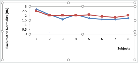
Table 2 shows the averages and standard deviations of flexion and extension measures in the trunk of visually impaired subjects. The ratio of these averages helped to detect a rachimetric abnormality with all participants. Most subjects B2 who were using their remaining vision certainly were able to limit the body decline which is generally source of stiffness of the spine. [9] However, these figures should be treated with great caution because an illustration of rachimetric normality in all groups (Figures 1-6) detects a rachimetric problem (TF/TE≠2), with almost all visually impaired subjects during the first test (T1). Young boys (visually impaired) and girls B1 have shown a loss of flexibility to the not significant threshold between 0.4 and 0.7 according to the gender, age and importance of the deficiency. This showed a loss of flexibility with these visually impaired subjects. When it comes to extensibility, only boys B1 of age groups of 15-19 years and 20-24 years have shown a loss of extensibility of the trunk. The threshold is only significant at the rate of 5% depending on the importance of the impairment. It remained insignificant according to gender and age at respective rates of 0.2 and 0.4. This extensibility of the lumbar spine was well noticed with girls B1 (7.56cm) and groups of partially sighted boys aged 15-19 years (6.70cm) and 20-24 years (7.60cm). The observed differences are not significant at the threshold of 5% regarding to the gender. Despite this relative extensibility observed with younger partially sighted boys (TE < 8 cm), the ratios trunk flexion/trunk extension during the first test (Table 2) are, on average, significantly different from 2 in all participants, and reflect a rachimetricabnormality, confirming the hypothesis which state that there are dysfunctions in the lumbopelvic statics with the visually impaired subject (dysfunctions highlighted by the Schöber test).
Table 2: Comparison of average measurements of the subjects’ flexibility before and after the Isostretching practical sessions.

Evaluation of Muscular Endurance
The boys B1 from the age group of 20-24 years old have shown better endurance of the flexor and extensor muscles of the trunk (65.70 ± 26.44 seconds and 57.70 ± 28.30 seconds) compared to their counterparts aged 15-19 and 25-39 years to the insignificant threshold of 0.1 ≤ P ≤ 0.4 during the first test (Table 3).This imbalance of muscle tone was very noticeable with all subjects B1 with a ratio F/E that varies between 1.01 and 2.50 predisposing thus this category of visually impaired to a chronic low back pain (Table 3). After all, the rachimetric abnormality detected with all the visually impaired subjects during the first assessment associated with the imbalance of muscle tone between the flexors and extensors highlighted through the ratio F/E not between 0.70-0.8 confirms that there are dysfunctions between the flexor and extensor muscles of the trunk with visually impaired subjects, dysfunction shown by Shirardo-Ito and Sorensen tests [13], which justifies our intervention plan made up mainly of isostretching exercises.
Assessment of the Program Effects
a) Muscle Flexibility
The effectiveness of isostrtching exercise program can be assessed by the return of physical and functional parameters’ values of the visually impaired subjects to some values close to healthy populations and the improvement or maintaining of these values over time.
The results obtained in the short term, related to flexibility and lumbopelvic extensibility parameters are shown in Figures 1 - 4 and in (Table 2).The flexibility results are significantly better at the end of the Isostretching exercises program for younger boys B1 aged from 15 to 19 years and for girls B1 aged from 20 to 24 years (p <0.05) from an average respectively 14.87±0.85 cm to 16.50±0.57 cm (±11%), and 14.12±2.32 cm to 15.37± 0.78 cm (±8.9%).The improvement of the trunk flexibility and/or extensibility is accompanied by a line displacement at T3 to the line y=2 (Figures 1-4).
b) Muscular Endurance
The improvement of strength endurance is confirmed during isometric musclar testing (Shirado-Ito and Sorensen tests). The maintaining time of the partial recovery position (curl-up) during the Shirado-Ito test and the horizontal position during the Sorensen test are significantly higher at T3than T1 (p<0.05), respectively from 65.70±26.44 seconds to 127.57±48.31 seconds (±94%) and 52.53±22.65 seconds to 100.91±55.15 seconds (±92%) with B1 aged from 20 to 24 years (Table 3).We sought to determine if there were an efficient exercise program for Isostretching related to the gender during the third test. The analysis of the effect gender of such short-term results for the physical parameters shows a better improvement, not significant (p <0.05) of trunk flexion in the range of 8.9% followed by a reduction of 0.8% of the extensibility of the trunk with girls B1compared to boys B1 of the same age group from 20 to 24 years (Table 2). As far as the endurance of the abdominal and para-vertebral muscles strength is concerned, a balance between these muscle groups seems to be established during the third test with girls B1, respectively from 56.87±26.44 seconds to 99.87±48.31 seconds (±75%) and 58.62±39,25 seconds to 98.37±60.48 seconds (±67%) compared to their male counterparts (Table 3).
Table 3: Comparison of average measurements of the subjects’ muscular endurance before and after the Isostretching exercises.

Discussion
From our results, we found that the exercises Isostretching provides two types of effects with visually impaired subjects:
a) Short-term beneficial effects on the flexibility of the trunk and the isometric muscular endurance;
b) Adverse effects on the extensibility of the trunk. The rachimetricabnormality found during the first test firstly falls in these adverse effects, consequences of mental decline, so bodly [9] and, secondly, in long past periods of inactivity [14], or subsequent to the impairment recognized as harmful [15]. Living with Blindness or low vision, the visually impaired subjects tend to limit their activities whether they are physical or professional [16, 17]. In view of dysfunctions in flexion, in extension, highlighted by a rachimetric abnormality and an imbalance observed between the endurance of the back and abdominal muscles with all visually impaired subjects during the first test, the proposed isostretching exercises aimed:
i. To fight against muscle contractures by backing up on a work of deep plan
ii. To encourage the postural activity of the visually impaired subject
iii. To develop a voluntary respiratory activity.
The latters have, in their heterogeneity, got benefits from the isostretching exercises proposed, by maintaining or by significantly improving their average flexibility which increased from 15.03cm to 16.20cm to the detriment of the average extensibility of their trunks that is reduced from 7.96 cm to 8.08cm (Table 2). This allowed them, as mentioned above, to establish on average rachimetric normality (2.00±0.09) (Table 2).The same rule is applied to partially sighted subjects aged between 20-24 years who have significantly improved their average flexibility and lost their extensibility in order to restore their average rachimetric normality (2.02). Comparing the average measurements of flexibility in our study population, we notice that all visually impaired subjects improved their flexibility with the highest average score in flexibility (16.50±0.57cm) carried out by boys B1 aged 15-19 years without being able to modify the extensibility of their trunk. The partially sighted boys aged 20- 24 also showed significant changes in their flexibility thanks to a significant improvement in their average flexibility and a reduction in their average starting extensibility. This has certainly helped in part the observance of the average rachimetric normality ±2 (Table 2) with these subjects that are supposed to use their “visual remaining” during practice sessions. Subjects B1 in the age group of 20-24 years have significantly improved their flexibility. But the extensibility of core muscles of all subjects B1 remains expressively better than that of other age groups of disabilities, even for girls. However, this improved extensibility and relative flexibility observed with the B1 didn’t allowed to establish the rachimetric normality because of a weakness of abdominal and back muscles. Specifically, girls B1 and partially sighted B2 aged 20-24 years, as well as the boys B1 aged 25-39 years, seems to have got à lot of benefit from the isostretching exercise program which goal was to improve the rachimetric normality (respectively equal to 2.00±0.20, 2.02±0.10 and 2.00±0.09 during the third test) by the improvement of the flexibility and the strengthening of the lumbopelvic complex (Tables 2 & 3). The rachimetric normality of boys B2 aged 15-19 years as well as the one of blinds aged 15-19 years and 20-24 years still remain to be improved because of dysfunctions in flexion and/or extension that persist in these groups, despite significant benefits provided by the Isostretching exercises that has tried a little to restore that rachimetric normality. Figures 1-4 illustrates the improvements produced in each age group according to the deficiency and the gender. The horizontality that is looking for around the number 2 during the third test is not synonymous with the disappearance of the normal curvature of the spine, but it is rather an expression of the equation TF=2 TE reflecting the rachimetric normality [18,19].
The improvements of the abdominal and back muscles maintaining times being also dependent on the initial level, it was therefore not necessary to determine a minimum or a maximum for our study population. On this basis, the overall average performance appears to be enhanced with age (Table 3), boys B1 aged 20-24 years paradoxically getting better results in abdominal endurance (127.57±48.31 seconds) followed respectively by the partially sighted of the same age bracket, subjects B1 of 25-39 years old and naturally girls (Table 3). As far the back muscles endurance is concerned, the partially impaired aged 20-24 years have excelled (125.40± 25.54 seconds), followed by girls and boys B1 of the same age bracket. A comparison by gender made disappear any difference between men and women at all levels of the program. However, age differences are significant at the threshold of 5% and each age group is significantly different from others. Whatever we are looking for in terms of balance between abdominal and back muscles, abdominal muscles seem to have a certain predominance over back muscles. This predominance already compromises the better distribution of muscle tone sought through the ratio F/E between 0.70 and 0.80 [20,21]. Nevertheless, this balance that we are looking for by proposing the isostretching exercises in a playful way on the Vogelbach’s Klein ball was obtained with boys B1 of the older group who presented a better distribution of the muscle tone between Flexors (F) and Extensors (E) of the trunk, with a ratio F/E = 0.79, which is actually between 0.70 and 0.80. This fact can be explained by a predominance of extensors tone (100.91±55.15 seconds) developed with age over the flexors (79.91±20.30 seconds) which are altered with age. Apart from girls and boys B1 aged 20-24 years with whom the exercise program did not produce the desired effect despite the improvement of the tone of the targeted muscles, all other age groups improved the distribution of initial muscle tone through the ratios F/E that come close to the range 0.70-0.80. These significant improvements obtained with the partially sighted (101.00± 25.31 seconds and 125.40±25.54 seconds) and girls B1 (99.87±42.76 seconds and 98.37±60.48 seconds) all aged 20-24 years, have contributed to the observance of the rachimetric normality mentioned above, in the three groups. That is to say the boys B1 aged 20-24 years and 25-39 years, and girls B1 aged 20-24 years (Tables 2 & 3). The comparison of our groups, in terms of physical performance, shows, for all subjects during the third test, significantly better results for the endurance of abdominal and paravertebral muscles: 129±50 against 92 ±38 seconds. The results of the Sorensen test obtained from the different groups during the third test seem to conform to normative values of healthy population (133 seconds) for the visually impaired (B2) aged from 20 to 24 years and conform to normative values of population suffering from low back pain (95 seconds) for the girls B1 and boys aged from 25 to 39 years [22]. These results confirm the important muscular deficit of visually impaired subjects during the first test. Our visually impaired subjects were more than 84% inactive before their treatment. This inactivity has caused a muscle atrophy and altered muscle composition [17]. Other studies carried on healthy volunteers have confirmed that the deconditioning affected more particularly the core muscles [23,24]. The trunk muscle deconditioning of visually impaired subjects of our study is mainly noted on the trunk flexors but is also noted on the trunk extensor.
On the Psychosocial Plan
The excitement, fun and the smile observed during and after the period of the experiment proves that the study has restored within the visually impaired population in general respect and the principle of equality and the equity which people with disabilities often suffer from. Equality has resulted in access to public services and availability of equal choice of training opportunities on technical equipment (cycle ergometer, treadmill, Klein balloon, gymnastic carpet) for all children of the society. This study helped participants to: Benefit a high access to resources (for example skills, abilities, social support) that helped them to control their own lives [25]. take responsibility for the changes leading to healthy and active life as well as a positive mental health [26,27].
Conclusion
The first comment to be made to conclude is that all of the literature on partial or total blindness, shows that learning physical skills is particularly difficult in the absence of visible pattern and marks. However, this study has helped to highlight the possibility to develop flexibility and overall endurance of the visually impaired subjects after revealing at first some flexion and/or extension dysfunctions and on the other hand a loss of balance between the endurance of back and abdominal muscles with all visually impaired subjects who took part in the study.
Limitations of the study
The excitement generated by the use of the cycle ergometer unknown by 72% of the study population and the implication of the subjects who find at 98% this study as an opportunity to improve their health and to overcome physical inactivity, couldn’t allow to constitute a witness group likely to accentuate another form of exclusion. Moreover, the study did not distinguish the subjects whose blindness appeared within 3 years and late B1 which may have different reactions to the training.
Acknowledgment
The completion of this study would not have been possible without the availability and support of several people including David and all his visually impaired friends who were willing to participate in the study, as well Mrs. GOUHOUE Marie, a teacher at CPSAA. My thanks also go to my supervisors, Professors Mansourou Mohamed LAWANI and Toussaint KPADONOU for agreeing to lead this work by giving me their full availability. Finally, my thanks to all those who have contributed to this work.
References
- Redondo B (2000) Isostreching : La gymnastique du dos. Paris: Chiron, France
- Asher R (1947) The danger of going to bed. Brit Med J 2(4536): 967-968.
- Alikhan R, Cohen AT, Combre S, Samama M, Desjardins L, et al. (2004) Risk factors for venous thromboembolism in hospitalized patients with acute medical illness. Arch Intern Med 164(9): 963-968.
- De Potter JC (2005) La psychomotricité des handicapés visuels. Acte du 1er congrès européen Activités physiques et sportives adaptées aux handicapés de la vue.
- Tremblay MS (2007) Major initiatives related to childhood obesity and physical inactivity in Canada: the year in review. Canadian Journal of Public Health, Revue canadienne de santé publique 98: 457-459.
- Brunet F (1999) Activités physiques adaptées aux personnes en grande situation de handicap:du modèle biomédical au modèle psychosocial et culturel In: Brunet F, Bui-Xuan G, Handicap mental troubles psychiques et sport.
- SIMARD C (1995) Impact du sédentarisme et de l’activité physique sur les fonctions mentales. Intégration avec la classification internationale des déficiences et handicaps (CIDIH), in Brunet F, Caouette M sous la direction, Corps et psychiatrie psychopédagogie des activités physiques et sportives Rennes. In Édition ENSP, pp. 130-137.
- OMS (2001) Classification internationale du fonctionnement, du handicap et de la santé. Genève p. 10-25.
- Bhambhani Y (2003) Principles of fitness assessment and training for wheelchair athletes. In: Steadward R, Watkinson J, Wheeler G, editors. Adapted Physical Activity. Edmonton (Canada): University of Alberta Press, pp. 511-535.
- Ludewig PM, Cook TM (2000) Alterations in shoulder kinematics and associated muscle activity in people with symptoms of shoulder impingement. Phys Ther 80: 276-291.
- Klein-Vogelbach S (1997) Gymnastique sur ballon. Masson.
- Badelon BF (1995) Analyse du complexe spino-pelvifémoral chez le sportif in Rachis et Sport. Masson, pp. 181-190.
- Ito T, Shirado O, Suzuki H, Takahashi M, Kaneda K, Strax TE (1996) Lumbar trunk muscle endurance testing: An inexpensive alternative to a machine for evaluation. Arch Phys Med Rehabil 77(1): 75-79.
- Koes BW, van Tulder MW, Ostelo R, Kim Burton A, Waddell G (2001) Clinical guidelines for the management of low back pain in primary care: An international comparison Spine 26(22): 2504-2513.
- Bortz WM (1984) The disuse syndrome. West J Med 141(5): 691-694.
- Mayer TG, Gatchel RJ (1988) Functional restoration for spinal disorders: The sports medicine approach. Lea and Febiger. Philadelphia 321: 544- 550.
- Verbunt JA, Seelen HA, Vlaeyen JW, Van de Heijden GJ, et al. (2003) Disuse and deconditioning in chronic low back pain: Concepts and hypotheses on contributing mechanisms. Eur J Pain 7(1): 9-21.
- Vaccaro P, Mahon A (1987) Cardiorespiratory responses to endurance training in children. Sports Med 4: 352-363.
- Fox E (1973) A simple accurate technique for predicting maximal aerobic power. J Appl Physio 35: 914-916.
- Astrand PO, Rodahl K. Textbook of work physiology. Med Sci Sports Exerc 19: 310-317.
- Taylor HL, Buskirk E, Henschel A (1955) Maximal oxygen uptake as an objective measure of cardiorespiratory performance. J Appl Physiol 8: 73-80.
- Latimer J, Maher CG, Refshauge K, Colaco I (1999) The reliability and validity of the biering-sorensen test in asymptomatic subjects and subjects reporting current or previous nonspecific low back pain. Spine 20: 2085-2089.
- Berry P, Berry I, Manelfe C (1993) Magnetic resonance imaging evaluation of lower limb muscles during bed rest-a microgravity simulation model. Aviat Space Environ Med 64: 212-218.
- Greenleaf JE (1997) Intensive exercise training during bed rest attenuates deconditioning. Med Sci Sports Exerc 29: 207-215.
- Gutierrez L (1994) Beyond Coping: An empowerment perspective on stressful life events. J Soc and Soc Welfare 21(3): 201-220.
- Hutzler Y, Sherrill C (1999) Disability, physical activity, psychological well-being and empowerment: A life span perspective. In: Lidor R, Bar Eli M [Eds.], Sport psychology: Linking theory and practice. Morgantown. Fitness Information Technology, pp. 281-300.
- Zimmerman M, Rappaport J (1988) Citizen participation, perceived control, and psychological empowerment. American Journal of Community Psychology 16: 725-750.

Top Editors
-

Mark E Smith
Bio chemistry
University of Texas Medical Branch, USA -

Lawrence A Presley
Department of Criminal Justice
Liberty University, USA -

Thomas W Miller
Department of Psychiatry
University of Kentucky, USA -

Gjumrakch Aliev
Department of Medicine
Gally International Biomedical Research & Consulting LLC, USA -

Christopher Bryant
Department of Urbanisation and Agricultural
Montreal university, USA -

Robert William Frare
Oral & Maxillofacial Pathology
New York University, USA -

Rudolph Modesto Navari
Gastroenterology and Hepatology
University of Alabama, UK -

Andrew Hague
Department of Medicine
Universities of Bradford, UK -

George Gregory Buttigieg
Maltese College of Obstetrics and Gynaecology, Europe -

Chen-Hsiung Yeh
Oncology
Circulogene Theranostics, England -
.png)
Emilio Bucio-Carrillo
Radiation Chemistry
National University of Mexico, USA -
.jpg)
Casey J Grenier
Analytical Chemistry
Wentworth Institute of Technology, USA -
Hany Atalah
Minimally Invasive Surgery
Mercer University school of Medicine, USA -

Abu-Hussein Muhamad
Pediatric Dentistry
University of Athens , Greece

The annual scholar awards from Lupine Publishers honor a selected number Read More...


