Lupine Publishers Group
Lupine Publishers
Menu
ISSN: 2643-6760
Research Article(ISSN: 2643-6760) 
Retroperitoneal encysted hematoma in the elderly: a case report Volume 4 - Issue 2
Ndong A1, Telnyaret A2, Tendeng JN1, Diao ML1, Diallo AC1, Dia DA1, Cissé M2, Dieng M2 and Konaté I1*
- 1Department of Surgery, Gaston Berger University, Saint-Louis, Senegal
- 2Department of General Surgery, Aristide Le Dantec Hospital, Senegal
Received: December 17, 2019; Published: January 16, 2020
Corresponding author: Ibrahima Konaté, Department of Surgery, Gaston Berger University, Saint-Louis, Senegal
DOI: 10.32474/SCSOAJ.2020.04.000182
Abstract
Background: Retroperitoneal encysted hematoma is rare. Early diagnosis is facilitated by computed tomography. The occurrence is often associated with an anticoagulant therapy. We report a case of retroperitoneal encysted hematoma in the elderly.
Case presentation: A 72-year-old man was admitted for a painless abdominal mass evolving for 6 months. In his history, he had an episode of ischemic stroke with antiplatelet treatment. Computed tomography showed a retroperitoneal encysted hematoma. Surgical resection was performed. Histopathology confirmed the diagnosis.
Conclusion: The diagnosis of retroperitoneal encysted hematoma should be considered in any patients who presents abdominal pain or mass with risk factors as anticoagulant therapy. There is no consensus in the treatment, but surgery has a key place, allowing to eliminate neoplasms
Keywords: Hematoma, retroperitoneum, cyst, computed tomography, anticoagulant
Background
Retroperitoneal hematomas are mainly caused by abdominal blunt traumas, vascular injuries during surgery or coagulation disorders [1]. This is a relatively rare clinical entity [2]. They can be diagnosed in the stage of encysted hematoma, caused by the long asymptomatic evolution due to the compliance of the space in which they develop. The occurrence of this disease in the elderly is often associated with a context of anticoagulant treatment [3]. When the context is suggestive associated with symptoms, early diagnosis is facilitated by CT scan [4]. We report a case of retroperitoneal encysted hematoma in an elderly.
Case Presentation
A 72-year-old patient was admitted for a painless abdominal
mass with feeling of heaviness evolving for 6 months. He had
no history of alcohol or tobacco use. In his history, he was
followed in Neurology for an ischemic stroke associated with a
subdural hematoma. Drainage was performed in association with
antiplatelet therapy (Aspegic 100 mg/day). He had a pulse at 80b
/ min, a blood pressure at 140/80 mmHg. Abdominal examination
showed a large, painless mass with hard consistency occupying the
left flank. Blood cell count showed thrombocytosis with platelet
concentration of 879,000/mm³ and anemia with hemoglobin rate
of 10.5g/dl. The rest of the biologic assessment was normal (white
blood count: 6700 / mm³, hematocrit:34.1%, Prothrombin: 91.5%
and glycaemia: 0.68g / l). Renal function was normal (urea at 0.21
g / l and creatinine at 10 mg / l). Serum levels of carcinoembryonic
antigen (ACE) (<0.50ng / ml) and Ca 19-9 (7.82U / ml) were normal.
Ultrasound showed a large, well-circumscribed homogeneous
mass, developed on the anterior surface of the left kidney with
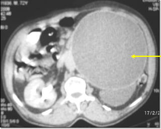
hydronephrosis. CT scan showed a left large cystic left mass
with ipsilateral uretero hydronephrosis (Figure 1) suggesting
an encysted hematoma or a cystic lymphocele. The exploratory
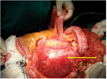
laparotomy found a rounded abdominal mass developed in the
retroperitoneum, with a diameter of 25cm pushing the root of the
mesentery and the first jejunal loop right (Figure 2). The mass
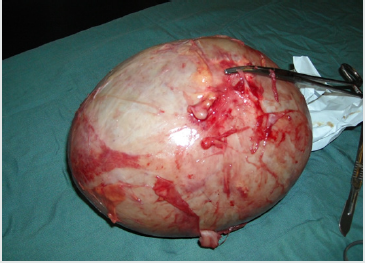
was resected (Figure 3) and the postoperative course was simple.
Bacteriology of the intracystic content showed no bacteria and the
culture was negative. Histopathology of the operative specimen macroscopically found a piece of 2500g, cystic, containing a liquid
substance of brown color. The wall of the cyst was thin (0.5 to 1cm
thick), without vegetation. Microscopy revealed a fibrous wall
infiltrated and bordered by polymorphic inflammatory cells rich
in plasma cells and neutrophils. Moreover, there was no tumor
proliferation. With a follow up of 7 years, the patient had no
complaints.
Figure 1: Abdominal computed tomography showing a hypodense mass pushing the mesenteric root (arrow).

Figure 2: Per-operative view of a retroperitoneal encysted hematoma pushing back the mesentery (arrow).

Discussion
Retroperitoneal hematomas are rare. Their incidence is
estimated between 0.1 and 0.6% [5]. Cystic transformation is due
to chronic evolution, often as a late consequence of an unnoticed
haemorrhage [2]. They are caused in 2/3 of the cases by hemostasis
disorders during an anticoagulant treatment. The other etiologies
are abdominal blunt trauma, surgical or instrumental treatment
in the retroperitoneum or, more rarely, aneurysms or hemophilia
[6]. In our observation, the occurrence of the hematoma would be
favored by antiplatelet therapy. Such treatment, like in our case, is
indicated after an ischemic stroke to reduce the risk of recurrence
[7]. The frequency of retroperitoneal hematomas has increased
in recent years, probably with the more frequent indications of
anticoagulation common in elderly patients with cardiovascular
comorbidities. This explains the more frequent occurrence in this
type of patient [8]. In the literature, the mean age of patients with
retroperitoneal hematomas related to antiplatelet therapy is 69
years [7].
This was the case in our observation who was elderly with a
history of stroke. The main clinical signs found in our case were
an abdominal mass with feeling of heaviness. The symptomatology
may be absent or minimal, explaining the chronic evolution towards
encysted stage. Therefore, the diagnostic of retroperitoneal
hematoma should be considered in any patients who presents
abdominal pain or mass with risk factors (anticoagulants,
antecedent of blunt trauma or coagulation disorders) [9]. In addition,
the anatomical configuration of the retroperitoneum with a rich
vascular network and good compliance facilitates the formation of
large hematomas in case of bleeding [10]. However, the hematomas
resorb more often. In rare cases, they can persist and increase in
size gradually before having an encysted appearance [1]. Diagnosis
at this stage can be difficult. Indeed, the differential diagnosis of
retroperitoneal cystic masses is not easy because of their rarity
and the multiplicity of etiologies. Thus, CT scan has an important
place in the management [11]. It is considered as the quickest,
safest, and most accurate modality for diagnosis compared to MRI
and ultrasonography [7]. It confirms the diagnosis and rules out
neoplasms especially in the elderly. In an evocative clinical context,
the encysted hematoma can be considered when the contents are
hypo dense with a thin wall without vegetations or partitions [4].
There is no consensus in the treatment of retroperitoneal
encysted hematoma [9]. Several options exist including conservative
treatment, interventional radiology or surgery [5]. In our case,
the chronic evolution posed the differential diagnosis with other
retroperitoneal cystic masses and prompted surgical exploration.
Retroperitoneal encysted hematomas may be difficult to
differentiate from soft tissue tumors, even with clinical and
radiological findings [1]. Histopathology of the operative specimen
is necessary to eliminate neoplasms [12]. Encysted hematoma is
confirmed by the presence of fibrous tissue in the capsule with
central liquefaction [2]. In our patient, we have a similar aspect
without malignant or benign cells. That’s why we considered the
diagnosis of encysted hematoma. A mortality of 20% is reported in
the literature, mainly conditioned by associated comorbidities and
the existence of active bleeding [9]. The favorable evolution in our
observation probably conditioned by the absence of active bleeding
and the realization of the adequate surgical treatment in time.
Conclusion
Encysted hematoma of the retroperitoneum is a rare condition. It often occurs in the context of hemostasis disorders during anticoagulant treatment. CT scan helps in the differential diagnosis with retroperitoneal cystic masses. There is no consensus in the treatment. However, surgery is still a good option because it allows to formally eliminate neoplasms.
Acknowledgment
None
Registration of research studies
Not applicable
Authors’ Contributions
All authors contributed to writing and editing the manuscript.
Availability of Data and Materials
Not applicable
Funding
None
Conflicts of Interest
“All authors declared that there are no conflicts of interest.”
References
- Syuto T, Hatori M, Masashi N, Sekine Y, Suzuki K (2013) Chronic expanding hematoma in the retroperitoneal space: a case report. BMC Urol 13(1): 60.
- Coley GM, Ottenheimer EJ (1963) Chronic retroperitoneal hematoma. Am J Surg mai 105(5): 696‑69
- Franjiae BD, Lovrièeviae I, De Syo D, Vukeliae M, Hudoroviae N, et al. (2007) Retroperitoneal hematoma following enoxaparin treatment in an elderly woman. Acta Clin Croat 46(4): 311-316.
- Yang DM, Jung DH, Kim H, Kang JH, Kim SH, et al. (2004) Retroperitoneal Cystic Masses: CT, Clinical, and Pathologic Findings and Literature Review. Radio Graphics 24(5): 1353‑13
- Chan YC, Morales JP, Reidy JF, Taylor PR (2007) Management of spontaneous and iatrogenic retroperitoneal haemorrhage: conservative management, endovascular intervention or open surgery: Management of retroperitoneal haemorrhage. Int J Clin Pract 62(10): 1604‑16
- Baekgaard JS, Eskesen TG, Lee JM, Yeh DD, Kaafarani HMA, et al. (2019) Spontaneous Retroperitoneal and Rectus Sheath Hemorrhage-Management, Risk Factors and Outcomes. World J Surg 43(8): 1890-1897.
- Ibrahim W, Mohamed A, Sheikh M, Shokr M, Hassan A, et al. (2017) Antiplatelet Therapy and Spontaneous Retroperitoneal Hematoma: A Case Report and Literature Review. Am J Case Rep 18: 85‑8
- Warren MH, Bhattacharya B, Maung AA, Davis KA (2019) Contemporary management of spontaneous retroperitoneal and rectus sheath hematomas. Am J Surg 10(19): 30097-30099.
- González C, Penado S, Llata L, Valero C, Riancho JA (2003) The Clinical Spectrum of Retroperitoneal Hematoma in Anticoagulated Patients: Medicine (Baltimore) 82(4): 257‑2
- Kasotakis G (2014) Retroperitoneal and Rectus Sheath Hematomas. Surg Clin North Am 94(1): 71‑7
- Zissin R, Ellis M, Gayer G (2006) The CT findings of abdominal anticoagulant-related hematomas. Semin Ultrasound CT MR 27(2): 117‑1
- Reid JD, Kommareddi S, Lankerani M, Park MC (1980) Chronic expanding hematomas. A clinicopathologic entity. JAMA 244(21): 2441‑244

Top Editors
-

Mark E Smith
Bio chemistry
University of Texas Medical Branch, USA -

Lawrence A Presley
Department of Criminal Justice
Liberty University, USA -

Thomas W Miller
Department of Psychiatry
University of Kentucky, USA -

Gjumrakch Aliev
Department of Medicine
Gally International Biomedical Research & Consulting LLC, USA -

Christopher Bryant
Department of Urbanisation and Agricultural
Montreal university, USA -

Robert William Frare
Oral & Maxillofacial Pathology
New York University, USA -

Rudolph Modesto Navari
Gastroenterology and Hepatology
University of Alabama, UK -

Andrew Hague
Department of Medicine
Universities of Bradford, UK -

George Gregory Buttigieg
Maltese College of Obstetrics and Gynaecology, Europe -

Chen-Hsiung Yeh
Oncology
Circulogene Theranostics, England -
.png)
Emilio Bucio-Carrillo
Radiation Chemistry
National University of Mexico, USA -
.jpg)
Casey J Grenier
Analytical Chemistry
Wentworth Institute of Technology, USA -
Hany Atalah
Minimally Invasive Surgery
Mercer University school of Medicine, USA -

Abu-Hussein Muhamad
Pediatric Dentistry
University of Athens , Greece

The annual scholar awards from Lupine Publishers honor a selected number Read More...


