Lupine Publishers Group
Lupine Publishers
Menu
ISSN: 2643-6760
Case Report(ISSN: 2643-6760) 
Median Cleft Lip: A Rare Malformation, Best Prognosis for the Isolated Form. Case Report Volume 4 - Issue 3
Raherison AR1*, Rasoaherinomenjanahary F2, Rabarikoto HF3, Andriamanarivo LRC1, Randrianarisoa FF1, Hunald FA1 and Andriamanarivo ML1
- 1Pediatric surgery unit, University hospital - Joseph Ravoahangy Andrianavalona – Antananarivo, Madagascar
- 2Digestive surgery unit, University hospital - Joseph Ravoahay Andrianavalona – Antananarivo, Madagascar
- 3Obstetricgynecology unit, Military hospital of Antsiranana, Madagascar
Received: February 04, 2020; Published: February 13, 2020
Corresponding author: Raherison Aristide Romain, Pediatric surgery unit, University hospital - Joseph Ravoahangy Andrianavalona, Antananarivo, Madagascar
DOI: 10.32474/SCSOAJ.2020.04.000187
Summary
The median cleft lip is a very rare form of cleft lip. It can be isolated or to be part of a complex malformation association which can involve the premaxillary bone, the nasal septum, or even the brain. In some cases, it is part of a syndrome. For the management of the median cleft lip, the excision with inverted-V incision or inverted-U incision is the most used. Muscle repair is the main step of surgery. We report the case of an incomplete and isolated median cleft lip in a 14-years-old boy. The inverted-V excision technique was used. The aesthetic result was satisfying. The isolated form has generally a good prognosis.
Keywords: Median cleft lip; Median cleft face syndrome
Introduction
The medial cleft lip is a very rare form of cleft lip [1]. It can be isolated, characterized by a midline vertical cleft of the upper lip. It can also involve the premaxillary bone, the nasal septum or even the central nervous system, entering into the framework of a syndrome in which the cleft is only one element [2]. The age and chronology of care must take into account any associated malformations. We report a case of isolated medial cleft lip diagnosed and treated at the age of 14 years old.
Case Report
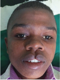
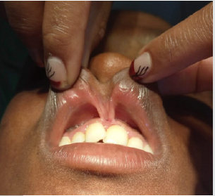
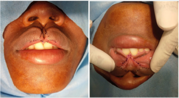
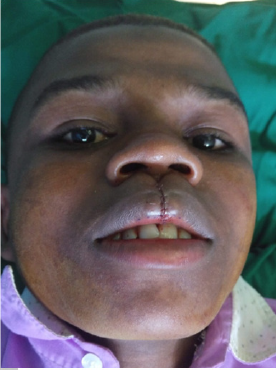
Our observation concerns a 14-year-old boy from a landlocked area sent by a missionary priest for a medial cleft lip. He presents an incomplete medial cleft lip with bifidity of the labial brake (Figures 1 & 2). The facial x-ray and the cranio-cerebral CT scan were normal. It was thus an isolated form. No similar family case was noted. The surgery was done under bilateral suprazygomatic maxillary nerve block supplemented by a labial infiltration of xylocaine-epinephrine 1%. The inverted V technique was adopted. The incision extended to the tops of each hemi-tubercle, cutting the crests of each labial hemi-brake before joining at the base of it (Figures 3 & 4). A complete disjunction of the orbicularis muscle has been observed. This muscle was dissected and sutured with Vicryl® 4/0. The aesthetic result was satisfying. We obtained an ad integrum reconstruction of the philtrum, cupid’s arch, lobule and respect for the alignment of the red lip-white lip junction.
Discussion
The medial cleft lip is an extremely rare malformation [1]. It
represents 0.4% to 0.7% of all cleft lips and affects around 1 case per
1,000,000 births [2,3]. Embryologically, during the fourth week of
pregnancy, the fusion at the midline of the distal part of the internal
nasal bud with the lateral nasal buds and the maxillary buds insures
the normal formation of the upper lip. The failure of this fusion is
responsible of the medial cleft lip [4,5]. There are three groups of
medial cleft lip: group I for the isolated form, group II for clefts with
craniofacial malformations and group III for forms associated with
extrafacial malformations [1]. This malformation can be part of a
syndrome. Medial facial cleft syndrome associates medial cleft lip,
nasal deformity, hypertelorism with or Pithou malformation of the
central nervous system [6]. Thus, the diagnosis must include the
systematic search for associated malformations. Facial x-ray and
craniofacial scan are of classic indication [3,6].
For the treatment, the age and the chronology of the surgery
depend on the possible associated malformations [7]. In case of
alveolar bone defect, an iliac graft is conventionally used [6,8]. For
the management of the medial cleft lip, several operating techniques
have been described. The most commonly used are inverted V and
U excision [4,6]. According to Millard, the use of inverted V excision
and 90° angle in the excision, 2 mm above the mucocutaneous white
roll on each side of the cleft which lengthened the skin in the center
of Cupid’s bow [4,9]. Muscle repair is the main step of treatment.
Even for incomplete forms, a defect in the orbicularis muscle is
almost constant [10,11]. Skin excision must be well calculated.
Excessive excision can lead to hypertrophic scarring, whereas
insufficient excision can lead to an unnatural depression on the
midline of the philtrum [7]. For the isolated form, the aesthetic
outcome is generally satisfying.
Conclusion
In case of a median cleft lip, the treatment must take into account any associated malformations. The inverted V excision gives an excellent aesthetic result for the reconstruction of the lip.
References
- Sarma VP (2019) Median cleft lip: an uncommon and unique anomaly. Int Surg J 6(8): 3035-3037.
- De Myer W (1967) The median cleft face syndrome: differentialdiagnosis of craniumbifidumoccultum, hypertelorism, and median cleft nose, lip and palate. Neurology 17: 961-71.
- Urata MM, Kawamoto HK (2003) Median clefts of the upper lip: are view and surgical management of a minor manifestation. J Cranio fac Surg 14: 749-755.
- Ichida M, Komuro Y, Yanai A (2009) Consideration of median cleft lip with frenulumlabii superior. J Craniofac Surg 20: 1370-1374.
- Mazzola RF (1976) Congenital malformations in the frontonasal area: their pathogenesis and classification. Clin Plast Surg 3: 573-609.
- Apesos J, Anigian GM (1993) Median cleft of the lip: its significance and surgical repair. Cleft Palate Craniofacial J 30: 94-96.
- Koh KS, Kim DY, Suk Oh T (2016) Clinical features and management of a median cleft lip. Arch Plast Surg 43: 242-247.
- De Boutray M, Beziat JL, Yachouh J, Bigorre M, Gleizal A, et al. (2016) Median cleft of the upper lip: a new classification to guide treatment decisions. J Cranio maxillofac Surg 44(6): 664-671.
- Millard DR (1977) Median cleft lip with hypertelorism. In Millard DR (Eds.), Cleftcraft: The evolution of its surgery: bilateral and rare deformities. Little, Brown, Boston, USA, pp. 727-768.
- Salampuria NR, Agrawal MB, Chugh A (2017) Median cleft lip: A case seriesfroma rural cleft center. J Cleft Lip Palate Craniofac Anomal 4: 198-200.
- Patel NP, Tantri MD (2010) Mediancleft of the upperlip: a rare case. Cleft Palate Craniofac J 47: 642-644.

Top Editors
-

Mark E Smith
Bio chemistry
University of Texas Medical Branch, USA -

Lawrence A Presley
Department of Criminal Justice
Liberty University, USA -

Thomas W Miller
Department of Psychiatry
University of Kentucky, USA -

Gjumrakch Aliev
Department of Medicine
Gally International Biomedical Research & Consulting LLC, USA -

Christopher Bryant
Department of Urbanisation and Agricultural
Montreal university, USA -

Robert William Frare
Oral & Maxillofacial Pathology
New York University, USA -

Rudolph Modesto Navari
Gastroenterology and Hepatology
University of Alabama, UK -

Andrew Hague
Department of Medicine
Universities of Bradford, UK -

George Gregory Buttigieg
Maltese College of Obstetrics and Gynaecology, Europe -

Chen-Hsiung Yeh
Oncology
Circulogene Theranostics, England -
.png)
Emilio Bucio-Carrillo
Radiation Chemistry
National University of Mexico, USA -
.jpg)
Casey J Grenier
Analytical Chemistry
Wentworth Institute of Technology, USA -
Hany Atalah
Minimally Invasive Surgery
Mercer University school of Medicine, USA -

Abu-Hussein Muhamad
Pediatric Dentistry
University of Athens , Greece

The annual scholar awards from Lupine Publishers honor a selected number Read More...


