Lupine Publishers Group
Lupine Publishers
Menu
ISSN: 2643-6760
Case Report(ISSN: 2643-6760) 
Lonely Pleural Fibrous Tumor: Case Report and Literature Review Volume 6 - Issue 2
H Sahli*, S Maalik, A El bakkari, H Jerguigue, R Latib and Y Omor
- Department of Radiology of the National Institute of Oncology in Rabat, Morocco
Received: April 26, 2021 Published: May 06, 2021
Corresponding author: Hind Sahli, Department of Radiology of the National Institute of Oncology in Rabat, Morocco
DOI: 10.32474/SCSOAJ.2021.06.000235
Summary
Patient of 65 years consultant for dry cough evolving for 1 year. The clinical examination found a dullness of the right hemi thorax. Chest x-ray objected to a right lower lobar opacity. The scanner showed a straight basal latero mass, connecting at an obtuse angle with the wall. This mass was tissue-enhancing heterogeneously after injection without net individualization of infiltration or invasion of adjacent structures. On the abdominal cuts there was a right adrenal nodule whose Wash out confirmed its adenoma some nature. There are no other remote lesions. A scan-mistbiopsy with anatomopathological and immunohistochemical study were made thus retaining the diagnosis of pleural solitary fibrous tumor. Pleural fibroid is rare. It is most often asymptomatic and accidental radiological discovery.
Keywords: Pleural Mass; Solitary Fibrous Tumor; CT Scan
Introduction
The solitary fibrous tumor of the pleura or benign mesothelium is a rare entity, is characterized by clinical latency and can manifest itself in irritative or compressive signs. It is often benign but malignant forms exist. The scanner helps guide the diagnosis, which will be confirmed by histology.
Observation
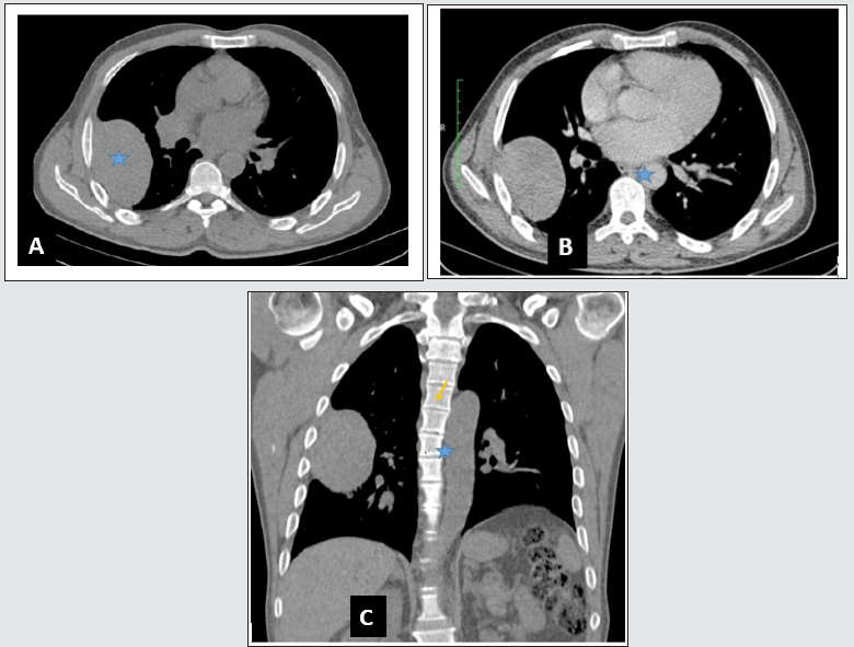
A 65-year-old man, a chronic tobacco man, had right chest pains that had been progressing for 1year with a dry cough. The whole thing was in a context of general state conservation. The physical examination regained a dullness of the right hemi thorax. A chest x-ray was done objectiving an opacity of the right lower lobe. The chest scanner with injection showed a fairly large right, oval, oval tissue chest mass, enhanced in a heterogeneous manner after injection of contrast product, without infiltration or invasion of adjacent structures. This mass connected with the wall at an obtuse angle (Figure 1). In addition, the extension balance sheet was negative. A scannoid biopsy puncture was made with anatomopathological study thus confirming the diagnosis of pleural fibroid. A complete resection of the mass was carried out with good evolution in postoperative. Anatomopathological examination of the surgical room with immunohistochemical supplement concluded that a solitary fibrous tumor had no signs of malignancy.
Discussion
Formerly described by “localized mesothelium” by Klemper and Rabin in 1931, the solitary fibrous tumor (TFS) took several names combining the terms mesothelial, pleural, fibrous, localized, benign, solitary [1]. Initially solitary fibrous tumours were considered mesothhelial in origin, hence the ancient benign mesothelium designation, but it has been demonstrated by immunohistochemical studies that they originate from pleural connective tissue [2]. These are mesenchymatous tumours, rare representing less than 10% of pleural tumors [1]. They develop at the expense of the visceral leaf of the pleura, but other locations have been described including pericardium, meninges, liver and pancreas [3]. They occur mainly after 50 years without predilection between the two sexes [4]. There is no proven relationship with tobacco or asbestos.
TFS is often asymptomatic in more than 50% of cases with a fortuitous radiological discovery, but sometimes it can manifest as compressive, irritative, hemothorax symptoms or paraneoplasic syndrome such as Peter Mary and Foix syndrome and Doege-Potter Syndrome [2]. Chest x-rays show a round or oval opacity, although limited, sometimes lobulated, of varying size, usually located at the base of the chest, connecting with the wall at an obtuse angle, may or may not be associated with a pleural effusion. This opacity is sometimes mobile with the patient’s position in pediculated fibroids [5]. In the scanner, the mass is tissue, although limited sometimes polylobed with homogeneous or heterogeneous content. Heterogeneity varies depending on the size of the tumor, the presence of necrotic or hemorrhagic reshufflings and calcifications (in 5% of cases) [1]. It can be scissural localization or develop at the expense of the costa, diaphragmatic or mediastinal [2] fold. The connection angle can also and frequently be acute or obtuse and acute at the same time, especially when it is a large orpediculated tumor. Direct visibility of the pedicule is rare [5]. The uplift is often intense and early given the large vascular contingent [4].
MRI can better characterize the internal structure of the mass and determine its relationship with the wall, apex and diaphragm [2,4]. The variable and often heterogeneous signal depending on the histological nature of the mass [4]. The malignancy of a TFS can be observed de novo or complicate a benign form [1]. There is no univocal criterion of malignancy, but a high mitotic index remains the best [2]. In imaging, malignancy is also evident in the event of invasion of adjacent structures and the existence of pulmonary nodules or other secondary locations [2]. The definitive diagnosis is histological, usually made on surgical procedure [5]. Differential diagnosis occurs with other causes of pleural tissue damage such as metastasis, localized mesothelioma, lymphoma, sarcoma, lipoma, desmoid tumor, hemangiopericytom and fibrous malignant histiocytoma [2]. Thymoma and neuroma may also pose a differential diagnostic problem with the solitary fibrous tumour if it originates respectively from the mediastle and costal [2] fold .
The reference treatment for TFS is broad surgical exeresis [1]. It may be preceded by neo-adjuvant chemotherapy in cases of large tumours that are not immediately resecable [2]. In aggressive forms, it can be supplemented by chemotherapy and/or radiotherapy, the benefits of which have not been proven [1]. Due to a difficult-toassess evolutionary profile, long-term clinical radio monitoring is recommended [1].
Figure 1: Thoracic scan in cross sections (A, B) and coronal (C): tissue process developed at the expense of the right, non-invasive, 6cm large axis, heterogeneously enhancing after injection after injection of contrast product (stars). Note the obtuse angle connection with the wall (arrow).

Conclusion
The pleural solitary fibrous tumor is a rare mesenchymatous tumor representing less than 10% of pleural tumors. Standard X-ray focuses on the diagnosis of a pleural tumor. The chest scan then allows to characterize this mass and to look for signs that plead towards a benign or malignant origin. The diagnosis will be confirmed by the anatomopathological and immunohistochemical study.
References
- Saint Blancard P, Bonnichon A, Margery J (2009) The solitary pleural fibrous tumor: About five observations. Rev Pneumol Clin 65(3): 153-158.
- Helage S, Revel Marie Stone (2012) Lonely fibrous tumors of the pleura: CT scans and major differential diagnoses.
- Birebent J, Durliat S, Driot D, Delabarre M, Rougé Bugat M (2018) A solitary fibrous tumor. Medicine 14(4): 164-165.
- Cannard L, Debelle L, Laurent V, Béot S. Thorax Pleural Tumors.
- Yangui I, Smaoui M, Daoud S, Msaad S, Kammoun S, et al. (2007) Lonely fibrous tumor of the pleura. Medical press 36(5): 811-812.

Top Editors
-

Mark E Smith
Bio chemistry
University of Texas Medical Branch, USA -

Lawrence A Presley
Department of Criminal Justice
Liberty University, USA -

Thomas W Miller
Department of Psychiatry
University of Kentucky, USA -

Gjumrakch Aliev
Department of Medicine
Gally International Biomedical Research & Consulting LLC, USA -

Christopher Bryant
Department of Urbanisation and Agricultural
Montreal university, USA -

Robert William Frare
Oral & Maxillofacial Pathology
New York University, USA -

Rudolph Modesto Navari
Gastroenterology and Hepatology
University of Alabama, UK -

Andrew Hague
Department of Medicine
Universities of Bradford, UK -

George Gregory Buttigieg
Maltese College of Obstetrics and Gynaecology, Europe -

Chen-Hsiung Yeh
Oncology
Circulogene Theranostics, England -
.png)
Emilio Bucio-Carrillo
Radiation Chemistry
National University of Mexico, USA -
.jpg)
Casey J Grenier
Analytical Chemistry
Wentworth Institute of Technology, USA -
Hany Atalah
Minimally Invasive Surgery
Mercer University school of Medicine, USA -

Abu-Hussein Muhamad
Pediatric Dentistry
University of Athens , Greece

The annual scholar awards from Lupine Publishers honor a selected number Read More...


