Lupine Publishers Group
Lupine Publishers
Menu
ISSN: 2643-6760
Case Report(ISSN: 2643-6760) 
Intestinal Obstruction by Blind Vove Iess Riobamba Hospital 2014 Volume 1 - Issue 2
Diego Fabricio Erazo Mogrovejo1*, Sandra Gómez2 and Andrea Montoya3
- 1Specialist in General and Laparoscopic Surgery, Medical treating Hospital IESS-Riobamba, Riobamba Ecuador
- 2General Doctor, Resident Hospital IESS-Riobamba, Riobamba Ecuador
- 3General Doctor, Riobamba Ecuador
Received: May 23, 2018; Published: May 31, 2018
Corresponding author: Diego Erazo Mogrovejo, Surgeon General of the IESS Hospital of Riobamba, General Surgeon and Bariatric Surgeon of RIO Hospital, Riobamba Ecuador
DOI: 10.32474/SCSOAJ.2018.01.000108
Summary
Introduction: Cecal volvulus is the second most common volvulus among colonic volvulus, usually presenting as bowel obstruction.
Case Presentation: A 65 year old female presented with abdominal pain, nausea, vomiting, abdominal distension and abscense of flatus and stool Erect Abdominal radiograph showed marked colonic distension with air-fluid levels. Urgent surgical treatment is decided.
Conclusion: Cecal volvulus should always be considered as a possible cause of mechanical ileus. Radiographs and CT are invaluable in the diagnosis of this condition.
Introduction
Intestinal volvulus is produced by the twisting of a mobile segment of the colon around its mesenteric axis. The location, in order of frequency, is: 80% in sigma, 15% in blind and 5% in transverse colon [1]. The volvulus of the right colon is rarer. They do not present a defined geographical distribution, as in the sigmoid volvulus, which would indicate that the external factors that produce intestinal diseases do not have a higher incidence [2- 3]. The colon, from the embryological, anatomical, functional and surgical point of view, is divided into a right sector (right colon) and another left (left colon) whose limit is a line that passes over the transverse colon to the left of the artery medium colic [4-5]. The volvules located in the right sector are mainly due to a congenital malformation. Those located in the left colon, always correspond to the sigmoid and recognize as an etiological cause an abnormally mobile loop added to diseases that dilate and lengthen the sigma (dolicomegasigma) [6]. The volvules located in the transverse are due to the exaggeration of a normal situation (garland colon) and are exceptional. Various causes have been implicated in etiopathogenesis: anatomical (long and redundant), alimentary (diet rich in residues), pathological (chronic constipation, laxative abuse, psychiatric and central nervous system diseases in 40% of cases: dystrophies) , Parkinson’s disease, Alzheimer’s disease, cerebrovascular accidents, etc.) [7,8], sex (more common in women) and age (over 70 years). Cecal volvulus is more common in women with a mean age of 50-60 years, with a history of previous episodes and a clinical picture of small bowel obstruction and abdominal pain [9-10].
Clinical Case
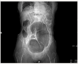
A 65-year-old female patient, with no relevant medical history. She went to the emergency department due to distension, abdominal pain and absence of bowel movements, which lasted 5 days. The examination revealed an important abdominal distension, with diffuse pain without signs of peritoneal irritation, percussion tympany and absence of peristalsis on auscultation. In the analytical study, only one leukocytosis of 13,000/μl was highlighted, with a deviation to the left of 84%. On plain abdominal radiography an image of great distension of the colon was identified (Figure 1).
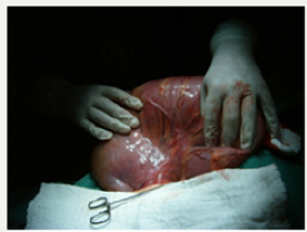
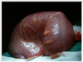
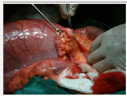
Given the suspicion of intestinal occlusion caused by a volvulus, urgent surgery was indicated. A midline laparotomy was performed, observing a cecal volvulus with adherence at the level of the peritoneum in union with the omentum (Figures 2-3). We performed, section and adherence ligature, blind developing, revision of intestinal loops and extraction of appendix (Figure 4). The pathological anatomy reported an initial acute appendicitis. The postoperative period was uneventful. The patient follows correct controls in external surgeries of surgery and develops a socio-labor activity according to her age up to now.
Discussion
The cecal volvulus supposes the torsion of a segment of the colon on its mesenteric axis, which causes strangulation, with the consequent occlusion of the two ends of the volvulated segment and with a compromise of the vascularization, all of which produces a closed loop obstruction [11-12]. To present a redundant colon is necessary, with excessive mobility of the caecum and the ascending colon caused by a lack of fixation to the lateral retroperitoneum, a narrow and short mesentery at its base, and distension with air [13]; it almost never happens when the colon is full of solid stool. Colonic volvulus usually occurs more frequently in the sigma, followed by the cecum, although it also occurs occasionally in the transverse colon and in the splenic or hepatic angle of the colon [14-16].
A series of factors that influence the development of colon volvulus (usually congenital in cecal volvulus and acquired in the sigmoid volvulus) have been described, such as: diet rich in residues, chronic constipation and abuse of laxatives (in patients hospitalized in centers psychiatric), Chagas disease, disabling neurological diseases, mental patients under treatment with psychotropic drugs (which contribute to constipation), pregnant women etc. [17,18]. The clinic is usually more or less acute, with the typical triad of abdominal pain, distension and constipation, followed by nausea and vomiting; there is usually tympany to percussion and absence of peristalsis to auscultation; If gangrene or perforation develops, it manifests as an acute abdomen with signs of peritoneal irritation and shock, and if it is evolved it can cause the patient’s death [19-21]. In almost half of the subjects there is a history of similar previous episodes, but of lesser intensity.
In the laboratory, there is leukocytosis with or without deviation to the left, in cases of ischemia and / or necrosis of the colonic wall [22]. Abdominal radiography shows a significant distention of the cecum, with the presence of hydro-aerial levels and dilatation of the small intestine; the “coffee bean” image can sometimes be observed. The opaque enema is usually difficult to interpret, but sometimes the sign of the “bird’s beak” appears. Abdominal TAC is usually very specific, identifying cecal volvulus and vascular compromise due to meso twisting and signs of ischemic colitis in the colon wall [23]. Diagnostic colonoscopy is sometimes useful and establishes the viability of the colonic mucosa [24].
The treatment can be conservative or with surgical measures. Return by colonoscopy or barium enema can sometimes be successful (endoscopic decompression is the procedure of choice in sigmoid volvulus prior to surgical approach in stable patients, without ischemic involvement of the colon or perforation [25], but colonoscopy is less effective in right and blind colon volvuli [26]). If there is ischemic compromise or intestinal perforation, resection is necessary, after the corresponding exploratory laparotomy, a right hemicolectomy is performed with or without primary anastomosis.
There are other surgical techniques that have been used occasionally, such as detorsion or simple reduction with or without cecopexy of the colon to the right retroperitoneum and cecostomy [27]. In recent years, minimally invasive techniques such as laparoscopic cecopexy28 have been described. Conservative treatment can sometimes overcome the acute period, recover the colonic mucosa and be able to perform a later definitive surgery with a lower morbidity and mortality than in the acute period and with greater success; but taking into account that endoscopic devolvulation has a recurrence rate of 30-50%. The definitive treatment, with better control of long-term symptoms, is right hemicolectomy with primary ileotransverse anastomosis [29].
Mortality of cecal volvulus varies from 10-15%, if the colon is viable, to 30-40%, if there is intestinal gangrene. The mortality of the sigmoid volvulus varies from 13%, if the previous endoscopic devolvulation is achieved and the clinical situation of the patient with delayed surgery is improved, to 30-70%, if urgent surgery is performed without previous attempt of conservative endoscopic management [30]. The high mortality rate in relation to urgent surgery is conditioned by being elderly patients, with important underlying pathology and serious deterioration of their general condition at the time of surgery, first episode of volvulation and more severe symptoms (strangulation, ischemic colitis and perforation).
References
- Zinner J (1998) Maingot Operaciones Abdominales. In: Editorial Médica Panamericana (10th edn) 2: 1310-1312.
- Goligher JC, Duthie HL, Nixon HH (1979) Cirugía del ano, recto y colon. Barcelona. Editorial Salvat 933-936.
- Sabiston DC (1998) Tratado de Patología Quirúrgica. Decimo tercera edición. México DF. Editorial Interamericana McGraw-Hill 1052-1056.
- Ferraina P, Oría A (2001) Cirugía de Michans. Quinta edición. Editorial El Ateneo 864-865.
- Baker RJ, Fischer JE (2004) El Dominio de la Cirugía Cuarta edición, Editorial Médica Panamericana 2: 1985-2003.
- Guller U, Zuber M, Harder F (2001) Cecum volvulus - A frequently misdiagnosed disease picture. Results of a retrospective study of 26 patients and review of the literature. Swiss Surg 7(4):158-64.
- Schumpelick V, Dreuw B, Ophoff K, Prescher A (2000) Appendix and cecum. Embryology, Anatomy, and surgical applications. Surg Clin North Am 80(1): 295-317.
- Machiels F, De Maeseneer M, Coen H, Desprechins B, Osteaux M (1999) Pediatric cecal malfixation. JBR-BTR 82(3):116.
- Catalano O, Grassi R, Rotondo (1996) A Intestinal malrotation in adults: Report of 2 cases studied with computed tomography. Radiol Med Torino 91: 821-3.
- Touloukian RJ, Smith EI (1998) Disorders of rotation and fixation. In: O’ Neill JA, Pediatric surgery. 5thedn. Baltimore: Mosby 2: 1199-1203.
- Steward DR, Colodny AL, Daggett WC (1976) Malrotation of the bowel in infants and children: a 15 year review. Surgery 79(6): 716-20.
- Snyder WH Jr, Chaffin L (1954) Embryology and pathology of the intestinal tract: presentation of 40 cases of malrotation. Ann Surg 140(3): 368-379.
- Ruiz Tartas A, Arizaga Rovalino P, Fernández Lobato R, Marín Lucas FJ, Jiménez Miramon FJ, et al. (1994) Intestinal malrotation in an adult. Rev Esp Enferm Dig 86: 701-702.
- Lin JN, Lou CC, Wang KL (1995) Intestinal malrotation and midgut volvulus: a 15-year review. J Formos Med Assoc 94: 178-181.
- Ford EG, Senac Mo Jr, Srikanth MS, Weitzman JJ (1992) Malrotation of the intestine in children. Ann Surg 215(2): 172-178.
- Von Flüe M, Herzog U, Ackermann C, Tondelli P, Harder F (1994) Acute and chronic presentation of intestinal nonrotation in adults. Dis Colon Rectum 37: 192-198.
- Guller U, Zuber M, Harder F (2001) Cecum volvulus-a frequently misdiagnosed disease picture. Results of a retrospective study of 26 patients and review of the literature. Swiss Surg 7(4): 158-64.
- Cózar A, Medina M, Del Olmo M, Moreno JM, Martínez G (2004) Vólvulo cecal en el síndrome de Cornelia de Lange. Rev Esp Enferm Dig 96(1): 85-6.
- Utrillas AC, López M, Rebollo J, Minguillón A, Moreno C, Del Vall JM (2003) Vólvulo de ciego: una rara causa de obstrucción intestinal. Cir Esp. 74(1): 120.
- Echenique M, Amondaraín JA (2002) Vólvulos de intestino grueso. Rev Esp Enferm Dig. 94: 201-205.
- Frizelle FA, Wolff BG (1996) Colonic volvulus. Adv Surg. 29: 131-139.
- Hiltunen KM, Syrja H, Matikainen M (1992) Colonic volvulus. Diagnosis and results of treatment in 82 patients. Eur J Surg 158(11-12): 607-611.
- Renzulli P, Maurer CA, Netzer P, Buchler MW (2002) Preoperative colonoscopic derotation is beneficial in acute colonic volvulus. Dig Surg 19(3): 223-229.
- Gupta S, Gupta SK (1993) acute caecal volvulus: report of 22 cases and review of literature. Ital J Gastroenterol 25(7): 380-384.
- Madiba TE, Thomson SR (2002) The management of cecal volvulus. Dis Colon Rectum. 45(2): 264-267.
- Rabinovici R, Simansky DA, Kaplan O, Mavor E, Manny J (1990) Cecal volvulus. Dis Colon Rectum 33: 765-769.
- Bhrandarkar DS, Morgan WP (1995) Laparoscopic caecopexy for caecal volvulus. Br J Surg 82(3): 323.
- Consorti ET, Liu TH (2005) Diagnosis and treatment of cecal volvulus. Postgrad Med J 81(962): 772-776.
- Gamblin TC, Stephens RE Jr, Johnson RK, Roth well M (2003) Adult malrotation: A case report and review of the literature. Current Surg 60(5): 517-520.
- Kelly MD, Bunni ND, Pullyblank AM (2008) Laparoscopic assisted right hemicolectomy for caecal volvulus. World Journal of Emergency Surgery 3: 4.

Top Editors
-

Mark E Smith
Bio chemistry
University of Texas Medical Branch, USA -

Lawrence A Presley
Department of Criminal Justice
Liberty University, USA -

Thomas W Miller
Department of Psychiatry
University of Kentucky, USA -

Gjumrakch Aliev
Department of Medicine
Gally International Biomedical Research & Consulting LLC, USA -

Christopher Bryant
Department of Urbanisation and Agricultural
Montreal university, USA -

Robert William Frare
Oral & Maxillofacial Pathology
New York University, USA -

Rudolph Modesto Navari
Gastroenterology and Hepatology
University of Alabama, UK -

Andrew Hague
Department of Medicine
Universities of Bradford, UK -

George Gregory Buttigieg
Maltese College of Obstetrics and Gynaecology, Europe -

Chen-Hsiung Yeh
Oncology
Circulogene Theranostics, England -
.png)
Emilio Bucio-Carrillo
Radiation Chemistry
National University of Mexico, USA -
.jpg)
Casey J Grenier
Analytical Chemistry
Wentworth Institute of Technology, USA -
Hany Atalah
Minimally Invasive Surgery
Mercer University school of Medicine, USA -

Abu-Hussein Muhamad
Pediatric Dentistry
University of Athens , Greece

The annual scholar awards from Lupine Publishers honor a selected number Read More...


