
Lupine Publishers Group
Lupine Publishers
Menu
ISSN: 2643-6760
Case Report(ISSN: 2643-6760) 
Initial Application of Ligasure in Head and Neck Surgery in Hue Hospital: is it Powerful? Volume 4 - Issue 5
Phan Huu Ngoc Minh*
- Department of Otolaryngology, Hue University of Medicine and Pharmacy, Hue University, Vietnam
Received: April 02, 2020; Published: April 22, 2020
Corresponding author: Phan Huu Ngoc Minh, Department of Otolaryngology, Hue University Hospital, Hue University of Medicine and Pharmacy, Hue University, 06 Ngo Quyen, Hue, 53000, Vietnam
DOI: 10.32474/SCSOAJ.2020.04.000197
Abstract
The application of Ligasure vessel sealing system in head and neck surgery has been currently published in many studies. The results published were reported to be reliable and safe. Since 2018, the Department of Otolaryngology - Head and Neck Surgery in Hue University of Medicine and Pharmacy Hospital performed applications of Ligasure in head and neck surgery for the first time and may have shown several advantages and benefits. In this presentation, we give a brief overview of first patients who underwent operation with the use of Ligasure. In our department, Ligasure is used for selective or modified radical neck dissection, thyroidectomy, and superficial parotidectomy.
Keywords: Ligasure; Neck dissection; Paroidectomy; Thyroidectomy
Introduction
Head and neck surgery is relatively complicated, involving
many important anatomical landmarks such as cranial nerves and
large vessels (carotid artery, jugular vein). Bleeding control is one
of the main goals during dissection. The application of Ligasure
vessel sealing system in head and neck surgery was reported to
be reliable and safe in several studies. The main advantages of
Ligasure are ability to achieve good hemostasis, reduce operation
time, relieve post-operative pain and recover quickly after surgery
[1,2]. The use of Ligasure in Otolaryngology-Head and Neck Surgery
may have several benefits over standard methods [3]. This device
was developed as an alternative to ligatures, hemoclips, staplers,
and ultrasonic coagulators for ligating vessels and tissue bundles.
Its generator is designed to produce high-current (4A), low-voltage
(200 V) output [1]. And it works by applying a precise amount of
pressure and energy to transform the collagen and elastin in vessel
walls to create a permanent seal for a width of several millimeters.
When Ligasure clamps into tissue bundle, it creates intracellular
friction, disrupts Hydrogen Bridge, denatures proteins and
performs coagulation. Despite the high degree of heat produced
between the jaws of the instrument, it does not produce a large
amount of lateral heat. It also reduces the smoke production and
adhesions, then damage to surrounding tissues is low. According to
study of Colella G., the Ligatures system can safely seal and divide
vessels that are up to 7mm in diameter. Meanwhile the conventional
bipolar are considered reliable only in vessels smaller than 2mm in
diameter [1].
Since 2018, the Department of Otolaryngology, Hue University
of Medicine and Pharmacy Hospital have used initially Ligasure for
selective neck dissection, superficial parotidectomy with preserved
facial nerve, thyroidectomy. The results have shown that Ligasure
provided sufficient hemostasis, reduced the operation time and
post-operative complications. It is, indeed, a reliable and safe
device. In this presentation, we give a brief description of first
patients performed surgical procedures with use of Ligasure vessel
sealing system in our department.
A couple of Cases Performed Surgical Procedures with Use of Ligasure
Case 1: Neck Dissection in Patient with Cancer of Tonsil
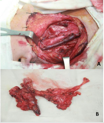
A 64-year-old female presented to our department of Otolaryngology, Hue University of Medicine and Pharmacy Hospital with dysphagia and a right-sided palpable cervical mass. She had no specific medical history. On clinical examination, she was detected with a mass on the right tonsil with a 13.8 x 28.8mm size on enhanced magnetic resonance imaging (MRI (Figure 1A). Additionally, she had multiple hard fixed cervical nodes involved at levels II, III on the right-sided neck with the largest 16.0 x 22.7 mm sized node (Figure 1B). Histopathological examination confirmed an epidermoid carcinoma of the right tonsil. According to the American Joint Committee on Cancer Staging, she was classified T2N2bMo, with stage IVA tonsillar carcinoma. Based on the National Comprehensive Cancer Network guideline for oropharyngeal cancer, with N2a or N2b should be performed resection of primary site in association with ipsilateral or bilateral neck dissection. Then, this patient underwent a surgery for wide excision of tonsillectomy and ipsilateral selective neck dissection (level IIA, IIB, III, IV, V, VI) with preservation of internal jugular vein, common carotid artery, spinal nerve and phrenic nerve in August 2018 (Figures 2,3). The estimated operative time was under one hour. Total postoperative neck drainage was less than 20ml. She was removed from 5mlhemovac drainage on the second postop day. Her status was stable. She was discharged on the 7th postop day and followed up at the Department of Oncology for chemo concurrent radiotherapy.
Figure 1: Measured size in diameter and contrast absorption of right-sided tonsillar mass and cervical lymph node.
A. Tonsillar mass measuring 13.8 x 28.8mm in diameter.
B. Largest lymph node at level II, III measuring 16.0 x 22.7mm in diameter.
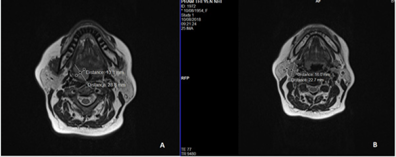
Figure 2: Anatomical landmarks during surgery.
A. Incision for right-sided selective neck dissection at level IIA, IIB, III, IV,V, VI.
B. Lymph node level II-III adhesion to jugular vein .
C. Preservation accessory nerve.
D. Preservation phrenic nerve.
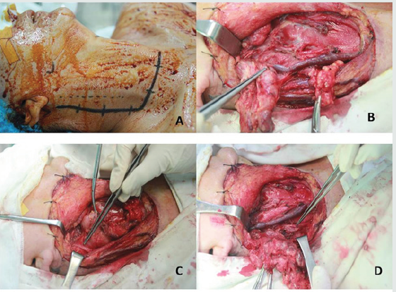
Figure 3: Operative champs after selective neck dissection with use of Ligasure.
A. Selective neck dissection with preservation CCA, IJV, spinal and phrenic nerve.
B. Removal of cervical lymph node en bloc.
Case 2: Superficial Parotidectomy with Preservation of Facial Nerve
A 58-year-old male patient presented to ENT Department, Hue University of Medicine and Pharmacy Hospital with a two-year gradually palpable mass at the left angle of mandibular. He had 20packs.year history of smoking. On clinical examination, he had an enlarged 30mm sized, tender, painless and mobile mass. On nasopharyngoscopy, he was found to have a normal nasopharynx. Neck sonography (13.02.2019) revealed parotid and submandibular glands were normal, suspicion of abnormal left-sided angle area. Computed tomography (CT) with contrast injection noted lymph nodes at the angle of mandibular with clear contour and limit, no sign of adjacent invasion (Figure 4). Histological findings revealed Warthin’s tumor. Thus, he underwent a surgery for superficial parotidectomy with facial nerve preservation (Figure 5). Permanent biopsied findings showed enlarged salivary glands and reactive proliferative lymph nodes with no malignant tissue. Postoperative findings noted no facial palsy, neck drainage was removed on the second postop day with < 5ml amount of fluid. He was discharged on the seventh postop day.
Case 3: Thyroidectomy with recurrent laryngeal nerve and parathyroid glands
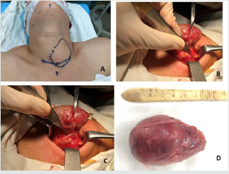
a. A 53-year-old female was admitted to our department with a discomfort feeling on anterior neck which leads to mild dyspnea and dysphagia. Neck sonography showed a cystic mass measuring 50 x 90 mm in diameter on the left lobe of thyroid gland. Amounts of fT3,fT4, TSH were in the normal limit. She had performed left thyroid lobectomy and preserved recurrent laryngeal nerve and parathyroid glands with the use of Ligasure in november 2019 (Figure 6). The operative time was less than 30 minutes. Preliminary postop assessments were no hoarseness, no hematoma, mild postop pain and good recovery after surgery.
Figure 4: Computed tomography with contrast injection revealed lymph node at the angle of mandibular with clear contour and limit, no sign of adjacent invasion, on axial and coronal coupe.
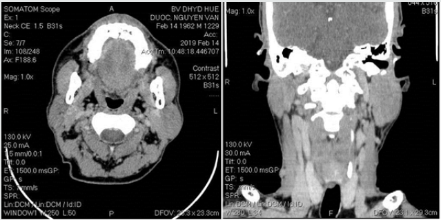
Figure 5:A,B: Trunk and branches of facial nerve shown.
C: The first post op day noted no facial palsy.
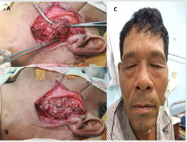
Figure 6: Anatomical landmarks during surgery thyroidectomy with use of Ligasure reducing bleeding.
A. Incision and palpable mass.
B. Preservation left-sided superior and inferior parathyroid glands.
C. Preservation left-sided recurrent laryngeal nerve.
D. Removal of cystic mass on the left thyroid measuring 55x 85 mm in diameter.
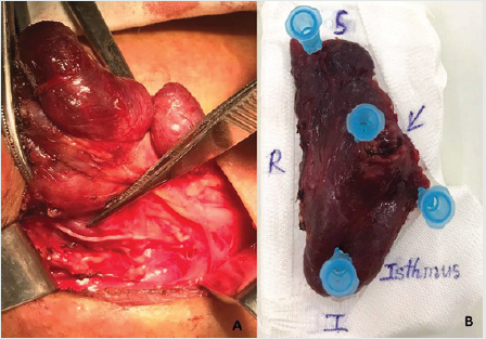
b. A 37 year-old man presented to our department with a mass on the right-sided thyroid gland measuring 15x11x18mm in diameter (TiRAD IVb). Amounts of T3, fT4, TSH were in the normal limit. Sonography and cervical computer tomography revealed no neck metastasis. He underwent right lobectomy and isthmusectomy (Figure 7). Histological findings revealed well-differential papillary carcinoma. Postop status were no hoarseness, no hematoma, no postop pain.
Figure 7: Anatomical landmarks during surgery thyroidectomy.
A. Preservation right-sided recurrent laryngeal nerve and parathyroid glands.
B. Removal of right lobe and isthmus of thyroid.
We would like to give a short communication about the first patients undergoing surgery with the use of Ligasure in our Department of Otolaryngology from 2018. In fact, Ligasure has considerably higher cost, there were limitations of amount patients used. Also, postoperative evaluation was on each patient who underwent a surgery with the use of Ligasure, the sample size was thus not representative to be able to statistic.
Conclusion
Through the case summarized above, in the initial application of Ligasure in head and neck surgery, we noted that Ligasure was a reliable, safe and power device. It had several beneficial results. This is an advance in the field of head and neck surgery, which needs to be widely applied.
Conflict of Interest Disclosures
None of the authors has any conflict of interest, financial or otherwise. This article does not contain any studies with human participants or animals performed by any of the authors.
References
- Colella G, Giudice A, Vicidomini A, Sperlongano P (2005) “Usefulness of the ligasure Vessel Sealing System During Superficial Lobectomy of the Parotid Gland”, Arch Otolaryngol Head Neck Surg.,131(5): 413-416.
- Ozturk K, Kaya I, Turhal G, Ozturk A.(2016) “A comparison of electrothermal bipolar vessel sealing system and electrocautery in selective neck dissection”. Eur Arch Otorhinolaryngol 273(11): 3835-3838.
- Prokopakis EP, Lachanas VA, Vardouniotis A (2010) “The use of the Ligasure vessel sealing system in head and neck surgery: a report on six years of experience and a review of the literature”. B-ENT 6(1): 19-25.

Top Editors
-

Mark E Smith
Bio chemistry
University of Texas Medical Branch, USA -

Lawrence A Presley
Department of Criminal Justice
Liberty University, USA -

Thomas W Miller
Department of Psychiatry
University of Kentucky, USA -

Gjumrakch Aliev
Department of Medicine
Gally International Biomedical Research & Consulting LLC, USA -

Christopher Bryant
Department of Urbanisation and Agricultural
Montreal university, USA -

Robert William Frare
Oral & Maxillofacial Pathology
New York University, USA -

Rudolph Modesto Navari
Gastroenterology and Hepatology
University of Alabama, UK -

Andrew Hague
Department of Medicine
Universities of Bradford, UK -

George Gregory Buttigieg
Maltese College of Obstetrics and Gynaecology, Europe -

Chen-Hsiung Yeh
Oncology
Circulogene Theranostics, England -
.png)
Emilio Bucio-Carrillo
Radiation Chemistry
National University of Mexico, USA -
.jpg)
Casey J Grenier
Analytical Chemistry
Wentworth Institute of Technology, USA -
Hany Atalah
Minimally Invasive Surgery
Mercer University school of Medicine, USA -

Abu-Hussein Muhamad
Pediatric Dentistry
University of Athens , Greece

The annual scholar awards from Lupine Publishers honor a selected number Read More...


