Lupine Publishers Group
Lupine Publishers
Menu
ISSN: 2643-6760
Case Report(ISSN: 2643-6760) 
Diagnosis of Uterine Metastasis of Lobular Carcinoma of Breast Prior to Breast Cancer Detection in Imam Reza Hospital- Tehran Volume 5 - Issue 4
Maryam Khayyat Khameneie1 and Nahid Arian pour2*
- 1Department of gynecology, Imam Reza hospital, Tehran, Iran
- 2Department of Microbiology, AJA Univ Med Sciences, Tehran,Iran
Received: August 07, 2020; Published: August 18, 2020
Corresponding author: Nahid Arian pour, Department of Microbiology, AJA Univ Med Sciences, Tehran, Iran
DOI: 10.32474/SCSOAJ.2020.05.000216
Abstract
Metastasis of the breast cancer to the uterus happens in 8% of cases. In most of the reported cases, metastasis is detected
following diagnosis and treatment of the primary cancer. Detection of metastases before diagnosis of the primary cancer is quite
rare. Here, we report a case of invasive lobular breast carcinoma metastasized to the uterus with initial presentation of uterine
myoma while she lacked any symptom related to breast cancer.
A 49 years old woman was admitted to Imam Reza hospital with a very large uterine leiomyoma or leiomyo-sarcoma. She was
diagnosed with osteo-metastasis based on CT scan, whole body bone scan and elevated tumor markers. The pathology report of
samples collected during laparotomy, subtotal hysterectomy, bilateral salpingo-oophorectomy, and colostomy revealed involvement
of the uterus and leiomyoma with invasive lobular carcinoma of breast.
Keywords: Breast cancer, Invasive lobular carcinoma, uterine metastasis, Large uterine myoma
Introduction
Breast cancer is the most common cancer affecting women all over the world with 14% mortality rate, accounting for 23% of all cases [1]. Invasive ductal carcinoma (IDC) is the most prevalent type of breast cancer (%86) and invasive lobular carcinoma (ILC) is the next (%5- %15) [2]. Both types of cancer metastasize to other organs with almost the same rate [3]. Here, we report a case of invasive lobular breast carcinoma metastasized to the uterus and adjacent tissues with initial presentation of uterine myoma without any sign related to breast cancer i.e. cancer in its primitive site is diagnosed following diagnosis of the metastasized site.
Case Presentation
The patient is a 49 years old woman with three pregnancies and one miscarriage (G3 P2 Ab1). She suffered headache, fatigue, weakness, scapula and back pain for 7 months prior to admission. Three months before admission, her white blood cells count (WBC) was 6600, hemoglobin (Hg) 10.7 and erythrocyte sedimentation rate (ESR) was 95. Abdomen and pelvis sonography revealed normal abdomen while a large heterogeneous mass (13 cm) was present in the uterus and ovaries (Figure 1).
In axial spiral computed tomography (CT) scan, a very large soft mass was seen in the uterus measuring 23x 19x 11 centimeter (cm). The right ovary was enlarged without definite mass lesions. Multiple sclerotic lesions were seen in the most vertebrae and both innominate bones. In addition to the presence of large uterus myoma, bone metastasis was also diagnosed (Figure 2).Patient was admitted to the oncology ward of the Imam Reza hospital- Tehran- Iran and bone marrow aspiration was performed. Her consultation was performed in the gynecology ward. Breast examination performed soon after was normal. Colonoscopy was also normal.
Although breast sonography was reported normal but multiple
adenopathy with central echogenicity, probably reactive type, were
seen. Whole body bone scan indicated multifocal osteo-metastasis.
Next laboratory tests measured Hg as 7.8, blood hematocrit (Hct)
27.2, C-reactive protein (CRP) +2, erythrocyte sedimentation
rate (ESR) 125, Creatinine 1.5, cancer antigen (CA)19-9 75,
CA125 758, CA15-3 > 1000, and carcinoembryonic antigen (CEA)
was 47.Sonography and bone marrow aspiration performed
after admission to the oncology ward indicated bilateral hydronephrosis,
mild ascites, increased bladder wall thickness in trigone
area, large uterus with large myoma (12x15 cm) and one cyst with
thick septum in the right ovary (78x92 mm). Breast examination
was normal except for right nipple retraction, which she claimed it
was present for more than last 20 years. In abdominal examination
we detected a large mass, extending up to the umbilicus. In vaginal
examination with speculum, cervix and vagina were normal.
Pap smear was examined and no epithelial cell abnormality was
observed. Bimanual examination was also performed and there was
a large mass occupying the entire pelvis up to above the umbilicus.
Breast examination after gynecology consultation was normal.
During laparotomy, uterus was found as a large mass with
adhesion to posterior and lateral walls of the pelvis. A rigid
adhesion between the cervix and the bladder was noticed.Bone
marrow aspiration following surgery revealed ILC metastasis and
the pathology report of surgery showed extensive involvement of
uterus and myoma-like mass with metastatic carcinoma suggestive
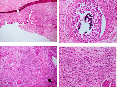
of ILC of breast origin (Figure 3).The immunohistochemistry (IHC)
analysis showed metastasis of ILC of breast origin to the uterine
body. Estrogen receptor (ER) and progesterone receptor (PR) tests
were positive (Figure 4) while CerB2 test was negative.
Her general condition worsened, level of serum creatinine increased to 3.9 mg/dl and her Hg decreased to 6.6 g/dl. In addition to nausea and vomiting, she suffered bowel obstruction, for which laparotomy was performed. Subtotal hysterectomy, bilateral salpingo-oophorectomy and colostomy were also performed. On breast re-examination, no mass was detected. The patient’s condition deteriorated after a month and she expired before mammography could be performed.
Discussion
Lobular carcinoma is the common type of breast cancer in
women and in 60% of patients suffering invasive lobular carcinoma
metastasis happens to peritoneum, ovary, and gastrointestinal
tract at the time of diagnosis [4,5].The rate of metastasis of
invasive ductal carcinoma (IDC) and invasive lobular carcinoma
(ILC) to gynecologic organs is about %0.8 and %4.5 respectively
[3]. Lobular carcinoma is the type of breast carcinoma that
frequently metastasize to gastrointestinal, gynecological and
peritoneal organs. The identification of unusual metastasis is the
characteristics of invasive lobular carcinoma [1].
Metastasis of cancer to ovaries happens in about 75.8% cases
followed by involvement of vagina in 3.4% cases while uterine
corpus is involved in 4.7%, cervix in 3.4%, vulva in 2% and salpinx
in 0.7% cases. Uterine metastasis usually originates from the
ovaries and its metastasis from the extra-genital sites are rare
[6]. Data presented by MAZUR et al. indicated that involvement
of only myometrium accounts for 63.5% of the total, followed by
myometrium and endometrium (32.7%) while endometrium
only is involved at the rate of 3.8% [7].In the present case also,
myometrium is involved. Invasive lobular carcinoma (ILC) has a
desmoplastic reaction which sometimes is not very clear or there is
no desmoplastic reaction at all [8]. The present case who was at the
last stage of the disease, with involvement of the organs to which it
was metastasized, the primary site lacked any sign of involvement.
On her breast examination also, nipple retraction was observed
though she stated that it was so since long time ago.
Breast sonography has proved to be more sensitive for diagnosis
of lobular breast carcinoma as up to %97.8 of patients had posterior
shadowing [9]. As accuracy of breast sonography is high, we also
advised it for diagnosis of probable abnormality for our case. In the
present case, breast cancer metastasized to the uterus while she
lacked symptoms in the breast. Her cancer propagated not only
to the uterus but also it involved ovaries and adjacent bones. Its
propagation exceeded much beyond the level of its propagation in
the breast which was the source of uterine carcinoma.
Conclusion
Low incidence rate of invasive lobular carcinoma of breast metastasized to myometrium, poses a significant diagnostic challenge. It is important to note that in cases of endometrial or uterine cancer, metastasis from breast cancer should be kept in mind. Lack of any sign related to breast cancer does not not exclude the possibility of breast cancer with a history of uterine cancer; rather it may be suggestive of endometrium metastatic disease. A precise diagnosis should be based on clinical history and immunohistochemistry to differentiate a metastatic breast cancer. To confirm the diagnosis, rapid endometrial sampling is to be performed. As sensitivity of breast sonography is high in detecting invasive lobular carcinoma of breast, it is to be performed for diagnosis of any abnormality of breast.
References
- McGuire A, Brown ALJ, Malone C, McLaughlin R, Kerin MJ (2015) Effects of Age on the Detection and Management of breast Cancer. Cancers 7: 908-929.
- Lobular Breast Cancer: Causes, Tests, and Treatment (2019)
- Gomez M, Whiting K, Naos R (2020) Lobular breast carcinoma metastatic to the endometrium in a patient under tamoxifen therapy: A case report. SAGE Open Medical Case Reports 8: 1-4.
- Arpino G, Bardou VJ, Clark GM, Elledge RM (2004) Infiltrating lobular carcinoma of the breast: tumor characteristics and clinical outcome. Breast Cancer Research 6(3): R149-R156.
- Bezpalko K, Mohamed MA, Mercer L, McCann M, Elghawy K, et al. (2015) Concomitant endometrial and gallbladder metastasis in advanced multiple metastatic invasive lobular carcinoma of the breast: A rare case report. International Journal of Surgery 14: 141-145.
- Kumar NB, Hart WR (1982) Metastases to the uterine corpus from extra-genital cancers. A clinic-pathologic study of 63 cases. Cancer 50(10): 2163-2169.
- Mazur MT, Hsueh S, Gersell DJ (1984) Metastases to the female genital tract: analysis of 325 cases. Cancer 53: 1978-1984.
- Porter AJ, Evans EB, Foxcroft LM, Simpson PT, Lakhani SR (2014) Mammographic and Ultrasound Features of Invasive Lobular Carcinoma of the Breast. Journal of Medical Imaging and Radiation Oncology. 58(1): 1-10.
- Johnson K, Sarma D, Hwang ES (2015) Lobular breast cancer series: imaging. Breast Cancer Research 17: 94.

Top Editors
-

Mark E Smith
Bio chemistry
University of Texas Medical Branch, USA -

Lawrence A Presley
Department of Criminal Justice
Liberty University, USA -

Thomas W Miller
Department of Psychiatry
University of Kentucky, USA -

Gjumrakch Aliev
Department of Medicine
Gally International Biomedical Research & Consulting LLC, USA -

Christopher Bryant
Department of Urbanisation and Agricultural
Montreal university, USA -

Robert William Frare
Oral & Maxillofacial Pathology
New York University, USA -

Rudolph Modesto Navari
Gastroenterology and Hepatology
University of Alabama, UK -

Andrew Hague
Department of Medicine
Universities of Bradford, UK -

George Gregory Buttigieg
Maltese College of Obstetrics and Gynaecology, Europe -

Chen-Hsiung Yeh
Oncology
Circulogene Theranostics, England -
.png)
Emilio Bucio-Carrillo
Radiation Chemistry
National University of Mexico, USA -
.jpg)
Casey J Grenier
Analytical Chemistry
Wentworth Institute of Technology, USA -
Hany Atalah
Minimally Invasive Surgery
Mercer University school of Medicine, USA -

Abu-Hussein Muhamad
Pediatric Dentistry
University of Athens , Greece

The annual scholar awards from Lupine Publishers honor a selected number Read More...


