Lupine Publishers Group
Lupine Publishers
Menu
ISSN: 2643-6760
Case Report(ISSN: 2643-6760) 
Cutaneous Breast Cancer Metastasis Volume 6 - Issue 3
Amani Saleh Hadi Saeed*
- Specialist of Clinical Oncology and Nuclear Medicine, National Oncology Center, Yemen
Received: May 14, 2021 Published: May 26, 2021
Corresponding author: Amani Saleh Hadi Saeed, Specialist of Clinical Oncology and Nuclear Medicine, National Oncology Center, Yemen
DOI: 10.32474/SCSOAJ.2021.06.000237
Introduction
Cutaneous metastasis (plural ‘metastases’) refers to growth of cancer cells in the skin originating from internal cancer, develops after the initial diagnosis of primary internal malignancy (such as breast, lung, melanoma, colon, stomach cancer, uterus, kidney and late in the course of disease. Many women diagnosed with breast cancer will achieve cure with surgery followed by adjuvant chemotherapy, hormonal therapy, or radiation therapy; however, some breast cancer survivors will develop locally recurrent disease. Skin metastasis are one of the most distressing presentations of locally recurrent breast cancer. The presence of skin metastasis signifies widespread systemic disease and poor prognosis [1]. Assessment of cutaneous metastatic disease after mastectomy can be perplexing because the clinical presentation appears similar to other skin disease such as cellulitis or lymphedema. Patient with breast cancer require differentiation between skin metastasis and benign dermatological disease. Differences between cutaneous metastasis and cellulitis or lymphedema were found most definitively on the histological study of tissue biopsy [2]. Excluding malignant melanoma, breast cancer has the highest incidence of cutaneous metastasis compared other solid malignancy [2]. Autopsy studies reported an estimated incidence of 24% [3].
The clinical presentation varies as 80% of the lesion are papules and nodules and 11% are telangiectatic carcinoma [4]. Other presentation includes eryspeliod carcinoma “en cuirasse carcinoma”, alopecia neoplastica, zoster form pattern, and melanoma like pigmented lesion [4,5]. Breast cancer is one of the most common malignancies to spread to the skin. the most likely site for cutaneous metastasis is the chest; less common sites include, the scalp, the neck, the upper extremities abdomen and back. Occasionally, patient metastatic breast cancer have a firm, scar like area in the skin. When this occurs on the scalp, hair may be lost, and the clinical appearance may mimic alopecia areata, except that the skin exhibits marked induration on palpation.
Case Presentation
The author report three Yemeni cases of skin metastasis of breast cancer with different clinical presentations
Case 1
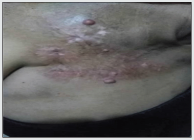
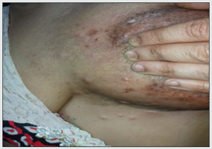
(Figure 1 and 2) was a 55-year-old Yemeni woman diagnosis as having infiltrating ductal carcinoma of left breast, she underwent lumpectomy followed by radiotherapy within 15 days, she developed metastasis to anther breast, treated with chemotherapy and hormonal than develop skin metastasis with crusted lesion and pink nodules pain less of various size in left breast.
Case 2
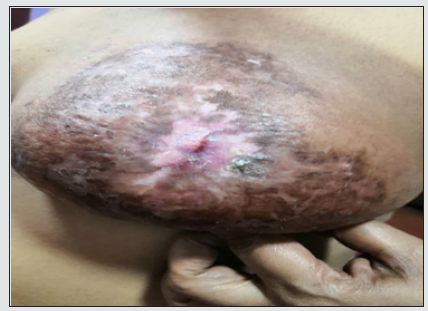
(Figure 3) was a 65 -year-old Yemeni woman diagnosed as having infiltrating ducal carcinoma of right breast underwent mastectomy with axillary clearance followed by chemotherapy and hormonal therapy, after a remission period of 18 months developed firm nodules pain less around the mastectomy scar in right chest wall.
Case 3
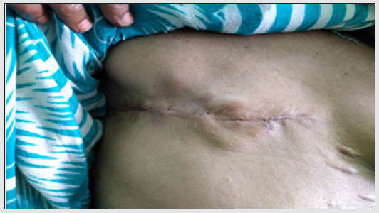
(Figure 4) was a 60 year -old-Yemeni woman diagnosed as having infiltrating ductal carcinoma of left breast underwent mastectomy with axillary clearance followed by chemotherapy and radiotherapy and hormonal. A remission period 3 months than develop painful erythematous lesion and ulcerated skin with a cellulitis-like appearance of left chest wall. A recurrence of ductal carcinoma was confirmed with skin biopsies, and patient were referred to oncology department for further investigation and appropriate management. Cutaneous metastases of breast cancer remain a therapeutic challenge and is assiocted with increase morbidity. progression of disease often results pain, chest wall ulceration, bleeding superinfection [6]. Therapeutic options are systemic agents and /or skin -directed treatments.
Effective treatment depends on treatment of underlying tumor. Palliative care is given if lesions are asymptomatic and the primary cancer is untreatable .This care includes keeping lesions clean and dry and debriding the lesions if they are bleeding or cursted. surgical care in many cases of cutaneous metastasis by excision and removal of metastases may be warranted to enhance the patient’s quality of life ,but excision of selected metastases dose little to increase survival. Simple excision is usually the treatment of choice. Short-wavelength radiation therapy may be helpful for providing symptomatic relief from painful lesion, using superficial electron beam therapy. carbon dioxide laser therapy [7,8]. liquid nitrogen cryotherapy with temperature probe control, and other treatment approach may also be of value [9]. Pulsed dye laser treatment, which can reduce blood flow to highly vascularized lesions, may be value. Intralesional chemotherapy and cytokines can also be helpful [10].
Figure 4: Clinical presentation as diffusely erythematous indurated and ulcerated skin with a cellulitis-like appearance of the left chest wall and pink nodules.

References
- Hussein MRA (2010) Skin metastasis: A pathologist's perspective. J Cutan Pathol 37(9): 1-20.
- Lund Nielsen B, Muller K, Adamsen (2005) Malignant wounds in women with breast cancer feminine and sexual perspectives. J Clin Nur 14(1): 56-64.
- Kalmykow B, Walker S (2011) Cutanouse metastases in breast cancer. Clin J Oncol Nurs 15(1): 99-101.
- Krathen RA, Orengo IF, Rosen T (2003) Cutanueous metastasis ameta-analysis of data. South Med J 96(2): 164-167.
- Mordenti CP, Concetta Fargnoli K, Cerroni M, Chimenti LS (2000) Cutanueous metastatic breast carcinoma
- Micallf RA, Boffa MJ, DE Gaetano J (2004) Melanoma like pigment cutaneous metastasis from breast carcinoma. Clin Exp Dermatol 29(2): 144-146.
- Sratt DE, Gordon Spratt EA, Wu S, et al. (2014) Efficacy of skin-directed therapy of cutaneous metastases from advanced cancer: A meta-analysis. J Clin Oncol 32(28): 3144-3155.
- Hill S, Thomas JM (1996) Use of the carbon dioxide laser to manage cutaneous metastasis from malignant melanoma. Br J Surg 83(4): 509-512.
- Lingam MK, McKay AJ (1995) Carbon dioxide laser ablation as an alternative treatment for cutaneous metastasis from malignant melanoma. Br Jsurg 82(10): 134446-134448.
- Kubota Y, Mir LM, Nakoda T, Sasagawa I, Suzuki H, et al. (1998) Successful treatment of metastatic skin lesions with electrochemotherapy. J Urol 160(4): 1426.

Top Editors
-

Mark E Smith
Bio chemistry
University of Texas Medical Branch, USA -

Lawrence A Presley
Department of Criminal Justice
Liberty University, USA -

Thomas W Miller
Department of Psychiatry
University of Kentucky, USA -

Gjumrakch Aliev
Department of Medicine
Gally International Biomedical Research & Consulting LLC, USA -

Christopher Bryant
Department of Urbanisation and Agricultural
Montreal university, USA -

Robert William Frare
Oral & Maxillofacial Pathology
New York University, USA -

Rudolph Modesto Navari
Gastroenterology and Hepatology
University of Alabama, UK -

Andrew Hague
Department of Medicine
Universities of Bradford, UK -

George Gregory Buttigieg
Maltese College of Obstetrics and Gynaecology, Europe -

Chen-Hsiung Yeh
Oncology
Circulogene Theranostics, England -
.png)
Emilio Bucio-Carrillo
Radiation Chemistry
National University of Mexico, USA -
.jpg)
Casey J Grenier
Analytical Chemistry
Wentworth Institute of Technology, USA -
Hany Atalah
Minimally Invasive Surgery
Mercer University school of Medicine, USA -

Abu-Hussein Muhamad
Pediatric Dentistry
University of Athens , Greece

The annual scholar awards from Lupine Publishers honor a selected number Read More...


