
Lupine Publishers Group
Lupine Publishers
Menu
ISSN: 2641-1644
Research Article(ISSN: 2641-1644) 
Synthesis and Microscopic Appearance of Thallus-Like Objects in Synthetic Dna (E. Coli) Crown Cells Created Using Monolaurin and Egg White Volume 1 - Issue 4
Shoshi Inooka*
- *Japan Association of Science Specialists
Received: February 20, 2023; Published:March 07, 2023
Corresponding author: Shoshi Inooka, Japan Association of Science Specialists
DOI: 10.32474/OAJRSD.2024.03.000157
Abstract
Synthesized DNA crown cells (artificial cells), which can be proliferate within egg white in vivo, can be prepared in vitro using sphingosine (Sph)-DNA-adenosine-monolaurin compounds. Synthesized DNA crown cells form assemblies and proliferate in the presence of monolaurin, both of which have been observed in cultures on agar plates. In further experiments with egg white, various type of cell-like organisms, some of which differed from the original cells, could be also observed in cultures on agar plates. In one of these cells, the proliferation of objects similar in appearance to thalli was observed. The microscopic characteristics of these thallus-like objects is described here.
Keywords: synthetic DNA (E. coli) crown cells; agar plate cultures; Sphingosine-DNA; cells; Objects like thallus; monolaurin; egg white
Introduction
Artificial cells that are covered with DNA are referred to as DNA crown cells [1-3]. Synthetic DNA crown cells can develop into DNA crown cells after culture in egg white and can be prepared using sphingosine (Sph)-DNA and adenosine-monolaurin (A-M) compounds. Such DNA crown cells formed assemblies or cells that proliferated in cultures to which monolaurin was added and could be cultivated in test tubes [4-6]. Assembly formation and cell proliferation (as a population) could also be observed in cultures using agar plates [7, 8]. In previous studies on proliferated cells, it was suggested that they differed from the original DNA crown cells and that they were regenerated cells [4-6]. In this study, various types of cell proliferation were observed when monolaurin-treated synthetic DNA (E. coli) crown cells were cultivated with egg white on agar plates. In one of these proliferated cells, thallus-like objects were observed. These objects were microscopically characterized and are described herein.
Materials and Methods
Materials
The materials used were the same as those employed in a previous study [9, 10]: Sph (Tokyo Kasei, Japan), DNA (Sigma-Aldrich), adenosine (Sigma-Aldrich; Wako, Japan), monolaurin (Tokyo Kasei), and adenosine-monolaurin (A-M), a compound synthesized from a mixture of adenosine and monolaurin [9, 10]. Monolaurin solutions were prepared to a final concentration of 0.1 M in distilled water. Agar plates, using standard agar medium (SMA) (AS ONE, Japan) were used. Egg whites were obtained from eggs bought at a local market.
Methods
Preparation of synthetic DNA (E. coli) crown cells: Synthetic DNA (E. coli) crown cells were prepared as described previously [9, 10]. Briefly, 180 μL of Sph (10 mM) and 90 μL of DNA (1.7 μg/μL) were combined, and the mixture was heated and cooled twice. A-M solution (100 μL) was added and the mixture was incubated at 37°C for 15 min. Next, 30 μL of monolaurin solution was added and the mixture was incubated at 37°C for another 5 min. The resulting suspension was used as the synthetic DNA (E. coli) crown cells.
Culture of monolaurin with DNA (E. coli) crown cells and incubation with egg white on agar plates was performed as follows:
a) 50.0 μL of sample was plated on an agar plate with a bacteria spreader.
b) Immediately, 1.5 mL of 0.1 M monolaurin was poured onto the agar plate.
c) After removing excess monolaurin, the plates were inverted and incubated for 3 h at 37°C.
d) Then, 1.5 mL of egg white was poured onto the plate and excess egg white was removed.
e) The plates were inverted and incubated for 7 days at 37°C and observed at 2, 4 and 7 days.
f) After 7 days of incubation, the plates were stored at 4°C.
Microscopic observations
Objects on plates was observed directly under a light microscope and with the naked eye.
Results and Discussion
(Figure 1) shows objects formed by synthetic DNA (E. coli) crown cells on the plates at 3h after monolaurin addition before the addition of egg white. Numerous acute rod-like objects of various sizes and shapes were observed (arrows a, and b). The object (arrow a) measured approximately 45-50 μm. (Figure 2) shows the microscopic appearance of synthetic DNA (E. coli) crown cells after 2 days of egg white culture. Numerous bar- or branch-like objects were observed (arrow a). White bristle-like objects were adsorbed onto the surface of these objects (arrow b). The diameter of the objects (arrow b) was approximately 45-50 μm. (Figure 3) shows the microscopic appearance of synthetic DNA (E. coli) crown cells after 4 days of egg white culture. Branch-like objects (arrow a), surrounded by a wide space (arrow b) were observed. A wide space without branch-like objects was also observed (arrow c). The length of the object indicated by arrow a was about 150-170 μm.
Figure 1: Synthetic DNA (E. coli) crown cells on agar plates 3 h after monolaurin addition. Acute rod-like objects (arrows a, and b). The object indicated by arrow a measured approximately 45–50 μm.
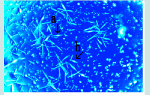
Figure 2: Microscopic appearance of synthetic DNA (E. coli) crown cells at 2 days after egg white addition. Numerous bar- or branch-like objects were observed (arrow a). Objects like white bristles adsorbed on the surface of these bar- or branch-like objects (arrow b). The diameter of objects (arrow b) is approximately 45–50 μm.
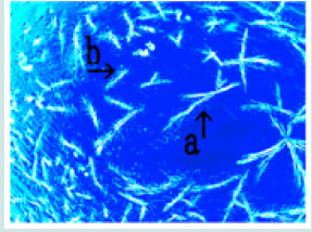
Figure 3: Microscopic appearance of synthetic DNA (E. coli) crown cells at 4 days after egg white addition. A branch-like object (arrow a) was encircled by a wide space (arrow b). Similar wide space (arrow c). The length of the object shown by the arrow a is about 150–170 μm.

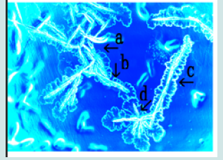
(Figure 4) shows the microscopic appearance of synthetic DNA (E. coli) crown cells after 4 days of egg white culture. Numerous objects appearing as crossed branches encircled by petal-like objects were observed (arrows a, b, and e). Objects possibly enclosed by petals were also observed (arrows c and d). The approximate diameter of these objects (arrow b) was 65-70 μm. (Figure 5) shows the microscopic appearance of synthetic DNA (E. coli) crown cells after 7 days of egg white culture. Large objects with several interlaced branch-like objects were observed (arrow a). In addition, objects with straight (arrow c) and crossed (arrow b) branches that were encircled by petal-like objects were observed. The approximate diameter of these objects (arrow c) was 200-210 μm.
Figure 4: Microscopic appearance of synthetic DNA (E. coli) crown cells 4 days after egg white addition. Crossed branch-like objects (arrows a, b, and e) and objects enclosed by petal-like objects (arrows c and d). The approximate diameter of objects (arrow b) is 65–70 μm.
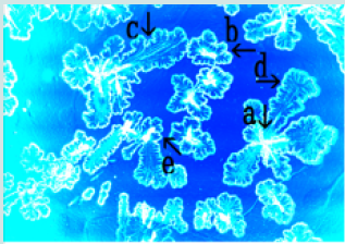
Figure 5: Microscopic appearance of synthetic DNA (E. coli) crown cells at 7 days after egg white addition. Large object (arrow a), crossed branch-like object (arrow b), and straight branch-like object (arrow c). The approximate size of objects (arrow c) is 200–210 μm.
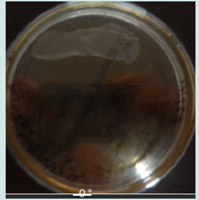
(Figure 6) shows the microscopic appearance of synthetic DNA (E. coli) crown cells after 7 days of egg white culture. Objects like cut branches, which were buried within transformed petal-like objects were observed (arrows a and b). Also, petal-like objects without cut branches were observed (arrow c). The diameter of the objects was approximately 65-70 μm (arrow b). (Figure 7) shows a photograph of agar plates of synthetic DNA (E. coli) crown cells after 7 days of egg white culture. A white region, visible with the naked eye, was observed in the upper part of the Petri dish, which had a diameter of 8.0 cm.
Figure 6: Photograph of agar plates used to culture synthetic DNA (E. coli) crown cells at 7 days after egg white addition. White areas (upper part) visible to the naked eye. The diameter of the dish is 8.0 cm.
Previously, the formation of assemblies and cell proliferation in synthetic DNA (E. coli) crown cell cultures to which monolaurin was added was demonstrated on agar plates [7]. Also, synthetic DNA crown cells or monolaurin-treated synthetic DNA crown cells could be cultivated in egg white, suggesting that a change from non-living to living occurred within egg white [11-14]. Hence, the present experiments examined whether similar growth dynamics were observed in cultures on agar plates and whether new evidence of a change from non-living to living cells was present. Numerous acute branch-like objects were observed 3 h after monolaurin addition (Figure 1) and many branch-like objects, comprising bristle-like objects were adsorbed on the surface of the most of objects. Such objects may be formed with the adsorption or formation of bristle-like objects on the surface of the branch-like objects. It was confirmed that the acute objects shown in (Figure 1) consist of Sph/DNA-adenosine-monolaurin, including related substances, as the objects were formed before the addition of egg white. Hence, objects with bristle-like objects (Figure 2) may be formed as a result of Sph/DNA-adenosine-monolaurin binding to the bristle-like objects.
It may be important to clarify the identity of the bristle-like objects in order to resolve the mechanisms associated with the change from non-living to living cells. However, these formation mechanisms could not be clarified in the present study. Hence, the mechanism proposed below is based on conjecture. Bristle or branch-like objects may belong to a category of firework- or spark-like objects that were often observed for objects consisting of Sph-DNA-adenosine- monolaurin or related components [15]. It was also assumed that some components of egg white were adsorbed by Sph-DNA-adenosine- monolaurin (bristle-like objects) and that new components, i.e., acute-like objects (tentatively referred to here as lifeborn basic substances (LBBS)) may be formed. Also, wide spaces ((Figure 3) arrows b and c) that enclosed acute branch-like objects were observed. These spaces may originate from LBBS, because LBBS adsorbed by the branch-like objects disappeared.
More large spaces may be formed when the spaces fuse, as shown in (Figure 6) (arrow c). Interestingly, in this case, acute branch-like objects may disappear, suggesting that the spaces contain Sph-DNA-adenosine-monolaurin or related components. Such objects (hereafter referred to as white spaces) were observed as aggregates, by eye, after 7 day of culture (Figure 7) and may be formed as an aggregate of several such objects (space), because such objects were observed to disperse after 4 days of culture. It was unclear whether objects like thalli were living organisms or non-living objects.
However, even if such objects were artifacts of egg white or monolaurin, they have a unique shape. Many objects with a unique shape may be found in future research. In the present study, synthetic DNA (E. coli) was used and various different kinds of celllike objects or organisms using different synthetic DNA crown cells, DNA crown cells or cultured DNA crown cells, may be found. Given the large number of such objects, it is considered important to order, or classify, such objects.
The author has therefore developed the following nomenclature for objects observed to date:
Bacteria (E. coli) SDCCME (Thalli) E. coli (Name of DNA source), where SDCC refers to synthetic DNA crown cells), ME refers to monolaurin and egg white, and Thalli refers to the name of the characteristic structure; when the name of an object does not yet exist, the corresponding designation is used, for example (Ta).
Bacteria (E. coli) SDCCME (Thalli) E. coli (Name of DNA source), where SDCC refers to synthetic DNA crown cells), ME refers to monolaurin and egg white, and Thalli refers to the name of the characteristic structure; when the name of an object does not yet exist, the corresponding designation is used, for example (Ta).
References
- Inooka S (2012) Preparation and cultivation of artificial cells. App Cell Biol., 25: 13-18, ISSN 1881-0772.
- Inooka S (2016) Preparation of Artificial Cells Using Eggs with Sphingosine- J Chem Eng Process Technol 7: 277.
- Inooka S (2016) Aggregation of sphingosine-DNA and cell construction using components from egg white. Integrative Molecular Medicine 3(6): 1-5.
- Inooka S (2021) Microscopic observation of assemblies formed in mixtures of DNA (Human placenta) crown cells with Bacilus subtilius. An archive of organic and inorganic science 2637-4609 5(2): 1-7.
- Inooka S (2022) Assembly Formation of Synthetic DNA ( coli) crown cells with Salt Reconstruction and Regeneration of Synthetic DNA Crown Cells. American Journal of Biomedical Science ISSN 2642-1747 16(1):1-10.
- Inooka S (2022) Cell Proliferation from the Assembly of Synthetic DNA (E. coli) Crown Cells with Stimulation by Monolaurin Twice. Current Trends on Biotechnologyand Microbiology 3(3): 1-5.
- Inooka S (2024) Microscopic appearance of Cell Assembly and Proliferation in Agar Cultures of Synthetic DNA ( coli) Crown Cells with Monolaurin. Chemical & Pharmaceutical Research ISSN 2684-1050 6(1): 1-4.
- Inooka S (2024) Microscopic Apperaarance of Monolaurin-Treated Syntheticc DNA ( coli) Crown Cells in Agar Medium Supplemented with Egg White. Scientic Journal Biology and Life Science 3(3): 1-5.
- Inooka S (2017) Biotechnical and Systematic Preparation of Artificial Cells (DNA Crown Cells). The Global Journal of Research in Engineering 17(1).
- Inooka S (2017) Systenmatic Preparation of Artificail Cells (DNA Crown Cells). J Chem Eng Process Technol 8(2): 1-4.
- Inooka S (2022) Preparation of a DNA (E. coli) Crown Cell line in Vitro-Microscopic Appearance of Cells. Annals of Reviews and Research 8(1).
- Inooka S (2023) Preparation and Microscopic Appearance of a DNA (Human Placenta) Crown Cell Line. Journal of Biotechnology and Bioresearch 5(1):1-4.0
- Inooka S (2023) Preparation of a DNA (Akoya pearl oyster) crown cell line. Applied Cell Biology Japan 36: 33-42, ISSN 2433-5800.
- Inooka S (2023) Preparation of a DNA (HepG2 is a Hepatoblastorma-Derived Cell Line) Crown Cell Line. Journal of Tumor Medicine and Prevention 4(2): 1-6.
- Inooka S (2023) Formation and Microscopic Appearance of Fireworks-like Objects Created from Synthetic DNA ( coli) Crown Cells with Adenosine-DNA (E. coli). Annals of Review and Research 9(2): 1-6.

Top Editors
-

Mark E Smith
Bio chemistry
University of Texas Medical Branch, USA -

Lawrence A Presley
Department of Criminal Justice
Liberty University, USA -

Thomas W Miller
Department of Psychiatry
University of Kentucky, USA -

Gjumrakch Aliev
Department of Medicine
Gally International Biomedical Research & Consulting LLC, USA -

Christopher Bryant
Department of Urbanisation and Agricultural
Montreal university, USA -

Robert William Frare
Oral & Maxillofacial Pathology
New York University, USA -

Rudolph Modesto Navari
Gastroenterology and Hepatology
University of Alabama, UK -

Andrew Hague
Department of Medicine
Universities of Bradford, UK -

George Gregory Buttigieg
Maltese College of Obstetrics and Gynaecology, Europe -

Chen-Hsiung Yeh
Oncology
Circulogene Theranostics, England -
.png)
Emilio Bucio-Carrillo
Radiation Chemistry
National University of Mexico, USA -
.jpg)
Casey J Grenier
Analytical Chemistry
Wentworth Institute of Technology, USA -
Hany Atalah
Minimally Invasive Surgery
Mercer University school of Medicine, USA -

Abu-Hussein Muhamad
Pediatric Dentistry
University of Athens , Greece

The annual scholar awards from Lupine Publishers honor a selected number Read More...


