
Lupine Publishers Group
Lupine Publishers
Menu
ISSN: 2641-1709
Case Report(ISSN: 2641-1709) 
Use of 18F-Fdg Pet/Ct In the Staging of a Metastatic Nasopharyngeal Lymphoepithelioma: Case Report Volume 6 - Issue 2
Heloisa dos Santos Sobreira Nunes1*, André Marcondes Braga Ribeiro2 and Eduardo Nóbrega Pereira2*
- 1Department of Otolaryngology, Nucleo de Otorrinolaringologia, Cirurgia de Cabeça e Pescoço de São Paulo, Brazil
- 2Department of Nuclear Medicine, AC Camargo Center, São Paulo, Brazil
Received: March 26, 2021; Published: April 07, 2021
Corresponding author: Heloisa dos Santos Sobreira Nunes, Department of Otolaryngology, Nucleo de Otorrinolaringologia, Cirurgia de Cabeça e Pescoço de São Paulo, Brazil
DOI: 10.32474/SJO.2021.06.000234
Abstract
Nasopharyngeal lymphoepithelioma is a rare condition, especially when metastatic. The study of positron emission tomography / computed tomography (PET/CT) using fluoroxyglucose (FDG) labeled with Fluorine-18 (PET/CT- 18FDG) is well established for head and neck tumors. In this case report we identified, in a 20-year-old patient, multiple lymph node enlargements and bone lesions that complemented the staging and provided a targeted treatment for this metastatic scenario.
Keywords:Fluorodeoxyglucose F18; positron emission tomography computed tomography; nasopharyngeal neoplasms
Introduction
The nasopharynx consists of a trapezius, bounded superiorly
by the sphenoid bone; posteriorly through the vertebral bodies of
the atlas and axis and extends from the base of the skull to the soft
palate; before the choana; and inferiorly to the oropharynx and soft
palate [1]. Among malignant tumors, nasopharyngeal epithelial
tumors correspond between 75% to 85% of tumors, with the
remainder mostly lymphomas1.The most common nasopharyngeal
tumors are non-glandular and non-lymphatic epithelial neoplasms,
grouped as nasopharyngeal carcinoma (NPC). NPCs were
subdivided by the World Health Organization into three categories
according to their differentiation and keratin production:
a) Type I: Squamous Cell Carcinoma (25% of NPC).
b) Type II: Non- Keratinized Carcinomas (12% of NPC).
c) Type III: Undifferentiated carcinoma (approximately. 60%
of NPC).
Type III NPC is composed of several tumors such as
lymphoepithelioma, anaplastic and clear cell. Types II and III have a
positive serological profile for Epstein-Barr Virus (EBV) while type
I does not [1,2]. The standard exam for diagnosis of nasopharyngeal
carcinomas is nasal endoscopy because it is easy to perform and
the possibility of performing a biopsy at the same time. Computed
tomography (CT) is described as essential for the staging and
involvement of bone structures at the base of the skull. Magnetic
resonance imaging (MRI) has been shown to be superior to CT in
the assessment of soft parts, both of the superficial and deep parts
of the nasopharynx [1]. Another useful tool in the diagnosis and
staging of head and neck tumors is positron emission tomography
/ computed tomography (PET / CT) using fluoroxyglucose (FDG)
marked with Fluorine-18 (PET / CT-18FDG)3. This study allows
the analysis of the whole body, being possible to evaluate all the
organs and the bone framework. Thus, although the incidence
of lymphoepithelioma metastasis is only 6.25%, bone location
is the most frequent [3]. Considering the rare occurrence of
nasopharyngeal lymphoepithelioma, especially when metastatic,
the objective of this study is to present a case report focusing on the
usefulness of PET/CT-18FDG, in the evaluation of distant lesions
and prevention of possible risks related to them.
Case Report
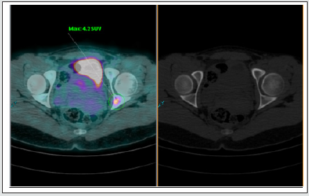
MLSC female, 20 years old, was admitted to the emergency room with a history of cervical nodules on the left for 2 months, with progressive growth, associated with radiated pain to the left shoulder. The patient did not show signs of epistaxis, dysphonia, dyspnoea or recent infections. On physical examination, multiple cervical nodules of up to 1 cm at levels II, III and V on the left, mobile, and the presence of lymph node conglomerate at levels IV and VI on the left, of approximately 10 cm, were hardened and painful. From then on, some exams were carried out to better evaluate these lymph node enlargements. Nasofibrolaryngoscopy showed a nodular lesion on the posterior wall of the nasopharynx, of approximately 1.5 cm and of lymphoid aspect. Neck CT showed multiple lymph node enlargements in the left supraclavicular region, as well as in the mediastinum, confluent and with intervening calcifications (Figure 1). Thus, a level II lymph node biopsy was performed on the left, which showed infiltration by undifferentiated carcinoma, suggestive of a nasopharynx primary. The complementary immunohistochemical study corroborated the diagnosis of undifferentiated carcinoma, type lymphoepithelioma, originating in the nasopharynx and with a positive EBV test. As it is an advanced disease and in order to complete the staging of this pathology, the patient underwent a PET / CT study with 18F-FDG that showed multiple confluent lymph nodes in the left cervical region, bilateral supraclavicular and mediastinal, with SUVs up to 11.0 (Figure 2). In addition, areas of anomalous 18F-FDG concentration were identified in the bone intramedullary regions, with no associated anatomical changes evidenced by CT, in the C7 vertebra with an SUV of 5.4, (Figure 3) in the head of the right femur with an SUV of 5, 8 (Figure 4) and in the left acetabulum with a 4.2 SUV (Figure 5). Therefore, systemic chemotherapy was performed for metastatic disease.
Figure 1: Axial sections of computed tomography showing cervical (left) and mediastinal (right) lymph node conglomerates.
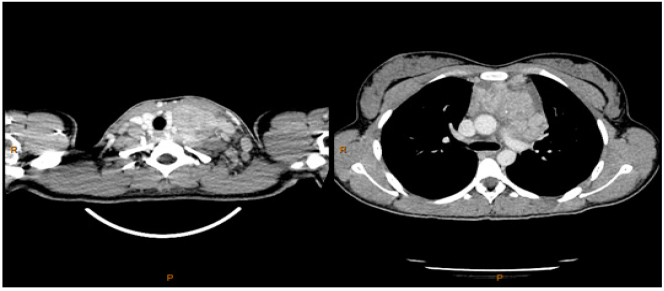
Figure 2: PET / CT fusion images showing multiple confluent lymph node enlargements in the left cervical, bilateral supraclavicular and mediastinal regions in the axial (left), sagittal (center) and coronal (right) sections.
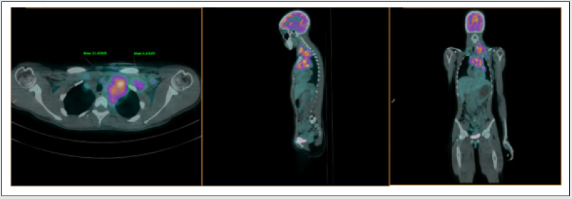
Figure 3: PET / CT fusion images showing multiple confluent lymph node enlargements in the left cervical, bilateral supraclavicular and mediastinal regions in the axial (left), sagittal (center) and coronal (right) sections.
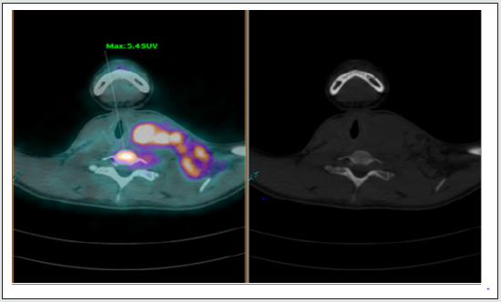
Figure 4: Axial sections showing an area of anomalous concentration of 18F-FDG in the head of the right femur (left) and without anatomical correspondence in the tomographic image (right).
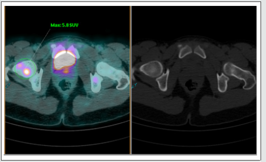
Figure 5: Axial sections showing an area of anomalous concentration of 18F-FDG in the left acetabulum (on the left) and without anatomical correspondence on the tomographic image (on the right).
Discussion
The nasopharynx has several types of epithelium (respiratory, scaly, transitional), as well as different tissues (glandular, connective, lymphoid), and for this reason it can harbor a wide variety of neoplasms [4]. The clinical picture of patients with lymphoepithelioma depends on the location of the primary tumor and the direction of its expansion. It can vary from a tubal dysfunction due to obstruction of the tube ostium in the nasopharynx, to more nonspecific symptoms such as nasal obstruction, epistaxis, tinnitus and frontal headache. In 70% of patients, however, the initial symptom is a cervical mass, as in this case. This is due to the weakness of the nasopharyngeal barriers, which allow a rapid dissemination of the parapharyngeal spaces. At the time of diagnosis, 80% of patients have lymph node involvement [1,5]. In this case, we would like to emphasize the importance of PET-FDG in the evaluation of distant metastatic lesions, which is already well established for head and neck tumors [3]. Although CT of the neck has already shown mediastinal lymph node enlargement, stage IVB1, and the therapeutic approach has already been defined, it was not able to show the intramedullary bone lesion in the C7 vertebra. This lesion did not present identifiable anatomical changes, but it did present abnormal uptake in the PET/CT-18FDG study. In addition, other intramedullary lesions were also identified in the right femur and in the left acetabulum, which would not be identified by other methods. In a general cancer scenario, the importance of correctly identifying metastatic bone lesions is the fact that a systemic treatment specific to each pathology is initiated. However, in this case report, PET-FDG was useful in identifying sites of bone injuries, not observed by other methods, which could cause serious consequences such as spinal cord compression and pathological fractures [6] .
Conclusion
Although metastatic nasopharyngeal lymphoepithelioma is a rare condition, bone metastases are the most common and an accurate diagnosis can help patients avoid possible complications of these injuries.
References
- de Almeida ER, Butugan O, Ling SY, Rezende VA, Miniti A (1992) Malignant nasopharynx tumors. (Epidemiology, histological type and treatment.). Braz J Otorhinolaryngol 58: 88-95.
- Yamashiro I, Souza RP de (2007) Diagnóstico por imagem dos tumores da nasofaringe. Radiol Bras 40: 45-52.
- Yoo J, Henderson S, Walker-Dilks C (2013) Evidence-based Guideline Recommendations on the Use of Positron Emission Tomography Imaging in Head and Neck Cancer. Clin Oncol 25: e33-e66.
- Romano FR, Mendonça ML, Cahali RB, Voegels RL, Sennes LU, et al. (2000) Linfoepiteliomas: Aspectos Clínicos e Terapêuticos. Int Arch Otorhinolaryngol 5: 93-97.
- Cotrim DP, Soares PBM (2012) Hypertrophic osteoarthropathy associated to lymphoepitelioma of the nasopharynx: a case report. Med (Ribeirão Preto) 45: 460-465.
- Meohas W, Probstner D, Vasconcellos RAT, Lopes ACDS, Rezende JFN, et al. (2005) Metástase óssea: revisão da literatura. Rev Bras Cancerol 51: 43-47.

Top Editors
-

Mark E Smith
Bio chemistry
University of Texas Medical Branch, USA -

Lawrence A Presley
Department of Criminal Justice
Liberty University, USA -

Thomas W Miller
Department of Psychiatry
University of Kentucky, USA -

Gjumrakch Aliev
Department of Medicine
Gally International Biomedical Research & Consulting LLC, USA -

Christopher Bryant
Department of Urbanisation and Agricultural
Montreal university, USA -

Robert William Frare
Oral & Maxillofacial Pathology
New York University, USA -

Rudolph Modesto Navari
Gastroenterology and Hepatology
University of Alabama, UK -

Andrew Hague
Department of Medicine
Universities of Bradford, UK -

George Gregory Buttigieg
Maltese College of Obstetrics and Gynaecology, Europe -

Chen-Hsiung Yeh
Oncology
Circulogene Theranostics, England -
.png)
Emilio Bucio-Carrillo
Radiation Chemistry
National University of Mexico, USA -
.jpg)
Casey J Grenier
Analytical Chemistry
Wentworth Institute of Technology, USA -
Hany Atalah
Minimally Invasive Surgery
Mercer University school of Medicine, USA -

Abu-Hussein Muhamad
Pediatric Dentistry
University of Athens , Greece

The annual scholar awards from Lupine Publishers honor a selected number Read More...


