Lupine Publishers Group
Lupine Publishers
Menu
ISSN: 2641-1709
Research Article(ISSN: 2641-1709) 
The Role of Ultrasonography and Fine Needle Aspiration Cytology in Thyroid Swellings Volume 6 - Issue 1
Afshan Fathima1*, Rehna Mohamed2, Havva Hanana2, Sandeep Jain A M2 and B Viswanatha3
- 1Senior Resident, Department of ENT, Bangalore Medical College & Research Institute, India
- 2Junior Resident, Department of ENT, Bangalore Medical College & Research Institute, India
- 3Professor & Head of Department of ENT, Bangalore Medical College & Research Institute, India
Received: March 10, 2021; Published: March 19, 2021
Corresponding author: Afshan Fathima, Senior Resident, Department of ENT, Bangalore Medical College & Research Institute, Bengaluru, India
DOI: 10.32474/SJO.2021.06.000230
Abstract
Introduction: Thyroid swellings are commonly encountered endocrine disorders. These disorders can be evaluated by thyroid function tests, ultrasonography (USG), fine needle aspiration cytology (FNAC), computed tomography (CT scan), histopathology etc. The present study was undertaken to evaluate the role of ultrasonography and fine needle aspiration cytology in the diagnosis of thyroid swellings. The sensitivity, specificity, positive predictive value, negative predictive value and diagnostic accuracy of both ultrasonography and fine needle aspiration cytology were evaluated and correlated with the histopathology diagnosis.
Materials and Method: 70 consecutive patients presenting with thyroid swellings were thoroughly examined in the department of otorhinolaryngology and details were documented. The patients were subjected to an USG and FNAC of the thyroid swellings to study the lesions from sonological and cytological point of view respectively. Patients were posted for thyroidectomy surgery and the thyroid specimen was subjected to histopathological examination. Data were collected and tabulated.
Result: A total of 70 patients were enrolled in the present study. 9 (12.86%) patients were males and 61 (87.14%) were females. Male to female ratio was 1:6.78. The most common age group noted was 31-40 years with 34 patients (48.57%). Histopathology was considered as the gold standard for diagnosis. Correlation of USG and FNAC diagnosis of thyroid swellings was made with histopathology by evaluating the sensitivity, specificity, positive predictive value, negative predictive value, and diagnostic accuracy. USG showed a sensitivity of 84.61%, specificity of 91.22%, positive predictive value of 68.75%, negative predictive value of 96.29% and accuracy 90% of in thyroid swellings. On the contrary, FNAC showed a sensitivity of 92.85%, specificity of 94.64%, positive predictive value of 81.25%, negative predictive value of 98.14% and accuracy of 94.29%.
Conclusion: USG and FNAC of thyroid swellings are simple, cost effective and yield quick results. Although histopathology is confirmatory and the gold standard for diagnosis, USG and FNAC in conjunction have reasonably good sensitivity, specificity, positive predictive value, negative predictive value and diagnostic accuracy for preoperative diagnosis of thyroid swellings.
Keywords: Ultrasonography; fine needle aspiration cytology; histopathology; thyroid swellings
Introduction
Thyroid gland swellings are one of the most commonly encountered endocrine disorders on a worldwide scale. According to the evaluation done by various studies on thyroid studies, an estimated 42 million people suffer from thyroid disorders in India [1]. Thyroid lesions can be evaluated preoperatively using parameters such as thyroid function tests, ultrasonography and fine needle aspiration cytology. The diagnosis of thyroid lesions using aspiration cytology was first reported by Martin and Ellis in 1930 [2]. The routine use of fine needle aspiration cytology (FNAC) in the assessment of thyroid nodules has reduced the number of patients subjected to thyroidectomy for benign diseases of the thyroid [3-5]. As a result, the incidence of malignancy at thyroidectomy has increased from 5-10% to 30-50% in the recent years [6]. This relatively simple procedure has assumed a dominant role in determining the management of patients with thyroid nodules [7,8]. Thyroid ultrasonography is one of the most popular radiological methods of diagnosing thyroid disease. Ultrasonography is commonly the first imaging modality after clinical examination in evaluation of thyroid swellings. Neck ultrasonography was first introduced in 1966-1967 [9]. It has been widely practiced since the 1970 and is now one of the most popular radiological methods of diagnosing neck disease [10]. It is an easily accessible, non-invasive method and helps to pinpoint a possible abnormality at an early stage. Rugu [11]and his associate Joham Vent were first to advocate surgical biopsy as an essential diagnostic tool. Currently this technique is practiced worldwide, and it is the investigation of choice in thyroid, lymph nodes and breast swellings. Histopathological evaluation is considered a vital modality in the diagnosis and management of thyroid lesions.
Aims & objectives
The present study was undertaken to evaluate the role of ultrasonography and fine needle aspiration cytology in the diagnosis of thyroid swellings. The sensitivity, specificity, positive predictive value, negative predictive value and diagnostic accuracy of both ultrasonography and fine needle aspiration cytology were evaluated and correlated with the histopathology report.
Materials and Method
The present study titled ‘The role of Ultrasonography and Fine Needle Aspiration Cytology in thyroid swellings’ was carried out in the department of Otorhinolaryngology, Bangalore Medical College & Research Centre between July 2018 and January 2020.
Mode of selection: 70 consecutive patients presenting with clinically palpable thyroid swellings were included in the present study.
Inclusion criteria: Patients consenting for the study, clinically palpable thyroid swellings, patients willing to undergo USG and FNAC and patients willing to undergo surgery for the thyroid lesions.
Exclusion criteria: Patients with clinically undetectable thyroid swellings, previous history of neck trauma or neck surgeries and patients with neck abscesses.
Study design: Prospective Observational study.
Methodology
A total of 70 consecutive patients presenting with thyroid swellings were thoroughly examined in the department of Otorhinolaryngology and details regarding history, clinical examination and clinical diagnosis were documented. The patients were subjected to an ultrasonography of the neck to study the thyroid swellings from sonological point of view. This was followed by subjecting the patients to fine needle aspiration cytology of the thyroid swellings which provided us information in terms of cytology. Patients were posted for thyroidectomy surgery and the thyroid specimen was subjected to histopathological examination. Data was collected and tabulated in an excel sheet. Results presented as percentages and proportions. A correlation of thyroid swellings using ultrasonography, fine needle aspiration cytology and histopathology was made. Statistical tests such as sensitivity, specificity and diagnostic accuracy were calculated accordingly.
Results
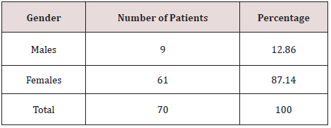
A total of 70 patients were enrolled in the present study. 9 (12.86%) patients were males and 61 (87.14%) were females (Table 1). The male to female ratio was 1:6.78. The most common age group noted was 31-40 years with 34 patients (Table 2). The most common clinical symptoms were swelling in the neck in midline with gradual increase in size. The thyroid swelling was most commonly noted in the right lobe in 42 (70%) patients. 10 (16.67%) patients presented with both lobes being involved. According to USG diagnosis, 57 (81.43%) patients were diagnosed with benign swellings such as solitary thyroid nodule (28 patients), multinodular goitre (13 patients), inflammatory lesions (10 patients) and benign neoplasms (6 patients). Malignant neoplasms were noted in 13 (18.57%) patients (Table 3). According to FNAC diagnosis, 56 (80%) patients were noted to have benign swellings such as solitary thyroid nodule (27 patients), multinodular goitre (12 patients), inflammatory lesions (11 patients) and benign neoplasms (6 patients). Malignant neoplasms were noted in 14 (20%) patients (Table 3). After the patients underwent thyroidectomy procedure, the specimen was subjected to histopathological diagnosis. According to it, 54 (77.14%) patients were diagnosed to have benign swellings such as solitary thyroid nodule (25 patients), multinodular goitre (13 patients), inflammatory lesions (11 patients) and benign neoplasms (5 patients). Malignant neoplasms were noted in 16 (22.86%) patients (Table 3). Histopathological diagnosis was considered as the gold standard. The correlation between USG versus histopathology and FNAC versus histopathology was made to determine the sensitivity, specificity, positive predictive value, negative predictive value, and accuracy of USG and FNAC in thyroid swellings (Tables 4 & 5). USG showed a sensitivity of 84.61%, specificity of 91.22%, positive predictive value of 68.75%, negative predictive value of 96.29% and accuracy 90% of in thyroid swellings. On the contrary, FNAC showed a sensitivity of 92.85%, specificity of 94.64%, positive predictive value of 81.25%, negative predictive value of 98.14% and accuracy of 94.29% (Table 6).
Table 3: Ultrasonography, Fine Needle Aspiration Cytology and Histopathology Diagnosis of Thyroid Swellings.

Table 6: Sensitivity, Specificity, Positive Predictive Value, Negative Predictive Value & Accuracy of USG and FNAC in Thyroid Swellings.

Discussion
The present study was done to correlate the ultrasonography and fine needle aspiration cytology of thyroid swellings with its histopathology. Ultrasonography is an important investigative modality in evaluating the thyroid swellings in terms of size, number, location, consistency, and morphology. It also helps in detecting impalpable thyroid swellings. FNAC helps in providing the most direct and specific information about the thyroid gland. It is a simple method of detecting thyroid swelling pathology. The present study was done to determine the role of ultrasonography and FNAC in thyroid swellings. Out of 70 patients, females (61 patients) outnumbered the males (9 patients). The most common age group affected was between 31-40 years. In a similar study done by Jain et al. [12], of the 110 patients enrolled in the study, 18 were males and 92 were females. The most common age group affected was between 31-40 years. These findings were in accordance with our present study. In our study, on ultrasonography of the thyroid gland, 57 swellings were diagnosed as benign and 13 were diagnosed malignant. Similarly, on FNAC, 56 swellings were diagnosed benign and 14 diagnosed malignant. Histopathology of the thyroid swelling was considered gold standard in diagnosis. On histopathological diagnosis, 54 thyroid swellings were diagnosed as benign and 16 diagnosed malignant. In a similar study done by Avinash et al.[13], of the 70 patients, 59 were diagnosed benign and 11 were diagnosed malignant on ultrasonography of the thyroid gland. These observations were similar to our present study. In our study, ultrasonography showed a sensitivity of 84.61%, specificity of 91.22%, positive predictive value of 68.75%, negative predictive value of 96.29% and accuracy of 90%. FNAC showed a sensitivity of 92.85%, specificity of 94.64%, positive predictive value of 81.25%, and negative predictive value of 98.14% and accuracy of 94.29%. Similar studies [14-16] done by other authors showed results which were in accordance with the present study (Tables 7 & 8).
Table 7: Sensitivity, Specificity, Positive Predictive Value, Negative Predictive Value and Diagnostic Accuracy of Ultrasonography of other authors.

Table 8: Sensitivity, Specificity, Positive Predictive Value, Negative Predictive Value and Diagnostic Accuracy of Ultrasonography of other authors.

Conclusion
Ultrasonography and FNAC are simple and cost-effective investigations in diagnosing thyroid lesions. They can be performed as outpatient department procedures and yield quick results. Both the procedures are effective modalities in making preoperative diagnosis in thyroid swellings. Although histopathology is confirmatory and the gold standard for diagnosis, ultrasonography and fine needle aspiration cytology in conjunction have reasonably good sensitivity, specificity, positive predictive value, negative predictive value, and diagnostic accuracy for preoperative diagnosis of thyroid swellings.
References
- Kirkup J (1995) The history and evolution of surgical instruments- VI The surgical blade: from fingernail to ultrasound. Ann R Coll Surg Engl 77: 380-388.
- Bashetty K, Nadig Kapoor S (2009) Electrosurgery in aesthetic and restorative dentistry: A literature review and case reports. Jrnl Cons Dent 12(4): 139-146.
- Hausner K (1986) Electrosurgery. Addison IL Elmed, Inc p. 2-10.
- Sharma R (2017) Safety of colorado microdissection needle (stryker) for skin opening in craniomaxillofacial surgery. J. Maxillofac. Oral Surg 11(1): 115–118.
- Rideout B, Shaw G Y (2004) Tonsillectomy using the colorado microdissection needle: A prospective series and comparative technique review. Southern Medical Journal 97(1): 11-17.
- Farnworth T, Beals S, Manwaring K, Trepeta R (1993) Comparison of skin necrosis in rats by using a new microneedle electrocautery, standardized electrocautery, and the Shaw hemostatic scalpel. Ann Plast Surg 31(2): 164-167.
- Sheikh B (2015) Safety and efficacy of electrocautery scalpel utilization for skin opening in neurosurgery. Br J Neurosurg 18(3): 268-272.
- Kearns S, Conolly E, Mcnally S, McNamara D, Deasy J (2001) Randomised clinical trial of diathermy versus scalpel incisions in midline laparotomy. Br J Surg 88: 41-44.
- Madhukar KT, Ganesh MT (2016) Comparative study of safety and efficacy of electrocautery blade with cold scalpel blade for skin opening during fixation of fracture of forearm bone with plate and screws. International Journal of Health & Allied Sciences 1(3): 153-157.
- Margarita Peneva, Andrijana Gjorgjeska, Vladimir Ginoski, Hristina Breshkovska, Roza Dzoleva Tolevska (2018) Electro- surgical micro- needle versus scalpel skin incisions in the facial region. SANAMED 13(3): 1-8.
- Rampalli Viswa Chandra, Boya Savitharani, Aileni Amarender Reddy (2016) Comparing the outcomes of incisions made by colorado microdissection needle, electrosurgery tip, and surgical blade during periodontal surgery: A randomized controlled trial. J Indian Soc Periodontol 20: 612-622.
- Yonca O Arat, Almila S Sezenoz, Francesco P Bernardini, Mark A Alford, Merih Tepeoglu, et al. (2016) Comparison of Colorado Microdissection Needle Versus Scalpel Incision for Aesthetic Upper and Lower Eyelid Blepharoplasty. Ophthal Plast Reconstr Surg 13: 1-6.

Top Editors
-

Mark E Smith
Bio chemistry
University of Texas Medical Branch, USA -

Lawrence A Presley
Department of Criminal Justice
Liberty University, USA -

Thomas W Miller
Department of Psychiatry
University of Kentucky, USA -

Gjumrakch Aliev
Department of Medicine
Gally International Biomedical Research & Consulting LLC, USA -

Christopher Bryant
Department of Urbanisation and Agricultural
Montreal university, USA -

Robert William Frare
Oral & Maxillofacial Pathology
New York University, USA -

Rudolph Modesto Navari
Gastroenterology and Hepatology
University of Alabama, UK -

Andrew Hague
Department of Medicine
Universities of Bradford, UK -

George Gregory Buttigieg
Maltese College of Obstetrics and Gynaecology, Europe -

Chen-Hsiung Yeh
Oncology
Circulogene Theranostics, England -
.png)
Emilio Bucio-Carrillo
Radiation Chemistry
National University of Mexico, USA -
.jpg)
Casey J Grenier
Analytical Chemistry
Wentworth Institute of Technology, USA -
Hany Atalah
Minimally Invasive Surgery
Mercer University school of Medicine, USA -

Abu-Hussein Muhamad
Pediatric Dentistry
University of Athens , Greece

The annual scholar awards from Lupine Publishers honor a selected number Read More...


