Lupine Publishers Group
Lupine Publishers
Menu
ISSN: 2641-1709
Case Report(ISSN: 2641-1709) 
Surgical Management of Oral Mucocele: A Case Report Volume 4 - Issue 3
Esha Yadav, Richa S Gautam, Akhil K Padmanabhan* and Prabhuji MLV
- Department of Periodontology, Krishnadevaraya College of Dental Sciences and Hospital, India
Received:June 11, 2020; Published: June 24, 2020
Corresponding author: Akhil K Padmanabhan, Department of Periodontology, Krishnadevaraya College of Dental Sciences and Hospital, India
DOI: 10.32474/SJO.2020.04.000189
Abstract
The present case report describes the surgical removal of mucocele in a young female patient. A thirteen year old female patient was diagnosed clinically with mucocele of lower lip. A conventional surgical procedure was performed using scalpel and the excised lesion was sent for histological investigation. Histological investigation revealed well circumscribed cystic spaces filled with mucous clearly illustrating extravasation type mucocele. Complete regression of the lesion was seen with no recurrence in 6 months follow up. Surgical excision of the lesion with the involved minor salivary gland proved to give satisfactory results.
Keywords: Oral mucocele; diagnosis; excision; minor salivary gland.
Introduction
Oral mucocele are benign mucous filled cavities commonly found in oral cavity, paranasal sinuses and appendix [1,2]. It is the seventeenth most commonly found salivary lesion of the oral cavity [3]. The term mucocele was derived from Latin word (“muco”- mucous and “coele”- cavity) [4]. The characteristic features of this lesion is simple or multiple, soft, translucent and fluctuant [5]. It occurs due to accumulation of mucous due to trauma and obstruction of salivary gland duct [6,7]. There are two types of oral mucoceles generally seen in oral cavity named as, extravasation and retention type [8]. Extravasation type is most commonly found in children [9]. The most frequently affected site is lower lip and other sites include palate, buccal mucosa and floor of the mouth [10]. Although mucocele is painless, it may cause disturbances in speech and swallowing [11]. Various treatment strategies which includes surgical excision, cryosurgery, laser ablation, sclerotherapy and intralesional injection of sclerosing solutions [12]. The present study was undertaken to evaluate a case report of mucocele on lower lip treated by surgical excision using scalpel.
Case Report
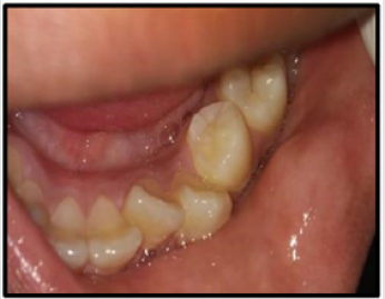
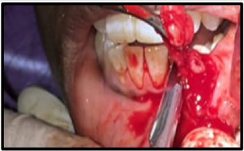
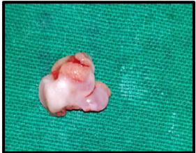
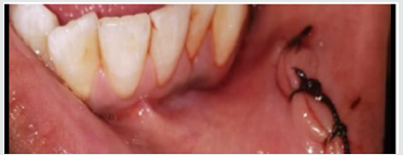
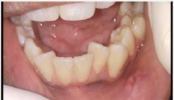
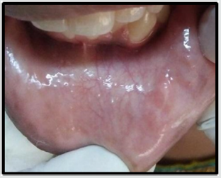
Thirteen year old female patients reported with the chief complaint of swelling on the inner aspect of her lower lip since 4 weeks. Swelling gradually increased in size and had difficulty in speech and swallowing. Patient also had a positive history of lip biting with no history of medical illness. On intra oral examination there was a small, round, fluctuant swelling seen on the labial mucosa of lower lip at the left premolar region. Swelling was 3-4 mm below the vermillion border of the lip measuring approximately 7-8 mm. Color of the swelling was similar to the adjacent mucosa (Figure 1). Based upon the clinical features and history of lip biting, the lesion was diagnosed as mucocele. The surgical procedure was carried under local anesthesia using scalpel by placing an incision circumferentially. Lesion was excised along with involved minor salivary glands (Figure 2 & 2a) and sent for histological investigations Interrupted sutures was placed (Figure 3) and removed after one week of follow up (Figure 4). Postoperative instructions were given and analgesics were prescribed. The diagnosis was confirmed as mucocele by histological report (Figure 5). Patient was followed up for 6 months and no history of recurrence was seen (Figure 6).
Histopathology
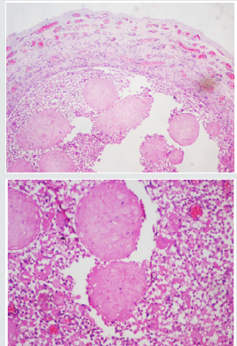
The histopathological features were suggestive of mucocele extravasation type confirming and correlating with the clinical diagnosis (Figure 5).
Figure 5: Histopathological picture (20X and 40X) showing small cystic spaces containing mucin and mucus-filled cells, areas of spilled mucin surrounded by a granulation tissue.
Discussion
Mucocele is the most common cystic swelling in the oral cavity with the incidence of 0.4 to 0.8% [13]. Traumas caused by lip biting or obstruction of duct are considered as critical factors in mucocele formation [14]. The present report illustrates a case of mucocele in lower lip which was surgically excised with the help of scalpel. The histological study revealed small cystic cavities filled with mucous enclosed by granulation tissue and inflammatory cells infiltrate. Although there are many treatment strategies for the management of mucocele conventional surgical removal is proven to be the most commonly used technique. The results were satisfactory with no signs of recurrence and minimal scar formation.
Conclusion
Although oral mucocele presents itself as an asymptomatic lesion can cause discomfort to the patient and therefore, requires treatment. Surgical excision of mucocele is a simple, cost- effective and the best alternative procedure.
References
- Baurmash HD (2003) Mucoceles and ranulas. J Oral MaxillofacSurg61(3):369-378.
- Ozturk K, Yaman H, Arbag H, Koroglu D, Toy H (2005) Submandibular gland mucocele: Report of two cases. Oral Surg Oral Med Oral Pathol Oral RadiolEndod100(6):732-735.
- Flaitz CM, Hicks JM (2015)Mucocele and Ranula. eMedicine.
- YagüeGarcía J, EspañaTost AJ, BeriniAytés L, Gay Escoda C (2009) Treatment of oral mucocele-scalpel versus CO2 laser. Med Oral Patol Oral Cir Bucal14(9):469-474.
- Hayashida AM, Zerbinatti DC, Balducci I, Cabral LA, Almeida JD (2010) Mucus extravasation and retention phenomena: A 24-year study. BMC Oral Health 10(1):15.
- Rao PK, Hegde D, Shetty SR, Chatra L, Shenai P (2012) Oral Mucocele - Diagnosis and Management. J Dent Med MedSci2(3):26-30.
- AtaAli J, Carrillo C, Bonet C, Balaguer J, Peñarrocha M, et al. (2010) Oral mucocele: Review of literature. J ClinExp Dent 2(1):18-21.
- CB More, KBhavsar, SVarma, M Tailor (2014) Oral mucocele: a clinical and histopathological study. J Oral MaxillofacPathol18(5):72-76.
- Bodner L, Manor E, Joshua BZ, Shaco Levy R (2015) Oral Mucoceles in Children - Analysis of 56 New Cases. PediatrDermatol32(5):647-650.
- Baurmash HD (2003) Mucoceles and ranulas. J Oral MaxillofacSurg61(3):369-378.
- Re Cecconi D, Achilli A, Tarozzi M, Lodi G, Demarosi F, et al. (2010) Mucoceles of the oral cavity: a large case series (1994-2008) and a literature review.A Med Oral Patol Oral Cir Bucal15(4):551-556.
- H Rodríguez, R De Hoyos Parra, G Cuestas, J Cambi, D Passali (2014) Congenital mucocele of the tongue: a case report and review of the literature. Turkish J Pediatr 56(2):199-202.
- Navya LV, Sabari C, Seema G (2016) Excision of Mucocele by Using Diode Laser: A Case Report, Journal of Scientific Dentistry 6(2):30-35.
- Gupta B, Anegundi R, Sudha P, Gupta M (2007) Mucocele Two Case Reports. J Oral Health Comm Dent 1:56-58.

Top Editors
-

Mark E Smith
Bio chemistry
University of Texas Medical Branch, USA -

Lawrence A Presley
Department of Criminal Justice
Liberty University, USA -

Thomas W Miller
Department of Psychiatry
University of Kentucky, USA -

Gjumrakch Aliev
Department of Medicine
Gally International Biomedical Research & Consulting LLC, USA -

Christopher Bryant
Department of Urbanisation and Agricultural
Montreal university, USA -

Robert William Frare
Oral & Maxillofacial Pathology
New York University, USA -

Rudolph Modesto Navari
Gastroenterology and Hepatology
University of Alabama, UK -

Andrew Hague
Department of Medicine
Universities of Bradford, UK -

George Gregory Buttigieg
Maltese College of Obstetrics and Gynaecology, Europe -

Chen-Hsiung Yeh
Oncology
Circulogene Theranostics, England -
.png)
Emilio Bucio-Carrillo
Radiation Chemistry
National University of Mexico, USA -
.jpg)
Casey J Grenier
Analytical Chemistry
Wentworth Institute of Technology, USA -
Hany Atalah
Minimally Invasive Surgery
Mercer University school of Medicine, USA -

Abu-Hussein Muhamad
Pediatric Dentistry
University of Athens , Greece

The annual scholar awards from Lupine Publishers honor a selected number Read More...


