Lupine Publishers Group
Lupine Publishers
Menu
ISSN: 2641-1709
Research Article(ISSN: 2641-1709) 
Statistical Study of the Effectiveness of EAT Treatment for Chronic Epipharyngitis Volume 9 - Issue 3
Hirobumi Ito*
- Department of ENT, Ito ENT Clinic, Japan
Received: November 07, 2022; Published: November 17, 2022
Corresponding author: Hirobumi Ito, Department of ENT, Ito ENT Clinic, Japan
DOI: 10.32474/SJO.2022.09.000311
Abstract
Introduction: However, few studies have compared the outcomes of EAT to those of conventional blind epipharyngeal abrasive therapy (B-spot therapy).
Subjects and Methods: EAT was performed on 546 patients with chronic epipharyngitis who visited our clinic between August 2018 and August 2019. A retrospective observational study was conducted on 55 of these cases that received a cure decision. As evaluation indices, the bleeding score and the EAT score were used. Multiple regression analysis was used to see if a prognostic indicator could be constructed using age, gender, disease duration, gross histology, EAT score, and bleeding score as evaluation indices.
Results: Chronic epipharyngitis was more prevalent in middle-aged and elderly women, and Mf type was more prevalent in women. After about 2 months of treatment, the cure case group showed improvement in subjective symptoms, and after about 4 months of treatment, the bleeding findings improved, and a cure decision was made. Multiple regression analysis revealed that the Mf type had a shorter healing period, and the higher the EAT score at the initial visit, the more treatment sessions were required. Tissue type and EAT score at the initial visit were thought to be factors influencing healing time.
Discussion: At the initial visit, tissue type and EAT score were beneficial prognostic indicators. It is critical to treat patients based on changes in the bleeding and EAT scores.
Keywords: Chronic epipharyngitis; bleeding score; EAT score; prognostic indicators; multiple regression analysis
Abbreviations: EAT: Epipharyngeal Abrasive Therapy; NRS: Numerical Rating Scale
Introduction
Horiguchi et al. used blind epipharyngeal abrasive therapy (EAT) (B-spot therapy) for chronic epipharyngitis and reported that symptoms improved after three months of treatment once or twice a week [1]. The degree of improvement in subjective symptoms, the degree of improvement in bleeding findings during rubbing, etc. are used to determine treatment effectiveness. However, pain during rubbing varies greatly between individuals, and clear diagnostic criteria for determining subjective symptoms and bleeding findings have yet to be developed. Currently, it is thought necessary to develop a cure diagnostic standard. Tanaka published a study on endoscopic epipharyngeal abrasive therapy (EAT) combined with endoscopic diagnosis to improve diagnosis and cure rates [2]. Although the introduction of endoscopic diagnosis may have increased the diagnosis rate of chronic epipharyngitis, there are few reports on the outcomes of EAT treatment [3]. In this study, bleeding findings were scored as a measure of other findings at the time of EAT using the Numerical Rating Scale (NRS) EAT questionnaire, and the bleeding score (bleeding score) was defined as a measure of subjective symptoms at the time of, and the total score was used as a measure of subjective symptoms at the time of EAT. The total score was calculated as the EAT score (EAT score). To clarify the cure criteria, the bleeding and EAT scores were used, and the subjects were divided into three groups: cured cases, noncured cases, and interrupted cases who withdrew during treatment. The primary complaint, gender, age, disease duration, gross histological features, EAT score, and the bleeding score were calculated, and the characteristics of each group were described. The effectiveness of EAT treatment was also investigated in terms of treatment duration and number of treatments in the cured case group. Furthermore, multiple regression analysis was performed to determine whether a prognostic indicator could be constructed from the evaluation items used during the initial examination. This statistical study, we believe, will be useful in clarifying the pathogenesis of chronic epipharyngitis and improving the cure rate.
Subjects and Methods
The subjects were 546 patients (136 males, mean age 44.4 ± 18.4 years; 410 females, mean age 52.1 ± 15.4 years; male to female ratio 1:3.0) who visited our clinic between August 2018 and August 2019, diagnosed and treated as chronic epipharyngitis. Patients were treated and followed up on for up to a year, and their age, gender, chief complaint, disease duration, EAT score, gross histomorphology, and bleeding score were all examined retrospectively on a chart basis using the method described below. Fifty-five patients (10 males, mean age 60.7 ± 16.5 years, 45 females, mean age 59.6 ± 15.1 years, male to female ratio 1:4.5) were deemed cured using the criteria outlined below. In the non-core group, there were 314 patients (78 males, mean age 44.5 ± 18.7 years; 236 females, mean age 52.1 ± 14.9 years; male-female ratio 1:3.0) who could not be judged to be cured during the observation period. There were 177 patients in the group of interrupted cases who withdrew during treatment (48 males, mean age 41.0 ± 16.9 years; 129 females, mean age 49.6 ± 15.7 years, male-female ratio 1:2.7). According to Tomiyama’s diagnostic criteria [4], patients with acute nasopharyngitis who had been ill for less than one month were exempted from the current study based on the medical history questionnaire. Patients with chronic rhinosinusitis as the primary cause of posterior rhinorrhea or allergic rhinitis as the primary cause of symptoms as determined by sinus x-ray examination were also excluded from the study. The diagnosis and treatment of chronic epipharyngitis were made using A band-limited optical endoscope system was used to diagnose and treat chronic epipharyngitis (Pentax EPK-i7000 video processor, VNL11-J10 video scope with a tip outer diameter of 3.5 mm). The endoscopic diagnosis was made using Tanaka’s diagnostic criteria [2] during the initial examination and EAT was performed. Nasal abrasion with Rutze swabs soaked in 1% zinc chloride solution was used for EAT, followed by oral abrasion with Zermach pharyngeal crimp cotton swabs. The hemorrhage findings were assessed two-dimensionally in terms of the extent of the EATcaused hemorrhage area. The nasopharynx was divided into four regions, with the center being the central fossa of the pharyngeal tonsil. If the bleeding was discovered in three or four regions, it was scored as severe bleeding (3 points), if the bleeding was discovered in two or more regions, it was scored as moderate bleeding (2 points), if the bleeding was discovered in one region, it was scored as mild bleeding (1 point), and if the bleeding was not discovered even after rubbing, it was scored as no bleeding. No bleeding was scored as 0 if there was no bleeding or barely blotting. The score obtained was used as the bleeding score. Bleeding scores were assessed at the initial examination and at approximately one-month intervals during treatment [5].
Subjective symptoms were assessed using the EAT questionnaire in conjunction with the NRS. This questionnaire assessed both local and general symptoms of chronic epipharyngitis. The following symptoms were considered: posterior rhinorrhea (posterior rhinorrhea, etc.), nasopharyngeal irritation (abnormality, headache, stiff shoulders, etc.), nasal symptoms (rhinorrhea, nasal obstruction, nasal irritation, etc.), Eustachian tube symptoms (otorrhea, tinnitus, etc.), dizziness-related symptoms, voice-related symptoms (hoarseness, voice disorder, etc.), laryngeal irritation symptoms (cough, sore throat, etc.), sleep apnea-related symptoms on a 6-point NRS scale (snoring The 10 gastrointestinal symptoms (gastric acid reflux, lethargy, etc.) and autonomic nervous system symptoms (chronic fatigue, depression, etc.) were scored. The following scores were used: 0 for no subjective symptoms, 1 for slightly or rarely bothering, 2 for a little or sometimes bothering, 3 for always bothering, 4 for fairly or almost always bothering but tolerable, and 5 for very bothering and intolerable. The EAT score was calculated by adding the scores from each group, yielding a maximum score of 50 points and a minimum of 0 points. The EAT score was assessed during the initial visit and at approximately one-month intervals between treatments [5].
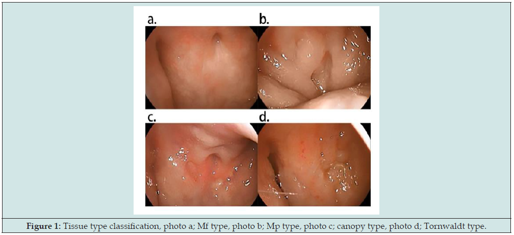
The time when both subjective symptoms and objective conclusions improved, i.e., when the bleeding score reached 0 and the EAT score was 10 or less, a defined cure. In terms of the gross histology of chronic epipharyngitis, Mogi classified it into Mycoplasma pneumoniae type (Mp type) and Mycoplasma fermentans type (Mf type) to Mycoplasma infection. Mycoplasma pneumoniae type (Mp type) and Mycoplasma fermentans type (Mf type) are the two histological types [6]. In this study, we used Mogidate’s criteria to divide the gross histology into two histological types for evaluation: Mp type and Mf type. Other gross histological features include canopy type, which is distinguished by edema and swelling of the mucosa near the canopy, and Tornwaldt type, which is distinguished by crust formation and adhesion, as well as mucosal swelling primarily around the site of Tornwaldt cyst development. Although the pathogenesis of these types differs from that of the Mf and Mp types, they were included in this study as subtypes of the Mf type (Figure 1).
Figure 1: Tissue type classification, photo a; Mf type, photo b; Mp type, photo c; canopy type, photo d; Tornwaldt type.
First, basic statistics for the chief complaint, sex, age, disease duration, tissue type, EAT score at the initial visit, and bleeding score at the initial visit were obtained for each case group to better understand the pathogenesis of chronic epipharyngitis. The healed and unhealed case groups were compared using Fisher’s direct establishment test and unpaired t-test. The healed case group was then divided into two tissue types, and basic statistics were computed. Unpaired t-tests were used to compare age, disease duration, and EAT scores between tissue types. The age was further subdivided into three groups: under 40 years, 40–60 years, and 60 years or older. The disease time was stratified into three categories: 1 month to 4 months, 4 months to 1 year, and 1 year or more. The first bleeding score was divided into four categories: 0–3 points. Next, the efficacy of EAT treatment in the cured case group was assessed in terms of treatment duration and number of treatments until the EAT score was 10 or less, the bleeding score was 0, and the EAT score was 10 or less and the bleeding score was 0 or less (at the cure determination time). First, Spearman’s rank correlation coefficient for treatment duration and number of treatments was calculated. The duration and number of treatments were then subjected to repeated measures of ANOVA (rm-ANOVA). Multiple comparisons were conducted using the Bonferroni method.
The frequency of treatment in the cured case group was then examined. Patients who visited the clinic more than twice a week were classified as high-frequency, patients who visited once a week were classified as intermediate-frequency, and patients who visited the clinic more than 2 weeks apart were classified as low-frequency. For each frequency group, a one-way analysis of variance (oneway ANOVA) was performed on the number of visits and the length of time between visits. Multiple comparisons were performed using the Bonferroni method. Next, multiple regression analysis was performed to develop a prognostic indicator using the duration and number of visits until the patient was deemed cured as the objective variables. Based on the information available at the time of the first visit, gender, age, disease duration, tissue type, EAT score at the first visit, and bleeding score at the first visit were used as explanatory variables. Gender and tissue type were dummy variables on the nominal scale. Gender was set to 0 for males and 1 for females. The tissue type was set to 0 for Mp and 1 for Mf. Age was divided into three categories: under 40 years, 40–60 years, and 60 years and older. The duration of the disease was divided into three categories: 1 month to less than 4 months, 4 months to less than 1 year, and more than 1 year. Multiple regression analysis was performed on all variables because there were no explanatory variables with strong correlations in the correlation matrix table.
The healed case group was then subjected to Cox proportional hazards regression analysis to investigate the effect of predictive indicators related to the length of hospital visits preceding the healing decision. The analysis utilized gender, age group, disease duration, tissue type, EAT score at the initial visit, and bleeding score at the initial visit as covariates, using the healing decision as the event. Kaplan-Meier curves were generated and analyzed to show the length of hospital visits until the cure. Kaplan-Meier curves were also generated and compared for each tissue type. The statistical software EZR version 2.6-2 was used for the analysis. A risk rate of less than 5% was outlined as statistically significant. This was a retrospective observational study that used existing data, not human samples. The study followed the Helsinki Declaration, and all patients provided oral and written informed consent. We took care to protect the subjects’ confidentiality when handling data and other materials related to the study, we did not include any information that could identify the subjects when publishing the study’s results. The Ethics Committee approved the conduct of this study. (Approval No. 20200625006 by the Ethics Committee of the Chiba Prefecture Health Physicians Association).
Results
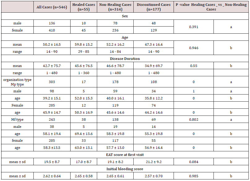
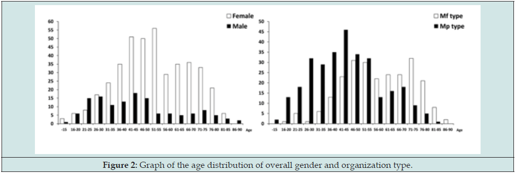
The most common initial complaint in all 546 patients was posterior rhinorrhea (45.2%), followed by a sense of abnormality (14.1%), sore throat (7.5%), hoarseness (7.5%), and cough (4.2%). Additionally, dizziness, tinnitus, otorrhea, stiff shoulders, headache, and general malaise were reported. The gender distribution of all patients’ ages revealed that women were older than men at the time of consultation. The Mf type had a higher average age at presentation than the Mp type (Figure 2). Among the 55 patients in the cured group, the most common subjective symptoms were posterior rhinorrhea (43.3%), abnormality (14.9%), sore throat (7.1%), nasal obstruction (5.8%), hoarseness (5.8%), headache (3.7%), and cough (3.4%). The distribution of chief complaints followed the same pattern as the overall case group. Tissue types exhibited the same distribution pattern as the overall case group. There were no significant differences in gender, age, duration of illness, EAT score at the initial visit, or bleeding score at the initial visit between the cured and non-healed groups, but Mf type was significantly more common in the cured group. When comparing ages by tissue type, the cured group had significantly older Mf and Mp types (Table 1).
Data display: mean ± SD; range (min-max).
P -value: a: Fisher’s exact test; b: unpaired t test
Mp type: Mycoplasma pneumoniae type; Mf type: Mycoplasma fermentans type.
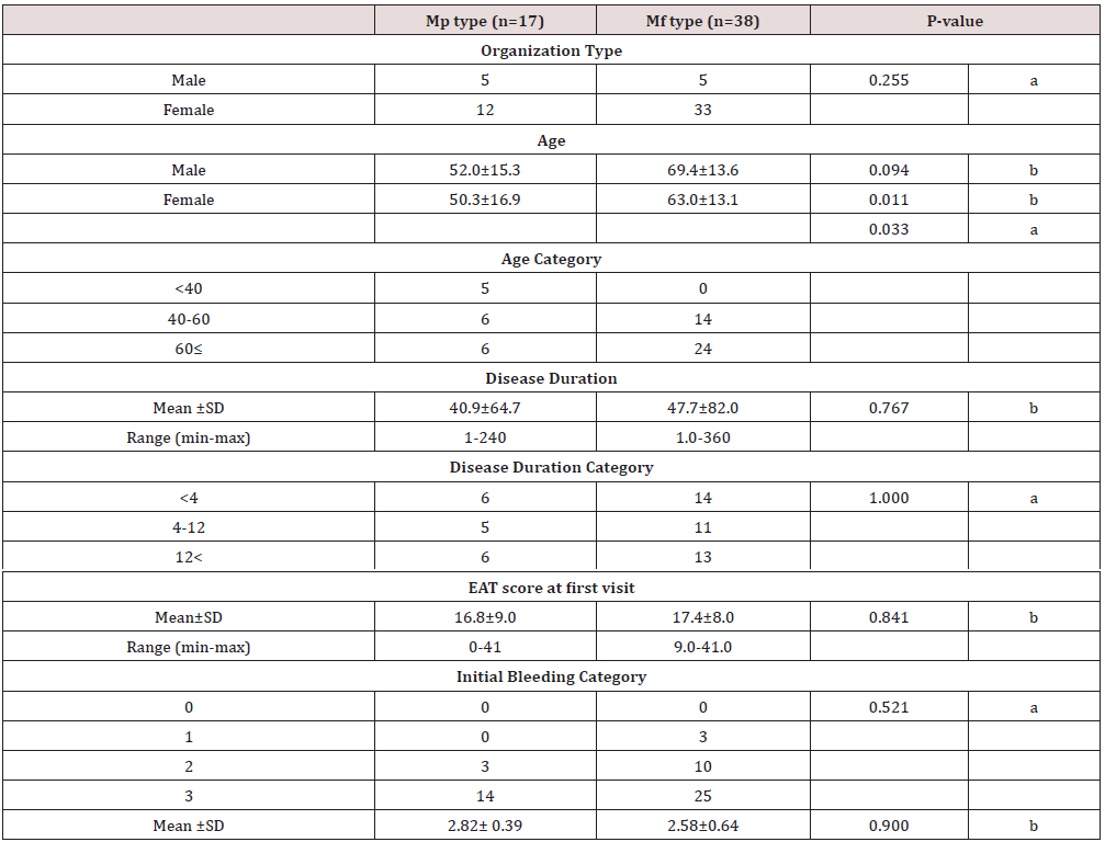
For within-group comparison, the cured case group was divided into two tissue types, Mp type, and Mf type. There were no statistically significant differences in disease duration, EAT score at the first visit, or bleeding score at the first visit, but female Mf type patients were significantly older (Table 2). Spearman’s rank correlation coefficients were calculated for the EAT score and bleeding score based on treatment duration and number of treatments. The test results revealed no significant difference in treatment duration (p = 0.415) and no correlation (r = 0.112). The number of treatments was significant at p < 0.001 with a correlation coefficient of r = 0.441, denoting that the EAT and bleeding scores varied with the number of treatments. Table 3 displays the findings of an analysis of the treatment duration and the number of treatments required until the EAT score was 10 or less, the bleeding score was 0, and the EAT score was 10 or less and the bleeding score was 0 or less (at the time of cure determination) in the cured case group. The EAT score improved the fastest during treatment, followed by the bleeding score and then the time of cure. The treatment period differed significantly between groups, according to rm-ANOVA. The number of treatments was lowest until the EAT score improved, then the bleeding score, and finally at the time of healing. The rm-ANOVA revealed a significant difference between the EAT and bleeding scores, and between the EAT score and time of cure (Table 3).
Data display: mean ± SD; range (min-max).
P -value: a: Fisher’s exact test; b: unpaired t test.
Table 3: Comparison of treatment duration and number of treatments until EAT score alone is cured, bleeding score alone is cured, and both EAT score, and bleeding score are cured.

Duration of Treatment (month± SD) number of Treatments (times ±SD) rm-ANOVA (Bonferroni).
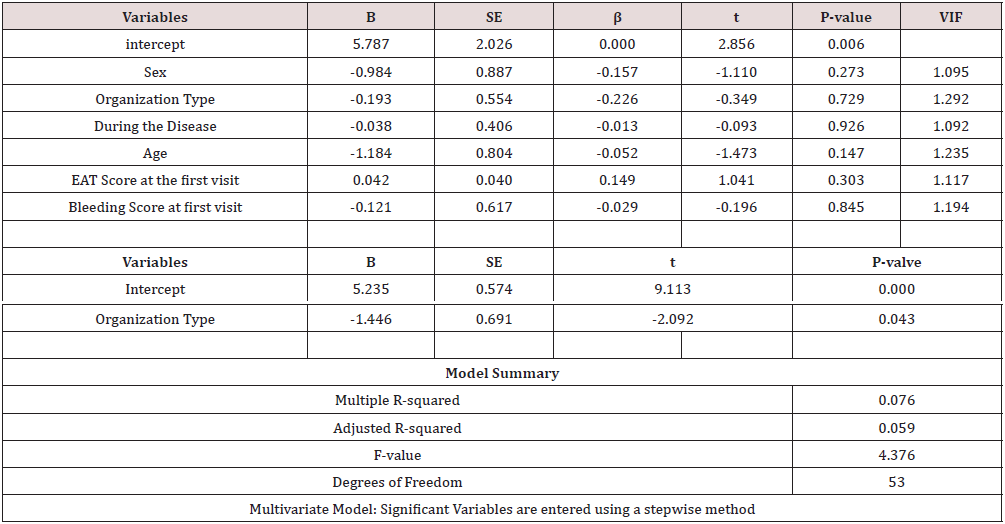
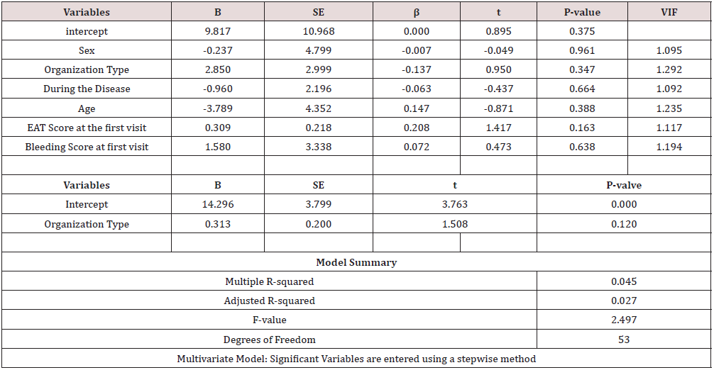
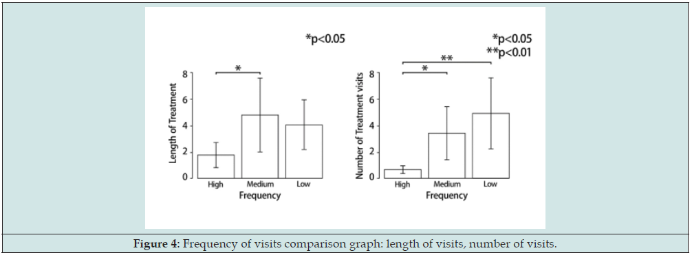
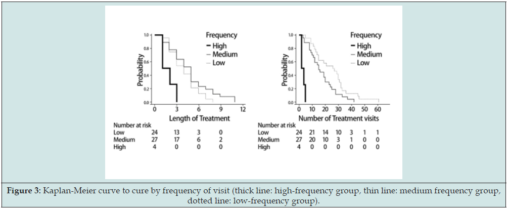
The frequency of treatment was examined in the cured case group. With cure as the outcome, the Kaplan-Meier curves were created and compared. The high-frequency group received treatment more frequently, followed by the intermediate-frequency group, and finally the low- frequency group (Figure 3). On the frequency of visits until cure and the duration of visits until cure, a one-way ANOVA was performed. Multiple comparisons revealed that patients who received treatment twice a week healed faster, but there was no discernible difference between the intermediate- frequency group (once a week) and the low-frequency group (once every two weeks or more). The frequency of visits differed significantly between the high-frequency, intermediate-frequency, and low-frequency groups. The more frequent the visits, the faster the healing time (Figure 4). Multiple regression analysis was done to investigate the factors influencing the duration of clinic visits. There were no explanatory variables found to be significantly related to healing time, and all VIFs were less than 10.0, indicating that multicollinearity was not an issue. When the explanatory variables were entered and analyzed stepwise, a significant association for the tissue type item was discovered. The analysis predicted that the Mf type would heal more quickly. However, the adjusted coefficient of determination for degrees of freedom was 0.059, which was low, and the model was deemed unfit (Table 4). At the time of the initial visit, a multiple regression analysis was performed on the factors influencing the number of hospital visits to cure in a group of cured cases. There were no explanatory variables that were significantly related to the number of hospital visits to cure, and all VIFs were less than 10.0, indicating that multicollinearity was not an issue. No explanatory variable was found to be significantly related to the number of hospital visits. The analysis revealed that the EAT score at the first visit was related to the number of hospital visits. A higher initial EAT score was expected to be associated with a higher number of hospital visits (Table 5).
B: partial regression coefficient; SE: Standard Error; β:standard partial regression coefficient; VIF: Variance Inflation Factor
B: partial regression coefficient; SE: Standard Error; β:standard partial regression coefficient; VIF: Variance Inflation Factor.
Figure 3: Kaplan-Meier curve to cure by frequency of visit (thick line: high-frequency group, thin line: medium frequency group, dotted line: low-frequency group).
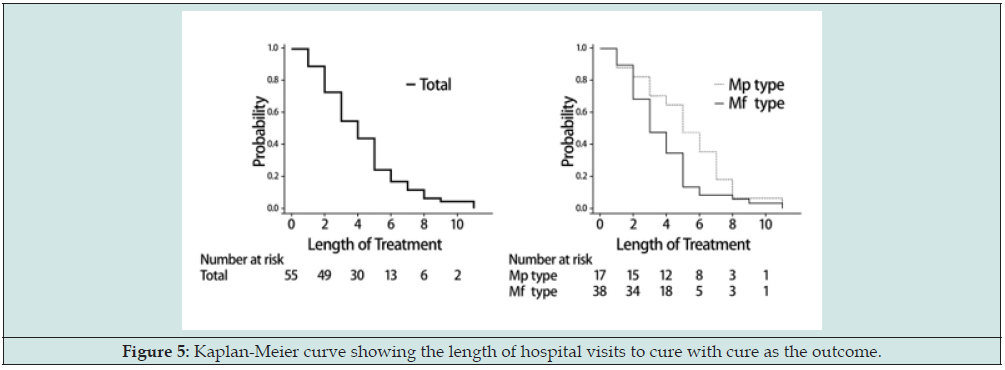
The Cox proportional hazards regression analysis was used to investigate the effect of predictive indicators on the number of hospital visits required to cure the group of cured cases. Gender, age group, disease duration, tissue type, EAT score at the first visit, and bleeding score at the first visit were used as covariates in the analysis, with cure as the event. The findings revealed an increase in sex, age group, disease duration, tissue type, and bleeding score at the initial visit all increased the hazard ratio, except an increase in the EAT score at the initial visit, which decreased the hazard ratio. In other words, having a higher EAT score at the first visit was associated with a longer healing time, whereas being female, older, having a longer disease duration, having a Mf type tissue type, and having a higher bleeding score at the first visit were associated with a faster healing time. There were no variables that independently affected healing time as a result of narrowing by P value in the multivariate model with forced entry, but Mf type tended to heal more easily and a high EAT score tended to make it harder to heal (Table 6). A Kaplan-Meier curve was created to depict the number of hospital visits required to make a cure decision, with cure as the outcome. For cured patients, the median survival time (MST, mean time to cure) for a 50% cure rate was 4 months. The average number of visits required to achieve a 50% cure rate was 16. The cumulative cure rate rises rapidly in the early stages of treatment before stabilizing after 5 months. The MST for the two tissue types, Mp type, and Mf type, was 5 months for the Mp type and 3 months for the Mf type, according to the Kaplan- Meier curve. The Mf type healed faster than the Mp type (Figure 5).
Table 6: Cox proportional hazards regression analysis of the impact of predictive indicators on the length of hospital visits leading to a cure decision.

Figure 5: Kaplan-Meier curve showing the length of hospital visits to cure with cure as the outcome.
Discussion
Chronic epipharyngitis has been linked to several diseases and manifests itself in a variety of ways [1,6-8]. Tanaka that EAT can be used to diagnose chronic epipharyngitis [2], but the criteria for determining a cure are still unclear. Ohno reported the cure rate based on EAT findings and the relative improvement of symptoms [3]. We performed a statistical analysis of the therapeutic effect of EAT in this study by defining the cure judgment based on the absolute improvement of subjective symptoms and bleeding findings at the time of abrasion by EAT and clarifying the judgment criteria. We also investigated the feasibility of developing a prognostic indicator using the initial examination evaluation items. We believe that this research will help to clarify the pathogenesis of chronic epipharyngitis and improve EAT cure rate. The average age of all chronic epipharyngitis patients who visited our hospital was 50.2 years, with a male to female ratio of 1:3. Ohno reported that the average age of chronic epipharyngitis cases was 42.9 years, with a male to female ratio of 1:3, with a female bias [9]. Ohno reported that 41.1% of patients had posterior rhinorrhea, 17.8% had abnormal pharyngeal sensations, and 8.2% had a sore throat. The chief complaint and average age of patients with chronic epipharyngitis who visited our clinic for the first time were nearly identical to those reported by Ohno. Therefore, the findings of this study may represent the fundamental statistics of chronic epipharyngitis cases seen in general otolaryngology clinics.
First, there is a discussion of the subjective symptom evaluation method used in this study. Tomiyama described a scoring system-based evaluation method for acute nasopharyngitis [4]. The degree of difficulty in daily living, sore throat, and fever were scored on a three-point scale of 0 to 2 points, and nasopharyngeal examination findings, such as redness and swelling of the nasopharyngeal mucosa and a thick plug of the nasopharynx, were scored on a three-point scale of 0 to 2. The study employed a NRS, Numeric Scale. In this study, the NRS was used as a 6-point EAT score to assess subjective symptoms caused by chronic epipharyngitis. Based on the incidence of subjective symptoms of chronic epipharyngitis, the EAT questionnaire used was designed to create a scale to quantitatively evaluate changes in subjective symptoms over time; inter-rater reproducibility of the NRS is said to be low, but intra-rater reproducibility is high in the same examinee [10]. The NRS may be useful as a self-assessment tool in determining the time course of symptoms. Because the visual analog scale (VAS) is influenced by psychogenic factors and responses tend to be concentrated in the middle [11], the NRS was used as an evaluation method in this study.
Ohno rated subjective symptoms on a 4-point scale before and treatment. The degree of improvement of findings such as redness, swelling, and posterior rhinorrhea of the nasopharyngeal mucosa based on imaging records was graded on a 5-point scale (marked improvement, improvement, slight improvement, unchanged, and worsening). EAT was done once or twice a week, and evaluations were done after about 10 treatments. As a result, he reported a 79.5% improvement rate for the chief complaint and an 87.7% improvement rate for the local findings [3]. Ohno reported each case’s relative improvement rate, whereas this study reported each case’s absolute improvement rate. The cure criterion in this study is a relatively simple method with the advantage of being able to confirm that symptoms and findings have improved on the spot. Horiguchi reported that the pain of EAT and local bleeding is crucial in the diagnosis of chronic
epipharyngitis, but the bleeding findings and pain vary greatly from person to person, making determining the degree of inflammation difficult [1]. Saito compared changes in local pain and bleeding findings to those of nasopharyngeal mucosal epithelial cell smear findings after B- spot therapy and discovered that the degree of epithelial cell detachment in the nasopharynx shifted, indicating a close relationship with the local inflammatory state. He reported that if the inflammation is reduced with abrasion therapy, the detached mucosa regenerates quickly, within two weeks, and regenerates as normal mucosa in about one month at the latest [12]. Ide reported that pain from abrasion and epithelial cell detachment are parallel, but the degree of inflammation is not [13]. From the preceding, the degree of pain may reflect the degree of exfoliation and nerve stimulation, but not necessarily the degree of inflammation. For these reasons, we did not use pain as an evaluation method for the degree of inflammation in this study, but rather subjective symptoms and hemorrhage findings during rubbing as evaluation criteria for local inflammation.
Tanaka proposed EAT, and this method allows for observation and recording of the nasopharyngeal canopy and lateral wall with moving and still images, which is insufficient in Horiguchi’s blind EAT, and treatment can be performed under direct observation. This is expected to boost the healing rate. However, because EAT is associated with a higher frequency of severe hemorrhage than conventional methods, the treatment period may be extended. Bleeding findings were used as an evaluation method for other sensory findings in this study. Horiguchi et al. reported that blind EAT (B-spot therapy) should be continued once or twice a week for about 3 months [1], and the results obtained in this study showed that when EAT was continued once a week in the cured case group, subjective symptoms improved after the first 2 months of treatment and after about 4 months of treatment. The bleeding findings improved after about 4 months of treatment, and the cure was determined after that, which was longer than that reported by Horiguchi et al. This could be because EAT allows for abrasion treatment to be performed on the inflammatory sites that remain in the conventional method, which takes longer for the mucosa to normalize and regenerate than the conventional method, thus prolonging the improvement of bleeding findings. The degree of bleeding at the initial visit did not differ significantly between the healed and nonhealed case groups. The possibility of healing is thought to be higher when the tissue is thoroughly abraded and bled during the initial treatment, but cases with less bleeding may not have sufficiently abraded the inflammatory site, i.e., the inflammatory site may have remained, or the tissue changes due to strong inflammation were so severe that the patient did not bleed much due to severe circulatory disturbance. Because of the high degree of circulatory disturbance caused by the strong tissue changes caused by the strong inflammation, the patient may not have bled much. Therefore, the severity of the lesion may not have been determined solely based on the bleeding findings at the initial visit. Because the bleeding score at the initial visit and the change in bleeding findings after the start of EAT (after the second EAT) may differ in what is evaluated, it was thought that the cure rate of chronic epipharyngitis could be improved by continuing with treatment while referring to improvement in subjective symptoms rather than focusing solely on the bleeding findings.
Because it was discovered that the cured case group was distinguished by histological type,
age, and gender, the histological type was investigated. Tissue types were categorized according to Mogidate’s classification. Mogi defined mycoplasmal infection’s basic pathogenesis as systemic microvasculitis and neuritis, and its relationship to chronic epipharyngitis. He reported that differences in the external histology of the nasopharynx are signs of local infection and circulatory disturbances of venous and lymphatic return [14]. All of the examined cases and the non-healed cases tended toward the Mp type, whereas the healed cases tended toward the Mf type. The Mp type was more common in males, while the Mf type was more common in females. Mp type was more common in younger patients, whereas Mf type was more common in middle-aged and elderly patients. Mp type predominates in younger male patients, Mf type predominates in middle-aged female patients, and Mf type predominates in healed cases. Mf type patients have thinner mucosa, which can lead to submucosal vascular injury and hemorrhage. A comparison of the two groups, however, revealed no difference in the amount of bleeding between the Mp and Mf types. There is a type of chronic epipharyngitis known as the Thornwald type, in which crusts tend to adhere primarily to the pharyngeal central fossa [15]. The Tornwald type is thought to be caused by secretory gland hyperfunction [16] in the local mucosa near the central fossa of the pharynx, or by secretion accumulation due to secretory gland obstruction or local infection. There is also a canalicular type which has significant mucosal edema and swelling near the nasopharyngeal canal. This type is based on the circulatory disturbance caused by cerebrospinal fluid congestion in the cerebral basal region and can be caused by impaired drainage of the nasopharyngeal venous and lymphatic systems or increased excretion due to increased fluid movement from the local capillaries to the interstitium, resulting in interstitial edema [17]. It is possible that the Tornwald and canalicular types have distinct pathogenesis mechanisms and will need to be studied separately in the future.
When the efficacy of EAT was investigated, it was discovered that the treatment duration and frequency did not differ significantly from that of conventional blind epipharyngeal abrasive therapy. When we examined whether the EAT score and bleeding score, the existing assessment indexes, tended to correlate with the duration of treatment or the number of treatments, we discovered that they did. The results revealed that increasing the number of treatment sessions leads to improvements in subjective symptoms and objective findings rather than extending the treatment duration to reach a cure decision. Because a longer treatment period is expected to increase the number of treatments, there is a possibility of early improvement even if the treatment period is short. Therefore, we conducted a frequency analysis to investigate the frequency of treatment. The frequency analysis revealed that patients who were treated twice a week or more frequently in the early stages of treatment were more likely to be cured. It was thought that with a treatment frequency of once a week or less, the treatment period could not be shortened and that the only way to shorten the treatment period was to increase the number of treatment sessions. This implies that increasing the frequency of treatment in the early stages, i.e., high-frequency treatment in the early stages will result in a shorter treatment period.
Next, we investigated the possibility of developing a predictive index based on findings at the initial visit, and the results of Kaplan-Meier curves and multiple regression analysis revealed that Mf type patients tended to heal faster and that the higher the EAT score at the initial visit (the stronger the subjective symptoms), the more frequent the number of hospital visits. A Cox proportional hazards regression analysis using the healing decision as the event revealed that a higher EAT score at the initial visit tended to result in delayed healing, whereas being female, being older, having a longer disease duration, having a Mf type tissue type, and having a higher bleeding score at the initial visit tended to result in faster healing. According to basic statistics, middle-aged and older women with Mf type and large bleeding tended to heal quickly, whereas younger men with Mf type and strong subjective symptoms tended to heal less easily. Treatment based on tissue type and EAT score at the time of initial diagnosis may be beneficial in improving the cure rate. Furthermore, it is thought necessary to increase the number of hospital visits (increase the number of EAT procedures) in the early stages of treatment to improve subjective symptoms as early on.
Chronic epipharyngitis has been linked to a variety of pathogenesis mechanisms and symptoms. There are no standardized diagnostic criteria or treatment evaluation methods for chronic epipharyngitis currently. Although the information obtained from the basic statistic data obtained in this study has limitations, the predictive indices used are methods for which information is readily available and relatively easy to evaluate. With the understanding that the EAT and bleeding scores do not accurately assess subjective symptoms or other findings, it is important to use the basic statistic as a reference to understand the pathophysiology of the disease and to determine a treatment strategy for the patient. The current study was conducted in the hope that providing predictive indices would benefit patients. The current study was a retrospective observational study, and no chronic epipharyngitis cases without EAT were used as controls. A prospective cohort study is considered necessary for future validation.
Conclusion
Chronic epipharyngitis is more common in middle-aged and elderly women, and the Mf type is also more common in women. In the healed case group, subjective symptoms improved after approximately two months of treatment with continuous weekly abrasion therapy, and bleeding findings improved after approximately four months of treatment, and the patient was determined to be cured. Increasing the number of treatment sessions rather than extending the treatment period leads to improvements in subjective symptoms and objective findings to reach a cure decision. To improve the cure rate, it was considered effective to provide treatment more frequently than twice a week in the early stages of treatment. Multiple regression analysis revealed that the Mf type had a shorter healing period, and the higher the EAT score at the initial visit, the more frequently treatment was administered. Tissue type and EAT score at the initial visit were thought to be factors influencing healing time.
Acknowledgements
We would like to express our sincere gratitude to Dr. Manabu Mogitate, Director of Mogitate Otorhinolaryngology, for his guidance and great advice during his busy schedule.
Appendix
The abstract of this paper was presented at the 33rd Annual Meeting of the Japanese Society of Oral and Maxillofacial Surgery. I have no conflicts of interest to disclose regarding this paper.
References
- Horiguchi S (1966) Systemic diseases and otorhinolaryngology, with special reference to nasopharyngitis. Nihon Jibiinkoka Gakkai Kaiho (Supplement 1): 1-78.
- Tanaka A (2018) Studies on band-limited light endoscopic diagnosis and endoscopic epipharyngeal abrasive therapy in chronic epipharyngitis. Stomato-Pharyngology 31(1): 57- 67.
- Ohno Y (2021) Classification of severity of chronic epipharyngitis and efficacy of epipharyngeal abrasive therapy. Stomato-Pharyngology 34(2): 163-172.
- Tomiyama M (2016) Treatment course of adult patients with severe acute nasopharyngitis treated with garenoxacin -A study using a scoring system - Nihon Jibiinkoka Gakkai Kaiho 62: 92-100.
- Ito H (2022) Effect of epipharyngeal abrasive therapy on long COVID with chronic epipharyngitis. Sch J Oto 8(5): 939-946.
- Mogitate M (2018) Chronic epipharyngitis and mycoplasma infection, Effect of Epipharyngeal Abrasive Therapy (EAT: Epipharyngeal Abrasive Therapy). Nihon Jibiinkoka Gakkai Kaiho 121(4): 575-576.
- Hotta O, Nagano C (2018) Consideration of various conditions suggested being associated with chronic epipharyngitis and epipharyngeal abrasive therapy. Stomato-Pharyngeal 31(1): 69-75.
- Mogitate M (2022) Epipharyngeal abrasive therapy for patients with immunoglobulin A nephropathy: a retrospective study. Sch J Oto 8(3): 874-881.
- Ohno Y (2019) Effectiveness of epipharyngeal abrasive therapy for chronic epipharyngitis. Stomato-Pharyngology 32(19): 33-39.
- David L, Kihara M. Kaji M, Kihara M (2016) Translation theories and applications of medical measurement scales: from validity and reliability to G-theory and item response theory. Medical Science International, Tokyo, Japan.
- Yamagiwa M, Sakakura Y (1994) Intensity of abnormal pharyngeal sensation evaluated by Visual Analogue Scale (VAS) and its influencing factors. Nihon Jibiinkoka Gakkai Kaiho 97: 67-74.
- Saito Y (1963) Clinical and cytological studies on the healing process of nasopharyngitis. Nihon Jibiinkoka Gakkai Kaiho 66(6): 814-828.
- Ide Y (1964) Smear cytological picture of nasopharyngitis, with special reference to the relationship between the pain to the touch and the epithelial picture of the hairline. Nihon Jibiinkoka Gakkai Kaiho 67(2): 153-158.
- Mogitate M, Matuda K (2017) A study on the clinical significance of mycoplasma lipid antigen-antibody assay in patients with chronic epipharyngitis. Stomato-Pharyngology 30(3): 444.
- Tasaka Y (1994) Nasopharyngeal cysts. Nihon Jibiinkoka Gakkai Kaiho 40: 109-113.
- Valstar MH, de Bakker BS, Steenbakkers RJHM, de Jong KH Smit LA, Klein Nulent TJW, et al. (2020) The tubarial salivary glands: a potential new organ at risk. Radiother Oncol 154: 292-298.
- Uebaba K (2017) A study on the mechanism of action of stabbing therapy Part 1: “Venous circulatory system hub hypothesis” and “venous congestive pain hypothesis”. Eastern Med 33(1): 63-77.

Top Editors
-

Mark E Smith
Bio chemistry
University of Texas Medical Branch, USA -

Lawrence A Presley
Department of Criminal Justice
Liberty University, USA -

Thomas W Miller
Department of Psychiatry
University of Kentucky, USA -

Gjumrakch Aliev
Department of Medicine
Gally International Biomedical Research & Consulting LLC, USA -

Christopher Bryant
Department of Urbanisation and Agricultural
Montreal university, USA -

Robert William Frare
Oral & Maxillofacial Pathology
New York University, USA -

Rudolph Modesto Navari
Gastroenterology and Hepatology
University of Alabama, UK -

Andrew Hague
Department of Medicine
Universities of Bradford, UK -

George Gregory Buttigieg
Maltese College of Obstetrics and Gynaecology, Europe -

Chen-Hsiung Yeh
Oncology
Circulogene Theranostics, England -
.png)
Emilio Bucio-Carrillo
Radiation Chemistry
National University of Mexico, USA -
.jpg)
Casey J Grenier
Analytical Chemistry
Wentworth Institute of Technology, USA -
Hany Atalah
Minimally Invasive Surgery
Mercer University school of Medicine, USA -

Abu-Hussein Muhamad
Pediatric Dentistry
University of Athens , Greece

The annual scholar awards from Lupine Publishers honor a selected number Read More...


