Lupine Publishers Group
Lupine Publishers
Menu
ISSN: 2641-1709
Research Article(ISSN: 2641-1709) 
Post Endodontic Pain Reduction using three Irrigants with Different Temperature Volume 2 - Issue 2
Jorge Paredes Vieyra1*, Julieta Acosta Guardado2, Fernando Calleja Casillas3 and Alan Hidalgo Vargas4
- 1 Autonomous University of Baja California, School of Dentistry, Mexico
- 2 Private practice in Endodontics, Mexico
- 3 Department of Biochemistry, Autonomous University of Baja California, School of Dentistry, Mexico
- 4 Clinical Assistant, Autonomous University of Baja California, School of Dentistry, Mexico
Received:May 13, 2019; Published:May 24, 2019
Corresponding author: Jorge Paredes Vieyra, Professor in Endodontics, Autonomous University of Baja California, School of Dentistry, Tijuana Campus, Mexico
DOI: 10.32474/SJO.2019.02.000134
Abstract
Objective: The purpose of this research was to evaluate whether meticulous irrigation with three different temperatures would help in a decrease dental pain.
Materials and Methods: All 120 patients had teeth chosen for conventional RCT for prosthetic reasons in teeth with vital pulps. All canals were cleaned and shaped with Reciprocal files. Final irrigation was done with cold saline solution (6 OC, 4 OC, and room temperature).
Results: A total of 120 of 135 patients (69 females and 51 male) were included whereas 15 were excluded as not achieving the necessities of the study. All patients presented with a vital upper or lower molar, premolar, or front teeth. No statistically major change (P>0.05) between the groups was found regarding the degree or duration of pain.
Conclusion: The approach in both selecting the patients participating in the research and analyzing the data in this research allows us to determine that cryotherapy is an aid of clinical procedures to clean and shape the canals to decrease the occurrence of post-endodontic pain and the need for medication in patients presenting with a diagnosis of vital pulp.
Keywords: Apical healing; Flare-ups; Pain; Post endodontic pain; Post-operative pain
Introduction
Post-endodontic pain is an undesirable sensation occurred in patients regardless of the preoperative periapical status of the tooth treated. Therefore, prevention and management of post endodontic pain are essential in endodontic practice [1]. Organic material, microorganisms, and irrigating solutions extruding beyond the apical constriction during root canal therapy (RCT) will originate inflammation and periodontal ligament complications, such as severe pain or flare-ups. It must be noticed that the amount of extruded material (debris and/or irrigate) varies widely in the reported studies which indicate problems and inconsistencies in treatment methodologies [2-4]. Recent literature has showed that keeping apical patency would not generate more postoperative difficulties [5-7]. A recently issued in vitro study showed that intracanal delivery of cold irrigating solution at 2.5 °C with negative pressure flushing reduced the external surface temperature to close 10 °C [8-10], would be enough to create a local anti-inflammatory beneficial consequence in peri radicular tissues. Cryotherapy proposes that using cold over some procedures may decrease the diffusion of nerve signs, bleeding, edema, and local inflammation and is therefore effective in the reducing of pain. Therefore, the purpose of this research was to evaluate whether meticulous irrigation with three irrigating practices with different temperature would help in a decrease of post-endodontic pain.
Three expert endodontists with a private practice of 17 years and skilled in the procedures and procedures studied were included in the research and performed 40 RCTs each (a total of 120) in upper/lower front or back teeth with irreversible pulpitis recognized by pulp sensitivity testing with hot and cold. Pulpal response tests were achieved by the main author, and a digital X ray diagnosis was documented by three certified clinicians. Additional clinical necessities for patients´ inclusion were as follows: Necessities of the research were agreed and spontaneously accepted, healthy patients were included, teeth with enough coronal structure and diagnosed with vital pulps, no previous RCT, and no analgesics or antibiotic consumption 7 days before the procedures. A total of 120 of 135 patients (69 females and 51 male) aged 18 – 60 years were referred and integrated in this research, whereas 15 were rejected as not accomplishing the necessities wanted. All participants showed with a vital upper or lower molar, premolar or front teeth designated for conventional RCT for dental rehabilitation reasons.
Methods
Dental procedures
Root canal treatment was done in one visit. Topical anesthetic (Anesthesia Topical, Astra, Mexico) was used. Patients received 2 carpules of articaine 2% with epinephrine 1:200,000 (Septodont, Saint-Maur des-Fosses, France). Situations in which supplementary anesthesia was needed, intra-ligamental anesthesia (2mL articaine 2%) was supplied. For the upper front teeth, the solution was administered by tender and slow local infiltration. For the lower teeth, one of the carpules was used for the lingual and alveolar nerve block, the other one for a moderate bucal infiltration nearby the tooth to be treated.
Irrigation protocols
Group 6 °C. The R25 (size 25/ .08) instrument was employed in tinny and curved canals, and R40 files (40/ .06) were used in broad root canals. Three in-and-out pecking series were employed with a fullness of not more than 3mm until getting the calculated WL. Patients allocated to this group receive a final irrigation with 5mL of cold (6 °C) 17% EDTA followed by 10mL of cold (6 °C) sterile saline solution dispensed to the WL using a cold (6 °C) metallic micro-cannula.
Group 4: Canals were instrumented as in group A. Patients allocated to this set received a final irrigation with 5mL of cold (4 °C) 17% EDTA followed by 10mL of cold (4°C) sterile saline solution dispensed to the WL using a cold (4 °C) metallic micro-cannula for 1 minute.
Group RT: The R25 (size 25/ .08) instrument was employed in tinny and curved root canals, and R40 files (40/ .06) were used in wide canals. Three in-and-out series were employed with a space of not more than 3mm until getting the calculated WL. Reciprocal instruments were used in one tooth only (single use). Participants allocated to this control group were treated similarly to the experimental groups, except that they received a final flush with 5mL (room temperature) of 17% EDTA followed by 10 mL (room temperature) of sterile saline solution delivered to the WL.
Statistical analysis
The related issues preoperatively recorded were integrated into the examination as follows: age and sex, occlusal contacts, and maxilla or mandibular teeth. Changes in the strength of pain among groups were studied using the ordinal (linear) X2 test. Variances in VAS-recorded standards after 24, 48, and 72 hours and in the quantity of analgesic intake among the two groups tested.
Results
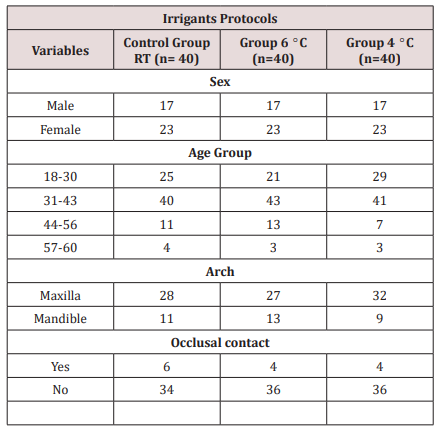
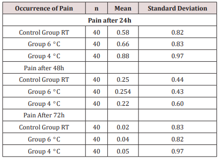
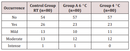
Table 1 displays the distribution of variables; a total of 120 participants took part in this study: 69 (57.5%) were women, and 51 (42.5%) were men. The ages fluctuated among 18 and 60 years; 87 (72.5%) were upper teeth, and 33 (27.5%) were lower teeth. The clinical management of the patients is showed in Table 1. No significant modification (P > 0.05) between the groups was encountered concerning the grade or period of pain. Rendering to the VAS examination, marks were seen 24 – 72 hours late in the 3 groups with a significant decline successively (Tables 2 & 3).
Discussion
Pain is tough to comprehend and calculate especially when it occurs unexpectedly in patients. The major trouble in knowledge painful and discomfort is the participant’s individual valuation and its dimension. For this objective, organization of the estimation form has to be entirely understood by participants. In our research, a simple spoken classification was followed in the feedback procedure with four classes: no pain, slight, modest, and intense pain. These classes were clearly comprehended by participants and were described by the occurrence or nonappearance of the necessity for pain-relieving treatment. Preoperative pain is one of the main predictors of post-endodontic pain [11-14]. Thus, only teeth with irreversible pulpitis indicated for RCT because of prosthodontic purposes were treated in this research. In our research, we reduced the variation in the procedures following protocols based on recommendations by authors and manufacturers. While successful endodontic treatment depends on various variables, an important point to consider in the shaping of the root canal system is the amount of the irrigating solution. Proper disinfecting and filling the root canal system is facilitated by the keeping of its original shape from the entrance to the apical third, without any iatrogenic event.
Conclusion
According to the conditions established for this study, there was no statistically significant difference between the instrumentation systems assessed.
References
- Ince B, Ercan E, Dalli M, Dulgergil CT, Zorba YO, et al. (2009) Incidence of postoperative pain after single- and multi-visit endodontic treatment in teeth with vital and non-vital pulp. Eur J Dent 3(4): 273-279.
- Tanalp J, Güngör T (2014) Apical extrusion of debris: A literature review of an inherent occurrence during root canal treatment. Int Endod J 47(3): 211-221.
- Ferraz CC, Gomes NV, Gomes BP, Zaia AA, Teixeira FB, et al. (2001) Apical extrusion of debris and irrigants using two hands and three engine-driven instrumentation techniques. Int Endod J 34: 354-358.
- Bürklein S, Schëafer E (2012) Apically extruded debris with reciprocating single-file and full-sequence rotary instrumentation systems. J Endod 38: 850-852.
- Torabinejad M, Kettering J, McGraw J, Cummings RR, Dwyer TG, et al. (1988) Factors associated with endodontic interappointment emergencies of teeth with necrotic pulps. J Endod 14: 261-266.
- Hülsmann, M, Peters O, Dummer P (2005) Mechanical preparation of root canals: Shaping goals, techniques and means. Endod Top 10: 30-76.
- Hülsmann M (2008) Extrusion von Debris und Spülflüssigkeiten während der Wurzelkanalbehandlung. Endodon 17: 353-354.
- Souza RA (2006) The importance of apical patency and cleaning of the apical foramen on root canal preparation. Braz Dent J 17: 6-9.
- Hülsmann M, Schäfer E (2009) Apical patency: Fact and fiction – a myth or a must? A contribution to the discussion Endo Endod Prac T 3(4): 285-208.
- Roane JB, Sabala CL, Duncanson MG (1985) The "balanced force" concept for Instrumentation of curved canals. J Endod 5: 203-211.
- Monsef M, Hamedzadeh K, Soluti A (1998) Effect of apical patency on the apical seal of obturated canals. J Endod 24: 284.
- Flanders D Endodontic patency: How to get it, how to keep it, why it is so important. NY State Dent J 68(3): 30-32.
- Yared G (2008) Canal preparation using only one Ni-Ti rotary instrument: Preliminary observations. Int Endod J 41: 339-344.
- Reddy SA, Hicks ML (1998) Apical extrusion of debris using two hands and two rotary instrumentation techniques. J Endod 24: 180-183.

Top Editors
-

Mark E Smith
Bio chemistry
University of Texas Medical Branch, USA -

Lawrence A Presley
Department of Criminal Justice
Liberty University, USA -

Thomas W Miller
Department of Psychiatry
University of Kentucky, USA -

Gjumrakch Aliev
Department of Medicine
Gally International Biomedical Research & Consulting LLC, USA -

Christopher Bryant
Department of Urbanisation and Agricultural
Montreal university, USA -

Robert William Frare
Oral & Maxillofacial Pathology
New York University, USA -

Rudolph Modesto Navari
Gastroenterology and Hepatology
University of Alabama, UK -

Andrew Hague
Department of Medicine
Universities of Bradford, UK -

George Gregory Buttigieg
Maltese College of Obstetrics and Gynaecology, Europe -

Chen-Hsiung Yeh
Oncology
Circulogene Theranostics, England -
.png)
Emilio Bucio-Carrillo
Radiation Chemistry
National University of Mexico, USA -
.jpg)
Casey J Grenier
Analytical Chemistry
Wentworth Institute of Technology, USA -
Hany Atalah
Minimally Invasive Surgery
Mercer University school of Medicine, USA -

Abu-Hussein Muhamad
Pediatric Dentistry
University of Athens , Greece

The annual scholar awards from Lupine Publishers honor a selected number Read More...


