Lupine Publishers Group
Lupine Publishers
Menu
ISSN: 2641-1709
Case Report(ISSN: 2641-1709) 
Old Penetrating Orbito-Infratemporal Wooden Foreign Body – “An Escape” Volume 8 - Issue 3
Abhinav Agarwal, Ravi Meher, Vikram Wadhwa, Vikas Kumar* and Jyoti Kumar
- Department of Otorhinolaryngology, Lok Nayak Hospital and Maulana Azad Medical College, India
Received: June 08, 2022; Published: June 23, 2022
Corresponding author: Vikas Kumar, Senior Resident, Department of Otorhinolaryngology, Lok Nayak Hospital and Maulana Azad Institute of Medical Sciences, India
DOI: 10.32474/SJO.2022.08.000290
Abstract
We report an interesting case of a foreign body lodged in right orbit and infratemporal fossa, which presented with a discharging sinus in the right infraorbital region. The patient gave history of history of road traffic accident six months back with facial trauma and penetrating injury with a wooden stick in infraorbital area. A long solid wooden stick was removed in toto using an external infraorbital approach. Here we discuss the various approaches to the infratemporal fossa and also review the literature of such type of foreign bodies.
Keywords: Infratemporal fossa; intraorbital; discharging sinus, wooden foreign body
Introduction
ENT surgeons commonly encounter unusual foreign bodies lodged in the aero digestive tract and sinonasal areas. Penetrating orbital and infratemporal fossa foreign bodies are rare. These foreign bodies certainly pose a challenge in diagnosis and removal because these areas are deep and poorly accessible. Even experienced surgeons can miss a foreign body lodged in such areas, hence there is a need for good radiological assessment. Here we present an interesting case of a non- healing discharging sinus in right infraorbital region. This was due to a neglected organic foreign body lodged in the right orbit and infratemporal fossa. In this case report we discuss the management and complications of such type of foreign bodies.
Case Report
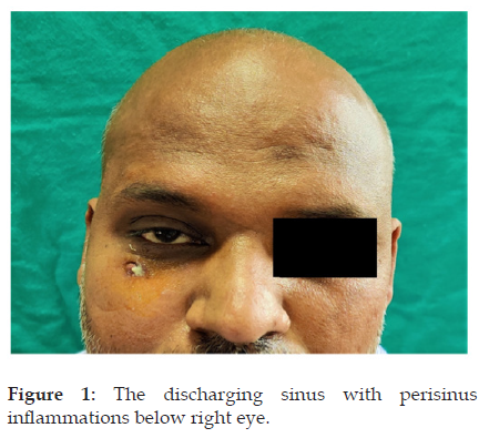
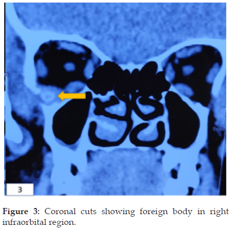
A 34-year-old male presented to ENT OPD with the chief complaint of pus discharge from right infra orbital region for five months. Patient had a history of road traffic accident around 6 months back in which he suffered facial trauma. He fully recovered from his injuries except for the small non-healing discharging sinus in the right infraorbital region. Before presenting to us, he was given multiple courses of oral and topical antibiotics, but the discharging sinus did not heal. He had no history of vision loss, trismus, facial paresthesia or numbness. The discharging sinus had peri sinus granulations with surrounding area of tenderness as shown in Figure 1. Patient’s general condition was good and eye movements were within normal limits. A computed tomography scan of the face showed a linear foreign body extending from infra orbital region reaching upto foramen lacerum through infratemporal fossa and inferior orbital fissure as shown in Figures 2 & 3. The patient was taken up for removal of foreign body in general anesthesia. Methylene blue dye was injected through the sinus and a 3 cm horizontal incision was given in the right infraorbital region incorporating the sinus opening. The sinus tract was followed and was seen to cross the inferior margin of the orbit and periosteum of the orbital floor. The external foreign body was located in the inferior part of the orbit, lodged in periorbita. After an incision was given on the periorbita, the tip of the foreign body was grasped with an artery forceps and pulled out completely under endoscopic guidance as seen in Figure 4 and the complete wooden stick after removal is shown in Figure 5. There was no bleeding after removal of the foreign body, but was followed by drainage of 1ml pus, which was sent for culture and sensitivity. The wound was closed in 2 layers. There were no post operative complications.
Figure 2: Axial cuts showing foreign body in right infratemporal fossa, reaching till right petrous apex.

Figure 4: Intraoperative picture of wooden stick foreign body retrieved by incising periorbita of right eye.

Discussion
As we reviewed the literature of foreign bodies in infratemporal fossa, facial trauma and orodental surgery are the most causes of foreign body entering in the infratemporal fossa [1]. These patients usually present with trismus and high clinical suspicion is required to suspect infratemporal fossa foreign body [2]. Ours was a unique case as the foreign body did not cause any trismus but there was a continuous discharging sinus without any other symptoms. Foreign body if left in infratemporal fossa can lead to grave complications like orbital abscess leading to loss of vision and even intracranial spread of infection, hence the need for early removal. Infratemporal fossa contains internal maxillary artery, pterygoid venous plexus of veins. Wulkan et al. [3] reported complications associated with extraction of such foreign bodies due to internal maxillary artery injury, which can be surgically uncontrollable and potentially fatal [4]. Some delayed complications have also been reported like aneurysm [5] and erosion of the vessel wall [6]. Hence, imaging in the form of computed tomography scan is very important in these type of cases as routine x-ray usually misses out on vegetative foreign bodies. As the sinus was non healing, we got a CT scan done which not only helped in clinching the diagnosis but also showed the exact location of the foreign body and we could decide how to approach the foreign body. Conventional approaches to infratemporal fossa include anterior (transoral, trans palatal, transmandibular transcervical and endoscopic), anterolateral (maxillary swing), lateral (infratemporal) or a combination of the three. But these approaches are rarely required for foreign bodies. In the past infratemporal fossa foreign bodies have been explored using pre auricular approach [7], intraoral approach [8], endoscopic intraoral approach [4], transmaxillary endoscopic approach [9] We explored the foreign body using infraorbital horizontal incision. As the periorbita was being raised from the orbital floor foreign body was palpated and confirmed using 0-degree endoscope and was pulled out under vision without any post operative complications. With the advancement of endoscopic and image guided surgery, the need for open radical surgeries should continuously decrease and minimal incision surgery causing little morbidity would be a routine surgical practice.
Conclusion
Foreign bodies in the orbit and infratemporal fossa can be tricky and difficult to diagnose and manage. Isolated Infratemporal fossa foreign bodies can have a delayed presentation with trismus or discharging sinus as in our case. Computed tomography scan should be advised in all cases having a penetrating head and neck injury because broken lodged foreign bodies can be easily missed on examination. Surgery should be done with the most meticulous approach and necessary multidisciplinary backup should be available in case of any complication.
References
- Ramdas S (2016) An unusual foreign body in the infratemporal fossa. Indian J Plast Surg 49(2): 275-278.
- Sajad M, Kirmani MA, Patigaroo AR (2011) Neglected foreign body infratemporal fossa, a typical presentation: A case report. Indian J Otolaryngol Head Neck Surg 63(Suppl 1): 96-98.
- Wulkan M, Parreira Jr JG, Botter DA (2005) Epidemiology of facial trauma. Rev Assoc Med Bras 51(5): 290-295.
- Tan SS, Yeo MS, Lee GH, Ho MS, Ang ML, et al. (2016) Penetrating foreign body in the masticator space with injury to the internal maxillary artery: a surgical challenge. The Annals of The Royal College of Surgeons of England 98(8): e194-196.
- Chedid MK, Vender JR, Harrison SJ, McDonnell DE (2001) Delayed appearance of a traumatic intracranial aneurysm. Case report and review of the literature. J Neurosurg 94: 637-641.
- Lahiri S, Ghosh S, Sengupta G, Bakshi U (2011) An unusual presentation of foreign body in the common carotid artery. Indian J Surg 73: 460-462.
- Hamdoon Z, Jerjes W, Al-Delayme RM, Upile T, Hopper C (2012) Glass displaced into the infratemporal region from submandibular injury: A case report. Hard Tissue 1: 6.
- Goswami S (2013) A bullet in the maxillary antrum and infratemporal fossa. Indian J Dent Res 24: 149.
- Meher R, Wadhwa V, Ali MR, Ahmad S (2021) Removal of Bullet from Infratemporal Fossa Through Endoscopic Approach. MAMC J Med Sci 7: 261-264.

Top Editors
-

Mark E Smith
Bio chemistry
University of Texas Medical Branch, USA -

Lawrence A Presley
Department of Criminal Justice
Liberty University, USA -

Thomas W Miller
Department of Psychiatry
University of Kentucky, USA -

Gjumrakch Aliev
Department of Medicine
Gally International Biomedical Research & Consulting LLC, USA -

Christopher Bryant
Department of Urbanisation and Agricultural
Montreal university, USA -

Robert William Frare
Oral & Maxillofacial Pathology
New York University, USA -

Rudolph Modesto Navari
Gastroenterology and Hepatology
University of Alabama, UK -

Andrew Hague
Department of Medicine
Universities of Bradford, UK -

George Gregory Buttigieg
Maltese College of Obstetrics and Gynaecology, Europe -

Chen-Hsiung Yeh
Oncology
Circulogene Theranostics, England -
.png)
Emilio Bucio-Carrillo
Radiation Chemistry
National University of Mexico, USA -
.jpg)
Casey J Grenier
Analytical Chemistry
Wentworth Institute of Technology, USA -
Hany Atalah
Minimally Invasive Surgery
Mercer University school of Medicine, USA -

Abu-Hussein Muhamad
Pediatric Dentistry
University of Athens , Greece

The annual scholar awards from Lupine Publishers honor a selected number Read More...


