Lupine Publishers Group
Lupine Publishers
Menu
ISSN: 2641-1709
Case Report(ISSN: 2641-1709) 
Miscellaneous Lesions of Trachea and Larynx: Case Report Volume 6 - Issue 2
Rahalkar M D1*, Anand M Rahalkar2
- 1Ex Consultant Radiologist, Sahyadri Hospital, India
- 2Ex Consultant Radiologist, Department of Radiology, Sahyadri Hospitals, India
Received: March 15, 2021; Published: March 22, 2021
Corresponding author: Rahalkar M D, Ex Consultant Radiologist, Sahyadri Hospital, India
DOI: 10.32474/SJO.2021.06.000231
Abstract
Selected images of trachea and larynx are presented. These are shown as congenital, acquired (infection, Laryngeal diverticula, Tumours, Extrinsic lesions causing tracheal compression, Jugular fossa causing paralysis of larynx and Tracheo-esophageal fistula.
Keywords: Tracheal lesions; TEF; tumours of trachea
Introduction, Materials and Methods
A variety of lions of trachea and larynx can be noted during routine radiographic study and CT of chest. It is important to know their cognisance and report accordingly.
Results and Observations
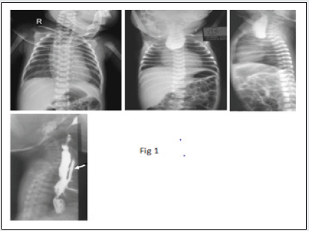
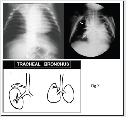
Congenital: TEF, tracheal bronchus. Tracheoesophageal fistula is an abnormal connection between the esophagus and the trachea. It is associated with different types of esophageal atresia [1] (Figure 1). This is an accessory bronchus with a small opening and is known to cuse recurrent pneumonia, as in this X ray [2] (Figures 2 & 3).
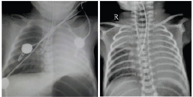
Figure 3: Wrong positions of ETT. Left image- The tip of ETT is in right lower lobe bronchus causing cut-off of left main bronchus and total collapse. After re-positioning, the position of ETT left lung opened up in a short time. Right image- The tip of ETT is further is right lower lobe bronchus causing collapse of right upper lobe.
Acquired (Figures 4-7)
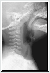
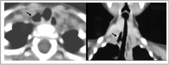
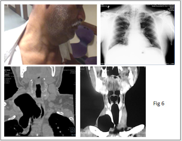
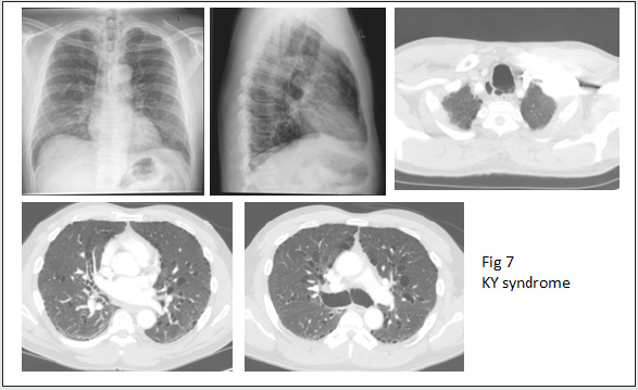
Lateral view of neck showing smooth narrowing of sub-glottic trachea due to viral infection called Croup (Figure 4). A small airfilled cavity is seen along the right lower wall of trachea due to a diverticulum. This was seen a chance finding on CXR [3] (Figure 5). A boggy swelling was noted in the root of neck on right in this male. CXR showed an air-filled lesion in the neck on right. CT revealed it to be a large cyst with a tiny communication with trachea in APO view. Paratracheal air cysts are not an uncommon incidental finding in routine thoracic imaging. They characteristically occur on the right side, in the region of the thoracic outlet. Occasionally they may mimic pneumomediastinum [4] (Figures 6 & 7).
Figure 7: Mounier-Kuhn syndrome, or tracheobronchomegaly, is a rare clinical and radiologic condition. It is characterized by dilation of the trachea/bronchi and multiple para-tracheal and lung cysts. Dilatation of trachea and cysts are better appreciated on CT.
Acquired lesions
a) Laryngeal diverticula
b) Tumours
c) Extrinsic lesions causing tracheal compression
d) Jugular fossa causing paralysis of larynx
e) Tracheo-esophageal fistula: acquired.
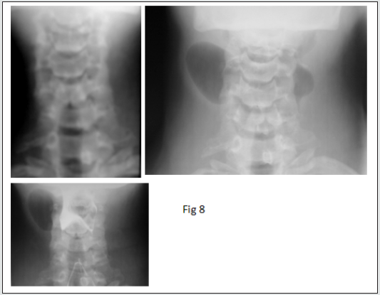
First image is at rest. On straining this young male showed
bilateral diverticula increasing in size. This is an external
diverticulum. The internal diverticulum remains inside thyrohyoid
membrane and confines to the parapharyngeal space. Barium
swallow study showed normal pyriform sinuses. g 9-Congenital
cyst in vestibule. An oval smooth soft tissue mass in vallecula is due
to a cyst [5,6] (Figures 8-13).
Figure 9: A lateral view of neck shows a benign-looking, soft oval tissue mass in the vallecular fossa due to congenital cyst.
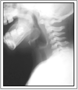
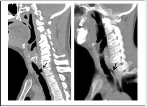
Figure 10: This middle-aged man had difficulty in berthing. CXR was normal. CT showed smooth narrowing of both walls of trachea, which proved to be due Ca (arrows). Primary tracheal tumors are very rare. Most (80-90%) are malignant.
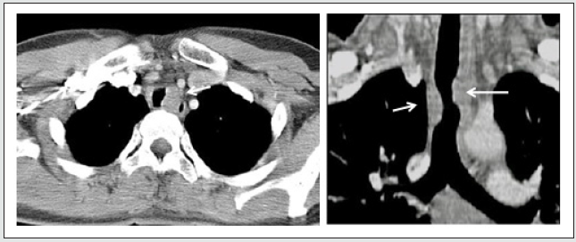
Figure 11: Extrinsic compression of trachea. This child had grunting due to a soft tissue mass of fluid (cystic hygroma). CXR reveals narrowing and deformity of trachea. CT shows this is due to cystic hygroma.
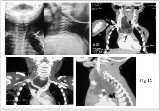
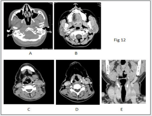
Figure 12: A male 45 years old presented with deafness, dysphagia and hoarseness of voice. This is a case of Vernet syndrome causing motor paralysis of 12th Vagus nerve. A) Glomus jugulare tumor b) B) Fatty atrophy of right half of tongue C) Thickened ipsilateral aryepiglottic fold on right D) Enlarged ipsilateral ventricle on right and E) Uvula displaced to left that is normal side.
Conflicts of interests
Source of Funding: Nil
References
- M D Rahalkar (1999) From book edited by Dr M D Rahalkar, Selected Topics from Pediatric Radiology. Chapter I, pp. 279.
- Ian Bickle (1999) Radiopedia. Endotracheal tube malposition (neonatal).
- Marina Pace, Annarita Dapoto, Alessandra Surace (2018) Tracheal diverticula: A retrospective analysis of patients referred for thoracic CT.Medicine (Baltimore) 97(39): e12544.
- Goo JM, Im JG, Ahn JM, Moon WK, Chung JE, et al. (1999) Right Paratracheal Air Cysts in the Thoracic Inlet: Clinical and Radiologic Significance. American Journal of Respiratory and Critical Medicine 173(1): 65-70.
- Harvey S Glazer, Matthew A, Dixie J Aronberg, Joseph KT Lee (1983) Computed Tomography of Laryngoceles. AJR 140(3): 549-552.
- Mukund Rahalkar, Anand Rahalkar (2020) Tracheo-Oesophageal Fistula (TOF)- A Late Complication of Tracheostomy. Otolaryngology Open Access Journal 5(2): 1-2.

Top Editors
-

Mark E Smith
Bio chemistry
University of Texas Medical Branch, USA -

Lawrence A Presley
Department of Criminal Justice
Liberty University, USA -

Thomas W Miller
Department of Psychiatry
University of Kentucky, USA -

Gjumrakch Aliev
Department of Medicine
Gally International Biomedical Research & Consulting LLC, USA -

Christopher Bryant
Department of Urbanisation and Agricultural
Montreal university, USA -

Robert William Frare
Oral & Maxillofacial Pathology
New York University, USA -

Rudolph Modesto Navari
Gastroenterology and Hepatology
University of Alabama, UK -

Andrew Hague
Department of Medicine
Universities of Bradford, UK -

George Gregory Buttigieg
Maltese College of Obstetrics and Gynaecology, Europe -

Chen-Hsiung Yeh
Oncology
Circulogene Theranostics, England -
.png)
Emilio Bucio-Carrillo
Radiation Chemistry
National University of Mexico, USA -
.jpg)
Casey J Grenier
Analytical Chemistry
Wentworth Institute of Technology, USA -
Hany Atalah
Minimally Invasive Surgery
Mercer University school of Medicine, USA -

Abu-Hussein Muhamad
Pediatric Dentistry
University of Athens , Greece

The annual scholar awards from Lupine Publishers honor a selected number Read More...


