Lupine Publishers Group
Lupine Publishers
Menu
ISSN: 2641-1709
Review Article(ISSN: 2641-1709) 
Ehlers-Danlos Syndrome (EDS) : A Common and often Disregarded Cause of Serious Gastrointestinal Complications in Children and Adults Volume 7 - Issue 2
Claude Hamonet1,2*, Mickael Delarue3, Jeremie H. Lefevre4, Jacques Rottembourg2 and Jean David Zeitoun5
- 1Department of Occupational Therapy, Faculty of Medicine, University of Paris-East-Creteil, Paris
- 2Centre medical ELLAsanté 28 bis rue d’Astorg, Paris
- 32 rue Grayan 33780, Soulac sue Mer, France
- 4Department of Digestive Surgery, University Hospital Saint Antoine, Paris
- 57 rue Chaligny 75012 Paris
Received:July 12, 2021; Published:July 20, 2021
Corresponding author: Claude Hamonet, Department of Occupational Therapy, Faculty of Medicine, University of Paris-East-Creteil, Avenue du General de Gaulle, 94000 Creteil, Paris
DOI: 10.32474/SJO.2021.07.000256
Abstract
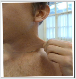
Ehlers-Danlos is a hereditary disease of the whole connective tissue initially described by dermatologists (Tscherchnogobov Moscow 1892, Ehlers, Copenhagen, 1900, Danlos, Paris, 1908). They emphasized the joint hyperlaxity and stretchiness of the skin which has long summed up the clinical expression of this entity. In recent decades, many other manifestations have been described and gradually identified, mainly by rheumatologists (Grahame, London, 1960). Several of them concern the digestive tract, mainly gastric reflux and constipation. They can be the cause of serious accidents: bronchial flooding by gastric reflux or aspiration, intestinal obstruction, hernial constriction, eventration, intestinal rupture, peritonitis of vesicular or appendicular origin, hemorrhages. It is important that gastroenterologists know how to link these manifestations to their etiology in order to adapt treatments, prevent iatrogenic accidents and direct the patients towards the treatment of other manifestations of Ehlers-Danlos disease. Nine clinical signs, including digestive manifestations, allow diagnosis by their significant grouping. The proof of hereditary origin is based on the identification of other identical cases in the family,, even if they are paucisymptomatic. A person affected by the disease systematically transmits the disease to all his children. We have verified this in all our patients.
Introduction
Ehlers-Danlos is an inherited disease of the common connective tissue but is often, mistakenly, considered rare. It is still poorly and non-completely described, more often than not it is not diagnosed at all, or it is diagnosed only after a long period of time and painful and dangerous medical wanderings for patients. A long list of diagnostic errors (Figure 1) causes iatrogenic effects as the disease is often wrongly diagnosed as fibromyalgia, multiple sclerosis, ankylosing spondylitis, Crohn’s disease, asthma, endometriosis, Lyme disease, vascular facial algia, Gougerot-Sjögren disease, Raynaud disease, osteoarthritis and above all, psychiatric disorders (depression, hysteria, autism, bipolar syndrome, schizophrenia). In our experience, based since 25 years on a cohort of 6,200 patients diagnosed with Ehlers- Danlos and, for a part of them, followed, we have observed a fragility of the connective tissue in numerous locations of the body: vessels, esophagus, stomach, intestine, abdominal-pelvic wall and pelvic floor, bladder, teeth and jaws, skin, muscles (diaphragm in particular) and tendons, bone system including cartilage, cardiovascular system, endocrine glands, bone marrow and blood including mastocytes, eyes, auditory and vestibular systems. This fragility of the connective tissue causes complications that can negatively affect the lives of the patients such as reflux or swallowing disorders, gastric or intestinal hemorrhage, lithiasis or gangrenous cholecystitis with peritonitis, acute pancreatitis, occlusions, intestinal rupture, acute intestinal invagination in infants, occlusions or swelling, rectal prolapse, aneurysm rupture. The clinical signs that usually alert of serious symptoms (abdominal contracture, pain, fever) can be absent and falsely reassuring. A wider knowledge dissemination of the particularities of this disease, which is easy to diagnose thanks to a combination of clinical signs, must be achieved with doctors (pediatricians in particular), surgeons and anesthetists to allow screening, recognize tissue fragility, avoid certain aggressive gestures (endoscopies) and adapt treatments, in particular surgical treatments.
Description of the digestive manifestations observed in Ehlers-Danlos disease
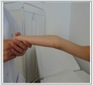
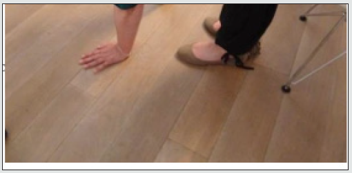
The manifestations of the disease appear in the entire digestive system from the mouth to the anus but also its “annexes”: the liver, the choledic canal and the vesicle, the pancreas, the ganglia, the peritoneum. But also, its “envelope”: the abdominal wall, the pelvic floor, the diaphragm and its costo-sternal attachments. Circulatory disorders (arterial, venous, lymphatic) and their effects on blood pressure and heart rate (POTS) also play a role. The interpretation of the mechanism of the symptoms is enriched by recent works [2-5] emphasizing the role of generalized dysproprioception, which is clearly evidenced by posturology (Figures 2-6) and by the study of the motor function of the “external” limbs or of the body, but which is also present in the “internal” body. These findings fit very well into the vision of the body in motion described by Alain Berthoz (6), with its three dimensions (internal, external and environmental). With the digestive tract, it is about the “internal” dimension. The nervous system, spared by genetic modifications, is programmed for a “normal” body, but it is confronted with a genetically modified body. The signals sent by their sensors are distorted either by exaggeration or, on the contrary, by reduction, and can even be totally lacking. For a better understanding of this complex symptomatology, it is also necessary to integrate biotope modifications that cause diet deficiencies as well as nutritional aspects. Appetite is often reduced [7,8] due to taste disorders often linked to changes in smell and difficulties in swallowing but on the opposite appetite can also increase (polyphagia) due to the loss of the feeling of satiety. Alterations of the sense of thirst are also possible with polydipsia which should trigger a search for a pituitary origin in the context of the diffuse attack of the connective tissue. Mastocytosis is one of the components of Ehlers-Danlos disease and may have a responsibility, difficult to identify in the midst of other digestive symptoms. It may also explain some of the common digestive intolerances to lactose, histamine, and the frequency of infections. Gluten is also poorly tolerated and promotes abdominal pain and bloating.
Ehlers-Danlos disease: manifestations in the mouth, jaw and teeth
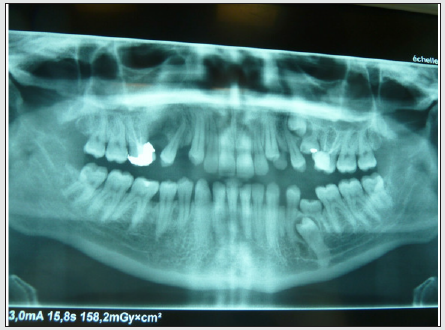
Gingival and oral mucous membranes are painful and hemorrhagic, justifying the use of xylocaine gel and adapted toothbrushes. A shift in the dental joints is often favored by the appearance of the palatine vault. The lower position of the tongue prevents it from playing its role in the palatine growth, which causes oral respiration. Early pediatric diagnosis would enable speech therapy associated with dental orthotics. The teeth grow anarchically in time (Figure 7), in the child, and in space with even possible horizontalization or inclusions. They are fragile with fractures, they are mobile because of the laxity of the dental ligament. Subluxations (cracks) and dislocations of temporomandibular joints are common during yawning, chewing or dental care. These joints are often painful and hinder chewing. Masticating muscles can also be painful, sometimes responsible for bruxism by dystonia. Dental anesthetics are often ineffective because of the rapid diffusion of products and require large doses associated with Adrenaline. The input of the gutters is essential here because they also contribute to the proprioception by sending a special signal on the position of the head through contact with the tongue. The tongue shows remarkable stretching possibilities enabling it to touch (Figure 8) with its end the tip of the nose (sign of Gorlin), it can also be twisted exaggeratedly on its axis and often suffers bites together with the lips. Some foods cannot be chewed or are badly crushed, which accentuates the difficulties of swallowing and digestion. These difficulties are accentuated by the usual blunders when eating food in the mouth. These ingestion difficulties also apply to beverages with swabbing, accidental overturning, or the release of a glass or bowl.Appreciable improvements have been reported with the treatment of dystonia (L-Dopa) ,local injections, transdermal application by gels or patches of xylocaine (temporomandibular joints, masseter), the wearing of a maxillary band in compressive cloth, the local installation of K- taping used by athletes and the wearing of a cervical collar.
Ehlers-Danlos disease: manifestations in swallowing
Difficulties in swallowing is related to the mouth, jaw and teeth manifestations described above but are also accentuated by the poor coordination of the muscles of the pharynx and the larynx. These can include blockages, burns and false roads. The blockages may concern certain foods that are particularly compact, insufficiently masticated, but also liquids. Improvements have been reported with the Ingestion of one spoonful of Xylocaine gel, followed by ingestion of one or more sips of water to prevent a false route. Also, in these cases L-Dopa has proven to be a useful tool. Esophageal burns due to hypersensitivity of the esophageal mucous membranes also benefit from xylocaine gel. Swallowing disorders are possible from birth and require caution when taking baby bottles or breast milk. Lactose intolerance is often associated and should lead to the use of lactose- free milk.
Ehlers-Danlos disease: manifestations in the stomach
The Stomach [9] is very widely affected by the disease because of the fragility of its mucous membrane, its flexibility with the possibility to expand and to slip into the spaces of the diagrammatic walls (reflux), or abdominal cavities (umbilical hernias or white line, eventration). A symptom in particular is very recurrent (in 74% of patients): the gastro- esophageal reflux, which we have made one of the 9 most evocative signs of Ehlers-Danlos [10]. This fragility of the stomach wall makes it vulnerable to antiinflammatory drugs (burning and risk of bleeding). However, they are often indispensable for treating pain, since they are one of the most effective treatments with Acupan, which means that a protective treatment of the gastric mucosa is required when taking these treatments. Scanners should absolutely be preferred to endoscopies as this latter will cause damage to a fragile gastric wall, pharynx and esophagus. This same fragility imposes the greatest caution in gastric surgery against reflux and bariatric surgery. Gastric wall protection and viscous Xylocaine also provide relief from gastric burns. Nausea, gastric intolerance, vomiting is very frequent and often appear as part of a backscene in this pathology with complex mechanisms, hindering the action of treatments with anti- nauseous drugs, but also of medications acting on the mastocytes or on the biotope.
Ehlers-Danlos disease: The intestine
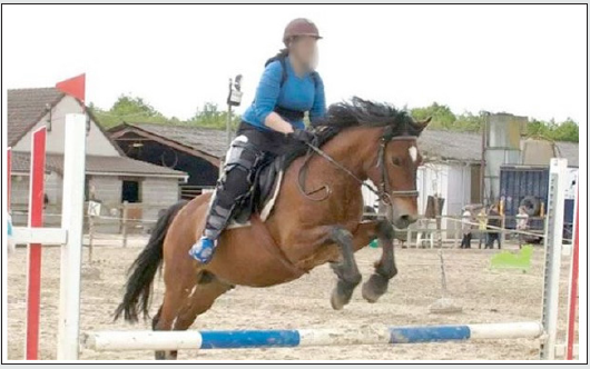
Constipation [11] is one of the most frequent manifestations of Ehlers-Danlos disease. The major complication is occlusion (Figure 9) with its corollary intestinal resection with often important consequences. It is accessible to the usual treatments of constipation with variable effects, sometimes requiring laxatives and intestinal enema. Intestinal massages, lactose-free and glutenfree diets, physical activity, wearing special compression clothing, lombo-abdominal belts and especially physical activity (walking, swimming, riding), all have positive effects. L-Carnitine acts on the mitochondria, improves the quality of muscle contraction, reduces cramps and increases intestinal transit. This treatment should be well dosed because it can cause diarrhea. These diarrhea can occur outside of any medication. These manifestations are often labeled as “irritable colon syndrome”, erroneously considered one of the symptoms of fibromyalgia that the French rheumatologist Marcel-Francis Kahn [12] thought to describe a “new disease” when examining women affected by Ehlers-Danlos. But he knew very little about it since he was convinced that this disease did not cause pain. This description however has been very successful, and more than half of our 6,200 patients were diagnosed with fibromyalgia prior to their Ehlers-Danlos disease diagnosis. This is an argument in favor of the high frequency (10 million people affected in the USA, according to Brad Tinkle [13] of Ehlers-Danlos disease.
The incidence of persons diagnosed with fibromyalgia is estimated by the French Academy of de medicine [14] at 2% of the French population, which is exactly the estimate of persons affected by Ehlers-Danlos [15] according to members of the same commission. The evidence of irritable colon syndrome appears therefore to be an argument for early detection of the disease. Ruptures in the intestine are possible but very rare, but it is very fragile during manipulations in surgery and during sutures. Diverticula are frequent, sometimes the seat of infections. Diarrhea is also a mode of expression and may require appropriate medication. Occlusion can be the cause of hernial strangulation, even from the beginning of life (acute intestinal invagination, umbilical hernia, crural or inguinal). Digestive hemorrhages are common and common with red blood if they are rectal or black if they are gastric or from small intestine. They may, by their importance or repetition, require compensation by globular caps or iron infusion. Iron is very poorly tolerated by the oral route. Endoscopic investigations should be avoided because of the risk of injury with hemorrhage or perforation. The presence of intestinal polyps is banal by excessive proliferation of connective tissue. Our experience is that they do not degenerate, they often disappear spontaneously or do not evolve, and they should not be withdrawn. The discovery of intestinal ganglia proceeds from the same mechanism, they also exist at the level of ganglionic areas of the underarms or crural. Mention should also be made of the fragility of the pharynx mucosa, which may be injured by intubation during a general anesthetic.
Ehlers-Danlos disease and the Anorectal region
The anorectal mucosa is particularly prone to injury and inflammation. It is fragile and cracks difficult to heal are frequent. Bleeding is common during defecation. They may be abundant in the case of external hemorrhoids that may require local treatment with caution. Rectal prolapse is difficult to access to surgery that carries the risk of failure or aggravation, as are rectoceles, often associated with hysterectomies in women. Non-surgical treatments (pessaries) are to be preferred. Botulinum toxin injections are ineffective. Proprioceptive rehabilitation with or without electrostimulation can be attempted.
Ehlers-Danlos disease and Gall bladder
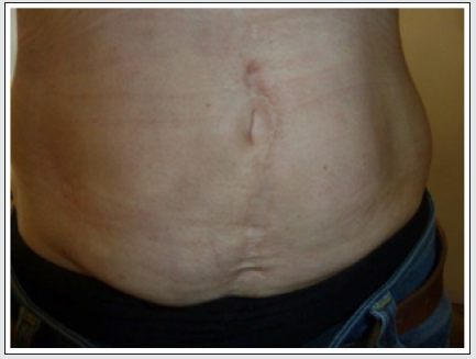
Gallstone in the gallbladder is particularly frequent in our experience but they have not been counted. We observed them in children whose youngest in age was seven years old. We therefore consider that from this age they should be systematically checked every year by an abdominal ultrasound. They can also sit in the choledoic channel. Their presence is probably the result of prolonged bile stasis in a distended and not very contractile vesicle. They can be very large or very numerous, up to 30 in one of our patients! The slimness of the vesicular wall, regularly described by the ultrasonists, confirms its fragility with the risk of perforation and peritonitis. Vesicular rupture is facilitated by the development of a purulent cholecystitis that can become gangrene. Its diagnosis is made difficult by the absence of abdominal defense, the atypical character of the pains which are not radiating towards the shoulder but in the back, or sometimes their total absence even with heavy palpation pressure, and the absence of fever. The only sure objective sign is hyperleukocytosis. Peritonitis diffusion in the peritoneum is difficult to treat because of the development of pus pockets in the numerous folds of a particularly distended peritoneum. Less frequently, flanges that favor secondary occlusions may develop. To avoid these complications, the removal of the vesicular stones, as soon as they are detected, must be systematic. Surgery should be performed according to certain rules for anesthesia and resuscitation (provide sufficient blood in case of hemorrhages, monitor general anesthesia by supplementing it in case of frequent early awakening) and should avoid traumatic manipulations of the abdominal organs that are fragile. In post-surgery, suture with non-resorbable threads and anticoagulants (hemorrhagic risk) should be avoided. The use of elastic contention stockings (Figure 10), active mobilizations and early lifting should be prioritized. Appendicitis can also be a cause of local, purulent complications that are difficult to detect by examining clinical signs at discretion. You should not hesitate to operate in case of doubt. This is what happened in one of our patients who insisted a lot on surgery, and the surgeon found much to his surprise an appendicular abscess ready to rupture.
Conclusion
Digestive manifestations, especially gastroduodenal, are constantly present in Ehlers- Danlos disease [16]. They are one of the major causes of the functional difficulties of these patients. They may also cause serious complications to the lives of these patients due to injury, medical or surgical treatments as a result of a misdiagnosis. Such dramatic situations are all the more shocking when one knows that the diagnosis of Ehlers-Danlos disease is very easy and quickly detectable when detecting certain signs such as diffuse joint manifestations (hyperlaxity, pain, instability), a very stretchy and fragile skin, stretch marks, skin hemorrhagic tendency with the presence of bruises, a diffuse and intense sensation of permanent fatigue, hypersensitivity (tact, noise, odors). This lack of diagnosis is widespread and has caused Doctor Rodney Grahame to write [17]: ” ...it is incomprehensible that the vast majority of Specialist and non-Specialist Doctors do not detect it, do not know how to diagnose it or, of course, how to treat it. Some even deny its existence, often leading the patient and his family into dramatic and unnecessary suffering”. Gastroenterologists do not escape this description. It is urgent that they update their knowledge and adapt because Ehlers-Danlos is a very common pathology that they all encounter in their daily practice.
References
- Beighton P (1970) The Ehlers-Danlos syndrome. William and Hellemann Medicine book ltd.
- Beighton P (1993) The Ehlers-Danlos syndrome in Mckusick’s Heritable Disorders of Connective Tissue (P Beighton) pp. 189-251. University of Cape Town Medical School, Cape Town, South Africa.
- Grahame R, Bird HA, Child A (2000) The revised (Brighton 1998) criteria for the diagnosis of benign joint hypermobility syndrome (BJHS). J Rheumat 27: 1777-1779.
- Bravo JF (2009) Ehlers-Danlos syndrome (EDS), with special emphasis in the joint hypermobility syndrome. January 2010. Revisa Medica de Chile 137(11): 1488-1497.
- Sabeeha M, Emma J Reinhold, Gemma S Pea RCE (2021) The Beighton Score as a measure of generalised joint hypermobility, Rheumatology Intern.
- Berthoz A (2013) Le sens du mouvement, Odile Jacob, Paris.
- Baeza-Velasco C, Van den Bossche T Grossin D, Hamonet C (2016) Difficulty eating and significant weight loss in Joint Hypermobility Ehlers– Danlos Syndrome, Hypermobility Type, Eat Weight Disord 21(2): 175-183.
- Baeza-Velasco C, Lorente S, Tasa-Vinyals E, Guillaume S, Soledad Mora M, et al. (2021) Gastrointestinal and eating problems in women with Ehlers-Danlos syndrome.
- Zeitoun JD, Lefevre JH, V de Parades, C Séjourné, I Sobhani, et al. (2013) Functional digestive symptoms and quality of life in patients with Ehlers-Danlos syndromes: results of a national cohort study on 134 patients 8(11): e80321.
- Hamonet C, Brissot R, Gompel A, Baeza- Velasco C, Guinchat V, et al. (2018) Ehlers-Danlos Syndrome - Contribution to Clinical Diagnosis - About 853 cases. EC Neurology.
- Castori M, Morlino S, Pascolini G, Blundo C, Grammatico P (2015) Gastrointestinal and nutritional issues in joint hypermobility syndrome/Ehlers-Danlos syndrome, hypermobility type. Am J Med Genet C Semin Med Genet 169C(1): 54-75.
- Kahn MF (1988) Syndromepolyalgique diffus, fibrosite, fibromyalgie. Doul Et Analg 1: 158-164.
- Tinkle B, Castori M, Berglund B, Cohen H, Grahame R, Kazkaz H, et al. (2017) Hypermobile Ehlers-Danlos syndrome (a.k.a. Ehlers-Danlos type III and Ehlers-Danlos syndrome mobility type) : clinical description and natural history, American Journal of Medical Genetics Part 4 Seminar in Medical Genetics 175(1): 48-69.
- (2018) Rapport 18-11 Bull Acad. Natle Méd 202(7): 1355-1370.
- Hamonet C, Manicourt D, Hermanns-Le T, Pommeret S (2018) Ehlers-Danlos. Clinical diagnosis, new fundamental perspectives. International Symposium on the Ehlers-Danlos Syndromes, Ghent, Belgium.
- Schneider V (2017) Prevalence and severity of digestive manifestations of Ehlers-Danlos syndrome, hypermobile. Master's thesis in medicine, Faculty of Biology and Medicine, University of Louvain, Belgium.
- Grahame R (2015) Introduction of Ehlers-Danlos. The disease forgotten by medicine. C Hamonet (Eds.), The Harmattan, Paris p. 20.

Top Editors
-

Mark E Smith
Bio chemistry
University of Texas Medical Branch, USA -

Lawrence A Presley
Department of Criminal Justice
Liberty University, USA -

Thomas W Miller
Department of Psychiatry
University of Kentucky, USA -

Gjumrakch Aliev
Department of Medicine
Gally International Biomedical Research & Consulting LLC, USA -

Christopher Bryant
Department of Urbanisation and Agricultural
Montreal university, USA -

Robert William Frare
Oral & Maxillofacial Pathology
New York University, USA -

Rudolph Modesto Navari
Gastroenterology and Hepatology
University of Alabama, UK -

Andrew Hague
Department of Medicine
Universities of Bradford, UK -

George Gregory Buttigieg
Maltese College of Obstetrics and Gynaecology, Europe -

Chen-Hsiung Yeh
Oncology
Circulogene Theranostics, England -
.png)
Emilio Bucio-Carrillo
Radiation Chemistry
National University of Mexico, USA -
.jpg)
Casey J Grenier
Analytical Chemistry
Wentworth Institute of Technology, USA -
Hany Atalah
Minimally Invasive Surgery
Mercer University school of Medicine, USA -

Abu-Hussein Muhamad
Pediatric Dentistry
University of Athens , Greece

The annual scholar awards from Lupine Publishers honor a selected number Read More...


