Lupine Publishers Group
Lupine Publishers
Menu
ISSN: 2641-1709
Research Article(ISSN: 2641-1709) 
Cupuloplasty for Mestizo Nose Volume 1 - Issue 3
Enrique Hernandez Vidal1*, Juan Eugenio Salas Galicia1 and Enrique Hernandez Del Moral2
- 1Department of Otorhinolaryngology and Head and Neck Surgeon, Mexico
- 2General Practitioner, Mexico
Received: December 05, 2018; Published: December 17, 2018
Corresponding author: Enrique Hernandez Vidal, Department of Otorhinolaryngology and Head and Neck Surgeon, Tabasco, Mexico
DOI: 10.32474/SJO.2018.01.000115
Summary
Background: The rhinoplasty techniques which are described by most authors are applied on leptorrhine-type noses and have some or no success at all in platyrrhine- and mesorrhine-type noses (Mestizo nose) as for the former reduction and removal techniques are used; whereas, in the latter, increase and elevation techniques are used. Objective. To provide an alternate surgical solution which offers proper results in patients with mesorrhine- or platyrrhine-type nose.
Materials and methods: Pre- and post-operative photographic records of 200 patients were utilized in this investigation. The same surgical technique was used in all cases, with variations related to the size and severity of the case.
Results: In a case in which no cardinal points were set, some loss of the nasal tip and the natural luminous points in it, as well as some upper depression of lateral crura, were noted. In a case in which no anterior elongated trapezoidal graft was placed, there was no adequate definition of the nasal tip in the natural form of its characteristic double fold.
Conclusion: Using this technique can help to define, thin, project and turn the nasal tip, give the height as desired, and lift the nasal dorsum when required. This is a highly accessible technique to lift the dorsum through osteo-cartilaginous or cartilaginous grafts with the anterior support (nasal tip) strengthened. This technique also works to increase the strength of tissue by providing an excellent structural support to the axis columella-alar-nose tip, without any elasticity or movement loss since the grafts are sufficiently thin.
Keywords: Septorhinoplasty; Platyrrhine; Mesorrhine; Mestizo nose
Introduction
The rhinoplasty techniques, described by most authors, are applied on leptorrhine-type noses, and have some or no success at all in platyrrhine- and mesorrhine-type noses which are very frequent in and a characteristic of world miscegenation. In the former type, reduction and removal techniques are used; whereas in the latter type, augmentation and elevation techniques are applied. Therefore, we come up with a surgical technique to functionally and aesthetically correct the nose in patients whose characteristics are those of the Latin, Asian, African and in mixed races; in other words, a technique which can be useful for the largest population of the world.
Objective
To provide an alternate surgical solution which offers proper results when a patient with mesorrhine- or platyrrhine-type nose requires augmentation of the anterior nasal spine, elongation and aesthetic alignment of columella, and thinning, elevation, rotation of the nasal tip and occasionally augmentation of the nasal dorsum.
Materials and Methods
During the years of medical surgical practice, in the hospital called “Institute of High Specialty In ears nose and throat”, located in the city of Villahermosa, Tabasco, Mexico. We have applied this technique to an undefined number of patients; however, 200-patient sample has been used in this study for scientific purposes. The same surgical technique was used in all cases, with variations related to the size and severity of the condition.
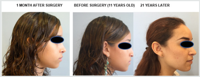
The 200 cases recorded were given a proper photographic preand post-operative follow-up. Cases were followed up for up to 24 years. 80% of patients were female and 20% were male. Age of patients where between 11 and 55 years. They all had mesorrhine or platyrrhine nose and their main signs included:
a) Lack of projection of the anterior nasal spine.
b) Short columella.
c) Poor ala nasal – columella ratio.
d) Wide tip.
e) Thick skin.
f) Wide base.
g) Lack of tip rotation and projection.
Surgical technique
Under general anesthesia and endotracheal intubation, with extreme care to ensure fixation of the catheter in the middle line of the chin, we infiltrated xylocaine 2% with epinephrine at 1x100, 000 in conventional points and the following measures were made:
a) Full transfixion at the caudal border of nasal septum.
b) Bilateral inter-cartilaginous incision.
c) Marginal incision (SLOT) at the caudal border of lower lateral cartilage up to the base of medial crus.
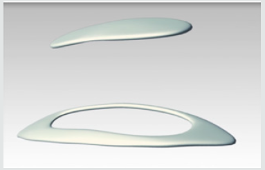
Figure 1: Preparation of grafts. 1. Intercrural cartilage bar. 2. Anterior elongated trapezoidal graft. 3. Cartilaginous bone graft for dorsum.
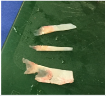
Figure 5: Application of the anterior elongated trapezoidal cartilage bar, which is 2 mm larger and is placed above the medial crura with strut already sutured (Hernández Vidal E & collaborators).
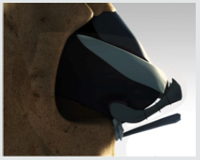
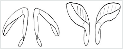
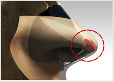
The septoplasty was made, and deviations were corrected, efforts were made to take advantage of them and obtain a cartilage bar according to the height, which was planned from the anterior nasal spine to the nasal tip (Figure 1) [1]. The shape of this cartilage was trapezoidal, very elongated, with blunt edges (Figure 2). When it was necessary to enlarge or make the dorsum regular, we took a bar of cartilage and bone with a strong supercut angled scissors in order to take the septum’s basal region without separating the bone from the cartilage (ethmoid bone perpendicular plate (LP) of the quadrangular plate or quadrangular cartilage (LC)). The length of this graft must be the same as the length of the nasal dorsum, and its shape was like a very long, narrow and thin rectangle (Figure 1). We then proceeded to valvuloplasty with resection of the return of the upper lateral cartilage and the cephalic border of lower lateral cartilage as required. It was extremely important that these dry fragments were kept separately in saline solution to use them afterwards as cardinal grafts. Once the lower lateral cartilage had been taken out and dried, we proceeded to identify with extreme care the dome which, in some cases, was non-existent, and must be assessed according to the desired height of the new nasal tip. We then carried out a cupulotomy (with skin) making a diagonal cut so that its limits end as a point thus achieve a higher projection and height of the tip (Figure 3) [2]. We then placeed a graft or cartilage bar (strut) between medial crura (Figure 4), we sutured with Dermalon 6-0 taking care of their stability in order to obtain satisfactory results. We then placed the elongated trapezoidal cartilage bar above the medial crura, taking care that it should be 2 mm higher than the height of medial crura which had been already sutured (Figure 5) [3].
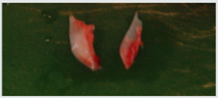
Figure 6: Creation of cardinal grafts from the fragments of the cephalic border of lower lateral cartilage.
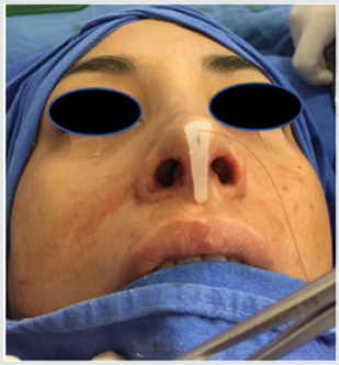
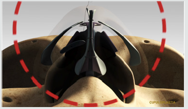
With the material removed from the cephalic border of lower lateral cartilage we proceeded to form two circular grafts (Figure 6), which were then fixed with Dermalon 5-0 suture, leaving the knot in the ventral face of the graft and the needle in the dorsal face (Figure 7), and these were placed going through the skin with the needle. Care must be exercised in order to prevent the knot remains in contact with the skin. These two grafts are placed where we want to make visible the two luminous points of the tip; that is, in this surgical moment we were reinstating the domes where we want them (Cupuloplasty). These were called the cardinal points (Figure 8). We proceeded to cut the thread without making a knot and leave a loose end of 3 cm approximately as a reference that cartilage remain in their place and are then fixed with tape (micro-pore) which was used to cover the nose at the end of the surgery. These stitches were cut seven or ten days in the postoperative period. If the anterior nasal spine is small or non-existent, we proceed to make a lateral incision and a blunt dissection in order to create a bag. All the remaining small fragments, including those of the bone, were placed as beads in a necklace in a Dermalon 5-0 suture with a large needle (Figure 9), and were inserted through the incision to be left in the desired area and give the volume required to the anterior nasal spine or subnasal expansion (Figure 10).
Figure 9: Preparation of the expander, with all the cartilaginous and bone fragments united with nylon 5-0 (prespinal expander grafts).
Osteotomy
When it is required, we proceed to carry out the needed osteotomy (lateral) in order to correct any defect in the skull. To augment the dorsum, a graft was used of septal cartilage. In cases where there is not enough material o cartilage, a graft composed of cartilage and bone from perpendicular plate is used. Septal cartilage can be used only if there is enough of it to harmonize the dorsum with the new height of the nasal tip [4]. In case it is required a higher elevation of the nasal tip, Dermalon 5-0 is used for this procedure, applying to the cephalic portion of the base of medial crura to the height desired in the middle to upper portion of the caudal border of septal cartilage (Figure 11). Endonasal incisions were sutured and Sarnoff sutures were applied to the nasal septum with Chromic Catgut 4-0 and the surgical procedure was hence completed.
Results
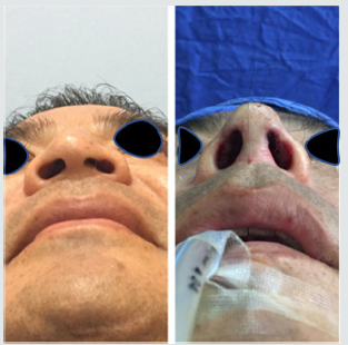
We evaluated in a retrospective manner the preoperative and postoperative control photographs (at 6, 12 months and up to 24 years in the first cases) of 200 patients who were given this procedure in a period of 24 years. In all cases it was truly evident the improvement in the facial aesthetic proportions. In all cases there was an augmentation of the nasolabial angle, 8 degrees in average, we managed to augment the height of nasal tip 3mm in average, there was an increase in the height of nasal dorsum in 30% of cases, while enlarging nasal angle 3 degrees in average. In a case in which no cardinal points were set, some loss of the nasal tip and the natural luminous points in it were noted, as well as some upper depression of lateral crura. In a case in which no anterior elongated trapezoidal graft was placed, there was no adequate definition of the nasal tip in the natural form of the double fold which characterizes it.
Conclusion
Using this technique can help to define, thin, project and turn the nasal tip and, in cases so requiring it, to lift the nasal dorsum. It is also useful to meaningfully increase the strength of tissue by providing an excellent structural support to the axis columellaalar- nasal tip and, since grafts are sufficiently thin, no elasticity or movement is lost, and we can prevent the effect of a “frozen” nose. Based on the observations and remarks made in relation to the patients referred to in this article, we are reporting a technique which meets many satisfaction requirements for both the patient and the doctor, which will always be influenced by the patient’s expectations and by the doctor’s skill to carry out the surgery. We make available to the otorhinolaryngologist an efficient and proven technique which will enable the doctor to decide how much he/ she wants to thin, sharpen, lift or turn the tip, or how much he/ she wants to lift the dorsum in relation to the doctor’s sense of aesthetics and beauty and the doctor’s knowledge and experience in the surgery of platyrrhine or mesorrhine nose.
Discussion
There is a number of techniques to lift the tip in nasal surgery. Rather than a technique, Goldman’s cupulotomy has become one step to complete the approach to relocate and reconstruct the domes (Cupuloplasty) which is attained by the relocation of the cardinal points, and it is the technique which yields better results in the mestizo, mesorrhine and platyrrhine nose, with hypoplastic lobules, bulbous tips with thick skin. These patients have small nasal projection and need additional tip grafts not only as a step in the resection but to complete it with a relocation, strengthening and reconstruction technique supported by the grafts, inter-crural and elongated anterior trapezoidal. We proceeded by approaching the lobule cartilages by using Goldman’s classic technique. A volume reduction is carried out as needed. The lateral crus of both lobule cartilages and the skin in contact with them are released through fine dissection. We then take 2 to 3mm of lateral crus with its vestibular skin attached, toward the dome and medial crura. This new position is fixed with nylon 5-0 suture, with support of septal cartilage in the middle of medial crura in order to obtain a higher elevation of the tip and better support. Further, we must place in front of the above an elongated trapezoidal cartilaginous graft 2mm higher than the sutured medial crura in order to improve the definition and elevation of the tip. The key component of our surgical technique is the use of cardinal grafts as follows: two of them at the level of the new dome (on the upper end the medial and lateral crus), this is the most frequent in 90% of the cases and, in cases so required by aesthetics, two between the cephalic border of lobule cartilage and caudal border of upper lateral cartilage. This surgical procedure prevents or minimizes the alar collapse, columella or excessive rotation of the lateral crus. None of the patients who was given this surgical procedure (the oldest was operated 24 years ago) suffered from distortion of nasal lobule after the operation. In patients with a low dorsum profile requiring augmentation, an excellent choice is to place a cartilaginous bone graft on the nasal dorsum (Figure 1).
The perpendicular plate of the ethmoid and the squared cartilage of the nasal septum are sectioned in one piece. The bone part of the graft is placed on top of the bone pyramid and the cartilaginous part of the graft is placed on the valve of this type. The layer-type graft or the cylinder-type graft (expansor) can be used to correct the pre-maxillary retraction in mesorrhine and platyrrhine patients. This composite graft contains fragments oflobule septal cartilage and of upper lateral cartilage, it even has bone from the nasal septum. We proceed with nylon 5-0 stitch, the graft is inserted in the nasolabial angle, under the columella (columellar labial angle). The stitch is fixed to the skin externally. This procedure provides additional support to the nasal tip, and it improves the shape of the columellar labial angle. Further, it makes rotation look controlled. Even with an excellent elevation of the tip of the mestizo nose, the width of nasal base is reduced, making the ratio dimensions of lobule and tip look proportionate because, when we make an elevation, we think the entire nasal lobule. The purpose of this procedure is to give a triangular shape from the basal view of the nose. In order to obtain the best result with this surgical technique, it is necessary the surgeon has a good aesthetic approach. We think this combination of various techniques is an excellent choice which must be included in the rhinoplasty procedures carried out in mesorrhine and platyrrhine noses.
References
- Rodolfo Nazar, Natalia Cabrera, Alfredo Naser (2013) Septoplastía endoscópica, Endoscopic septoplasty. Rev Otorrinolaringol Cir Cabeza Cuello 73(3): 100.
- Thiago Bittencourt Ottonide Carvalho, Emerson Thomazi, Leutz RP, Souza RP, Molina FD, et al. (2015) Gradual approach to refinement of the nasal tip. Brazilian Journal of Otorhinolaryngology 81(1): 31-36.
- Mahmoud F El Bestara, AbdAllah Y AlMahdyc, Fadi M Gharibb (2015) Assessment of nasal tip projection. Egypt Journal Otolaryngol 31(2): 105-110.
- Durbec M, Disant F (2014) Saddle nose: Classification and therapeutic management. European Annals of Otorhinolaryngology, Head and Neck diseases 131(2): 99-106.

Top Editors
-

Mark E Smith
Bio chemistry
University of Texas Medical Branch, USA -

Lawrence A Presley
Department of Criminal Justice
Liberty University, USA -

Thomas W Miller
Department of Psychiatry
University of Kentucky, USA -

Gjumrakch Aliev
Department of Medicine
Gally International Biomedical Research & Consulting LLC, USA -

Christopher Bryant
Department of Urbanisation and Agricultural
Montreal university, USA -

Robert William Frare
Oral & Maxillofacial Pathology
New York University, USA -

Rudolph Modesto Navari
Gastroenterology and Hepatology
University of Alabama, UK -

Andrew Hague
Department of Medicine
Universities of Bradford, UK -

George Gregory Buttigieg
Maltese College of Obstetrics and Gynaecology, Europe -

Chen-Hsiung Yeh
Oncology
Circulogene Theranostics, England -
.png)
Emilio Bucio-Carrillo
Radiation Chemistry
National University of Mexico, USA -
.jpg)
Casey J Grenier
Analytical Chemistry
Wentworth Institute of Technology, USA -
Hany Atalah
Minimally Invasive Surgery
Mercer University school of Medicine, USA -

Abu-Hussein Muhamad
Pediatric Dentistry
University of Athens , Greece

The annual scholar awards from Lupine Publishers honor a selected number Read More...


