Lupine Publishers Group
Lupine Publishers
Menu
ISSN: 2641-1709
Short Communication(ISSN: 2641-1709) 
Correlation Between Laboratory Diagnose of Trichomoniasis With Vascular Pattern of Cervix by Endoscopy Volume 6 - Issue 1
Osama Bakheet1 and Salwa Samir Anter2*
- 1Associate Professor of clinical Pathology, Military Medical Academy, Egypt
- 2Department of Obstetrics Gynecology, Cairo university, Egypt
Received: February 05, 2021 Published: February 15, 2021
Corresponding author: Salwa Samir Anter, Department of Obstetrics Gynecology, Cairo university, Egyptad and Neck Surgery, Abubakar Tafawa Balewa University Teaching Hospital Bauchi, Bauchi Nigeria
DOI: 10.32474/SJO.2021.06.000228
Abstract
Flexible endoscopy was used in examination cervix lesion the first was studied by Nishiyama et al was reported the used of endoscopy for diagnosing cervical neoplasms. The second study of K Uchita et al. study feature findings of high-grade cervical intraepithelial neoplasia or more on magnifying endoscopy with narrow band imaging Trichomoniasis is a common, worldwide, urogenital infection with Trichomoniasis vaginalis. It is a frequent cause of symptomatic vaginitis and a less common cause of nongonococcal urethritis in this study correlation were done by laboratory diagnosis of trichomoniasis with endoscopy finding.
Introduction
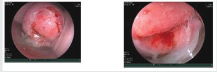
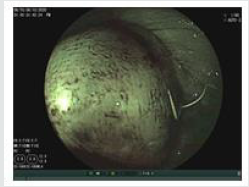
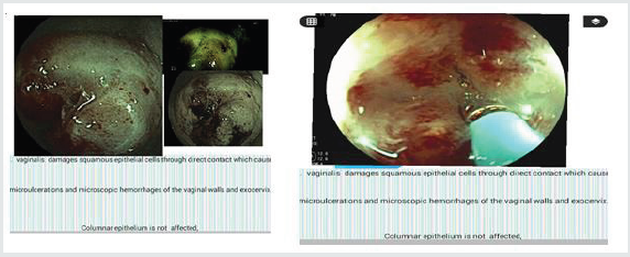
Flexible magnifying endoscopy is at present time used for the gastrointestinal tract and is tolerable for the diagnosis of GI neoplasms. Magnifying endoscopy with narrow band imaging can be used to clearly imagine the microstructures of the mucosal surface and interstitial capillaries. As regard of used of flexible endoscopy in examination cervix lesion the first was studied by Nishiyama et al was reported the used of endoscopy for diagnosing cervical neoplasms revealed micro-vascular pattern differences at different stages (Figure 1).The second study of K Uchita et al. study feature findings of high-grade cervical intraepithelial neoplasia or more on magnifying endoscopy with narrow band imaging. Trichomoniasis is a common, worldwide, urogenital infection with Trichomoniasis vaginalis. It is a frequent cause of symptomatic vaginitis and a less common cause of nongonococcal urethritis. Pathology T. vaginalis damages squamous epithelial cells through direct contact which causes microulcerations and microscopic hemorrhages of the vaginal walls and exocervix (Figure 2). Columnar epithelium is not affected, and thus trichomoniasis presents with vaginitis but not with end cervicitis. The simultaneous presence of an endocervical discharge should alert the clinician to the possibility of coincident infection with Neisseria gonorrhoeae, Chlamydia trachomatis, or Mycoplasma dentalium. Invasion of tissue does not occur. T. vaginalis is also isolated from the urethra in most infected women (Figures 3-5).
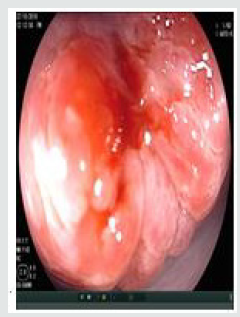
Figure 3: Congested with prominent but normal branching blood vessels. Inflammation of the columnar epithelium can give the cervix a beefy-red appearance.
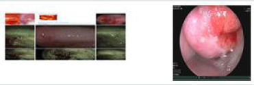
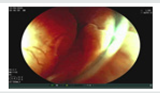
Figure 5: Sometimes staghorn-like small capillaries or prominent vessels are seen on the infected cervix.

Clinical Features
a) Vaginal Discharge
b) Vulva Irritation
c) Dysuria
d) Dyspareunia
e) Lower abdominal discomfort.
Physical Examination The vulva is erythematous on speculum examination, excessive discharge , yellow vaginal discharge, the classic signs of T vaginalis infection is colpitis macularis, or strawberry cervix, which describes a diffuse or patchy macular erythematous lesion of the cervix. This is a specific sign of trichomoniasis (Figure 6).
f) Complications
Premature rupture of the fetal membranes and pre-term delivery. Trichomoniasis, with its brisk inflammatory response and microulcerations of the genital epithelium, may increase the risk of acquiring HIV (Figure 7).
Figure 6: Infection with Trichomonas vaginalis may produce a “strawberry” appearance of the cervix because of focal round patches of dilated capillaries on the surface.

Figure 7: Inflamed cervix may have diffuse white patches after application of acetic acid. The white patches are thin with indistinct or irregular margins. Inflamed areas may bleed on contact because the epithelium is thinned.
Diagnosis
a. Determine the pH of vaginal secretion.
b. Normal vaginal pH of 4.7 or less.
c. Vaginal pH is elevated above 4.7 in most women with trichomoniasis.
d. Positive result of the whiff test is determined.
After the pH has been determined, several drops of 10% to 20% potassium hydroxide should be added to the discharge in the speculum. The clinician then seeks the elaboration of a pungent, fishy, amine like odor. Definitive diagnosis requires demonstration of the organism. Nucleic acid amplification techniques, vaginalis culture and antigen detection systems, which are less sensitive, The Pap smear can detect trichomonas infection, but the Gram stain is useless, wet mount. A swab of vaginal material can be agitated in about 1 mL of saline, and a drop is transferred to a microscope slide. Endoscopy finding of trichomoniasis corrected with laboratory diagnosis.
i. colpitis macularis, or strawberry cervix, which describes a diffuse or patchy macular erythematous lesion of the cervix.
This is a specific sign strawberry” appearance of the cervix because of focal round patches of dilated capillaries on the surface.
ii. Trichomoniasis may cause dilated capillary loops (strawberry spots) that can be interpreted as coarse mosaicism.
iii. Cervicovaginal inflammation – Inflamed cervix. The infected cervix is often tender on movement and is congested with prominent but normal branching blood vessels.
Inflammation of the columnar epithelium can give the cervix a beefy-red appearance.
iv. infected cervix is often tender on movement and is congested with prominent but normal branching blood vessels. infected cervix congested with prominent but normal branching blood vessels.
v. Staghorn-like small capillaries or prominent vessels are seen on the infected cervix.
vi. Infection with Trichomonas vaginalis may produce a “strawberry” appearance of the cervix because of focal round patches of dilated capillaries on the surface.
vii. Inflamed cervix may have diffuse white patches after application of acetic acid. The white patches are thin with indistinct or irregular margins. Inflamed areas may bleed on contact because the epithelium is thinned out.
viii. Inflammation is followed by repair of the damaged epithelium. During the reparative process, glycogen may be absent from the epithelium. As a result, patchy iodine -negative areas are seen after inflammation.
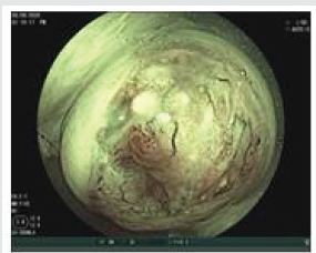
Application of Lugol’s iodine to an inflamed cervix sometimes produces the typical “leopard-skin” appearance because of multiple iodine-negative spots. Such changes are more commonly seen in trichomoniasis (Figures 8-10).
ix. Sometimes follicular (chronic) cervicitis is detectable as multiple small raised whitish areas on the squamous epithelium.
\x. T vaginalis damages squamous epithelial cells through direct contact which causes microulcerations and microscopic hemorrhages of the vaginal walls and exocervix. Columnar epithelium is not affected.
Figure 8: Application of Lugol’s iodine to an inflamed cervix sometimes produces the typical “leopard-skin” appearance because of multiple iodine-negative spots.


Top Editors
-

Mark E Smith
Bio chemistry
University of Texas Medical Branch, USA -

Lawrence A Presley
Department of Criminal Justice
Liberty University, USA -

Thomas W Miller
Department of Psychiatry
University of Kentucky, USA -

Gjumrakch Aliev
Department of Medicine
Gally International Biomedical Research & Consulting LLC, USA -

Christopher Bryant
Department of Urbanisation and Agricultural
Montreal university, USA -

Robert William Frare
Oral & Maxillofacial Pathology
New York University, USA -

Rudolph Modesto Navari
Gastroenterology and Hepatology
University of Alabama, UK -

Andrew Hague
Department of Medicine
Universities of Bradford, UK -

George Gregory Buttigieg
Maltese College of Obstetrics and Gynaecology, Europe -

Chen-Hsiung Yeh
Oncology
Circulogene Theranostics, England -
.png)
Emilio Bucio-Carrillo
Radiation Chemistry
National University of Mexico, USA -
.jpg)
Casey J Grenier
Analytical Chemistry
Wentworth Institute of Technology, USA -
Hany Atalah
Minimally Invasive Surgery
Mercer University school of Medicine, USA -

Abu-Hussein Muhamad
Pediatric Dentistry
University of Athens , Greece

The annual scholar awards from Lupine Publishers honor a selected number Read More...


