Lupine Publishers Group
Lupine Publishers
Menu
ISSN: 2641-1709
Clinical Presentation(ISSN: 2641-1709) 
Classical CT Findings in Cherubism Volume 8 - Issue 4
Kritika1, Tapendra N Tiwari2, Rajaram Sharma2*, Sunil Kast2 and Saurabh Goyal2
- 1Resident, Doctor, Department of Radio-Diagnosis, Pacific Institute of Medical Sciences (PIMS), India
- 2Assistant Professor, Radio-Diagnosis, Pacific Institute of Medical Sciences (PIMS), India
Received: June 21, 2022; Published: June 30, 2022
Corresponding author: Rajaram Sharma, Assistant Professor, Radio-Diagnosis, Pacific Institute of Medical Sciences (PIMS), Umarda, Udaipur, Rajasthan, India
DOI: 10.32474/SJO.2022.08.000291
Introduction
The condition known as ‘Cherubism’ is considered as a variant of fibrous dysplasia, but in reality, it is more likely a distinct entity. It is a benign disease of the pediatric age group. It is a fibro-osseous disease of childhood affecting only the jaws, is a self-limiting non-neoplastic autosomal dominant disorder and evident around the third or fourth year of life, rarely apparent before the age of two years. Here, we are presenting a case with typical presentation and age for this disease. Computed tomographic (CT) scan was done (in paediatric dose) in three dimensional plane to demonstrate all the features.
Clinical Presentation
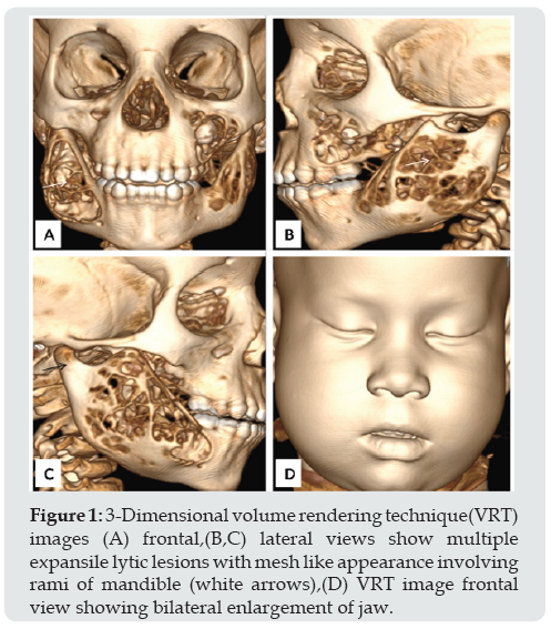
Figure 1: 3-Dimensional volume rendering technique(VRT) images (A) frontal,(B,C) lateral views show multiple expansile lytic lesions with mesh like appearance involving rami of mandible (white arrows),(D) VRT image frontal view showing bilateral enlargement of jaw.

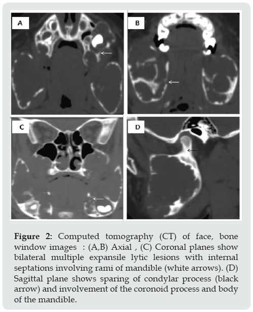
Figure 2: Computed tomography (CT) of face, bone window images : (A,B) Axial , (C) Coronal planes show bilateral multiple expansile lytic lesions with internal septations involving rami of mandible (white arrows). (D) Sagittal plane shows sparing of condylar process (black arrow) and involvement of the coronoid process and body of the mandible.

A 5-year-old male visited the outpatient department of our hospital. Parents were concerned about bilateral progressive swelling of the jawbones. The birth history was uneventful, and the patient didn’t show any abnormalities until two years of age. On examination, the patient was found to be well-built, active and mentally alert and showed asymmetry of the face with a gross chubby appearance. Deformities became more pronounced for the last two years. In addition, the patient also had upward gaze and bilateral submandibular lymphadenopathy. No abnormality was found on clinical examination of the chest, abdomen, central nervous and cardiovascular system. There were no cutaneous pigmentation or other congenital abnormality presents. After knowing these findings, we decided to perform a low dose computed tomography (CT) scan of facial bones. It was performed on a high-resolution CT scanner, and axial images were obtained in the region of facial bones. 3-Dimensional images were also reconstructed in the greyscale and colour rendered mode (Figures 1A-1D). Slices revealed multiple expansile osteolytic lesions involving the mandible and maxilla (Figures 2A-2D). There was marked thinning of the bony cortex in involved bones. The medial wall of the maxillary sinuses was spared on both sides. A visualized portion of the base of the skull appeared normal. The patient was diagnosed with grade III cherubism [1], as condyles of the mandible are spared. ( The lesions of cherubism can be classified according to their extent: grade I, bilateral involvement of the ascending rami of mandible; grade II, bilateral involvement of the ascending rami of the mandible and maxillary tuberosities; grade III, complete involvement of the maxilla and mandible compromising the coronoid processes and condyles).
Discussion
In 1933, Jones [2] first described the entity with its typical clinical features. Classically, the jaw lesions of cherubism are characterized by bilateral swelling of the lower face. Because of this prominence of the lower face, the patient gives an appearance reminiscent of the ‘cherubs’ portrayed in Renaissance art, hence the disease named ‘Cherubism’. It is one of the infrequent genetically determined osteoclastic lesions. Its gene is mapped on chromosome band 4p16.3 and is called SH3BP2 (for SH3 domain bind protein 2).
Radiographic features consist of multiple lucent expanded regions within the maxilla and mandible, giving a soap-bubble appearance. As the lesion ages, it often becomes sclerotic and reduces in size [2,3]. Despite the prominent features, the disease stabilizes and often regresses without treatment. Studies have shown a marked reduction in lesion size with Imatinib, a tyrosine kinase inhibitor [4].
Learning Points
a) Although radiological and histological characteristics of cherubism are not pathognomic, the overall presenting features are consistent, and with positive family history, we can consider painless progressive swelling in children as cherubism.
b) Due to its benign nature and self-limiting natural history, no definite treatment is usually required. It is a do not touch lesion and must be diagnosed accurately by the radiologist.
References
- Kalantar Motamedi MH (1998) Treatment of cherubism with locally aggressive behaviour presenting in adulthood: report of four cases and a proposed new grading system. J Oral Maxillofac Surg 56(11): 1336-1342.
- Tiziani V, Reichenberger E, Buzzo CL (1999) The gene for cherubism maps to chromosome 4p16. Am. J Hum Genet 65(1): 158-166.
- Larheim TA, Westesson PA (2009) Maxillofacial Imaging. Springer.
- Ricalde P, Ahson I, Schaefer ST (2019) A Paradigm Shift in the Management of Cherubism? A Preliminary Report Using Imatinib.Journal of Oral and Maxillofacial Surgery 77(6).

Top Editors
-

Mark E Smith
Bio chemistry
University of Texas Medical Branch, USA -

Lawrence A Presley
Department of Criminal Justice
Liberty University, USA -

Thomas W Miller
Department of Psychiatry
University of Kentucky, USA -

Gjumrakch Aliev
Department of Medicine
Gally International Biomedical Research & Consulting LLC, USA -

Christopher Bryant
Department of Urbanisation and Agricultural
Montreal university, USA -

Robert William Frare
Oral & Maxillofacial Pathology
New York University, USA -

Rudolph Modesto Navari
Gastroenterology and Hepatology
University of Alabama, UK -

Andrew Hague
Department of Medicine
Universities of Bradford, UK -

George Gregory Buttigieg
Maltese College of Obstetrics and Gynaecology, Europe -

Chen-Hsiung Yeh
Oncology
Circulogene Theranostics, England -
.png)
Emilio Bucio-Carrillo
Radiation Chemistry
National University of Mexico, USA -
.jpg)
Casey J Grenier
Analytical Chemistry
Wentworth Institute of Technology, USA -
Hany Atalah
Minimally Invasive Surgery
Mercer University school of Medicine, USA -

Abu-Hussein Muhamad
Pediatric Dentistry
University of Athens , Greece

The annual scholar awards from Lupine Publishers honor a selected number Read More...


