Lupine Publishers Group
Lupine Publishers
Menu
ISSN: 2641-1709
Case Report(ISSN: 2641-1709) 
A Giant Ranula: The Rima Oris Lantern – A Catch-All Approach Volume 7 - Issue 1
Belaldavar BP, Suhasini Hanumaiah* and Reshma Susan Mathew
- Department of Otolaryngology, Kaher University, India
Received:June 22, 2021; Published:July 02, 2021
Corresponding author: Suhasini Hanumaiah, Department of Otolaryngology, Kaher University, India
DOI: 10.32474/SJO.2021.07.000253
Abstract
Oral ranula is a retention cyst which arises from the sublingual salivary gland in the floor of the mouth due to ductal obstruction and fluid retention. Various surgical procedures have been quoted in the literature ranging from simple aspiration to complete or partial excision of the ranula. This paper highlights a case report of a huge impending plunging ranula in the floor of the mouth which has been successfully excised completely.
Keywords: Ranula; Marsupialisation; Pseudo-cyst
Introduction
The term ranula was derived from the Latin word Rana tigerina, symbolising its resemblance to a bulging frog’s underbelly.1 It classically presents as a soft submucosal swelling in the floor of the mouth. Ranula have a prevalence of about 0.2 cases/1000 persons and accounts for 6% of total sialo-cysts. Marsupialisation is still one of the attractive surgical modalities practiced avoiding injury to the surrounding anatomical structures, despite a reported recurrence rate of 61 to 89%.2 Hence, this surgery was designed to highlight the choice of an appropriate surgical technique for complete excision without any damage to surrounding anatomical structures.
Case Presentation
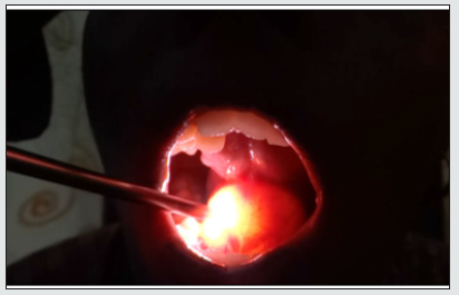
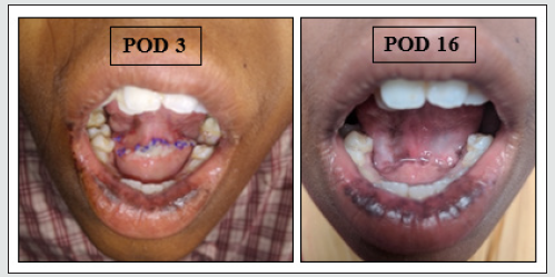
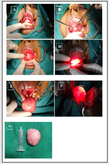
A 16-year-old girl reported with a 2-week history of swelling in left submandibular region. The swelling was completely asymptomatic only with a gradual increase in the size of the swelling. The patient’s health was satisfactory and had no history of any systemic disorder. On examination, a solitary, spherical swelling of 3 x 4cm, smooth surfaced, with well-defined margins was present in the floor of the mouth (Figure 1) which was cystic in consistency and was transillumination positive (Figure 2). After routine preoperative investigations, a contrast CT scan of the neck done revealed a well-defined non- enhancing cystic lesion in the floor of the mouth measuring 5.5 x 2.9 x 4.9cm with mild displacement of mylohyoid muscle with likely representation of a ranula (Figure 3). Under general anaesthesia, following local infiltration to the thinned out sub-epithelial plane, an elliptical incision was taken over the mucosa superior to the submandibular duct openings which were identified as small nodular papillae and the mucosa was excised. Meticulous dissection was carried out and the mucosa was carefully separated from the cyst wall superiorly, inferiorly and the sides. Inferiorly, mylohyoid muscle was visualised which was partially splayed by the cyst suggestive of an impending plunging. Indentation by the mylohyoid muscle was seen on the under surface of the cyst. The cyst was completely removed from its attachment and excised in toto (Figure 4). Care was taken not to injure the submandibular, sublingual salivary glands and their ducts, respectively. Wound was closed in layers. The case was followed up for two months at fortnightly interval. There is no recurrence of the lesion. Patient is still under follow up (Figure 5).
Figure 1: Solitary, spherical swelling arising from the floor of mouth with smooth surface and well-defined margins.
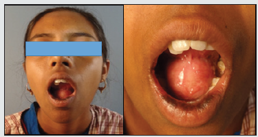
Figure 3: Well-defined, thin walled, non-enhancing cystic lesion in the floor of mouth. Superiorly pushing the tongue upwards, inferiorly limited to the sublingual space, and displacing the mylohyoid muscle.
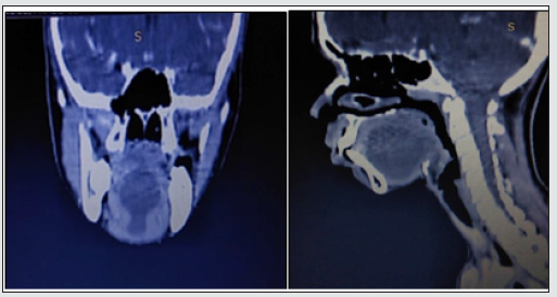
Figure 4: A&B: Elliptical incision taken. C: Mucosa around the cyst separated all around and outpouching of the cystic mass. D: Brilliantly trnsilluminant cystic mass. E: Splaying of the mylohyoid muscle inferiorly F: Lantern in the rima oris. G: Cystic mass measuring 5x6cm completely excised in toto.
Discussion
Oral ranulas are cystic lesions which are present in the floor of the mouth that is formed due to obstruction of the excretory duct of the sublingual gland. The rupture of the excretory duct causes extravasation of the mucus and accumulation of saliva into the surrounding tissue which forms a pseudo-cyst that lacks the epithelial lining [1]. On the basis of its formation, ranula is classified into two groups, simple (intraoral) and the plunging (cervical) type. Simple ranulas are more common than plunging type. A simple ranula is formed by localized collection of mucus within the floor of the mouth and may arise from the submandibular duct or from the body of the sublingual gland. In the plunging type, sufficient fluid pressure will be attained as to herniate through the mylohyoid muscle and produce swelling within the neck [2]. Dermoid cyst, lymphangioma, mucocele of the minor salivary gland, lymphoma and HIV-related lymphadenopathy are few differential diagnoses of ranula. CT and MRI imaging studies can be helpful in supporting a diagnosis and in determining the origin of the lesion [3]. Management of ranula is a polarising topic, with discordant evidence as to which treatment modality is best due to the existing gap in knowledge on the current concept of its aetiopathogenesis. The various procedures for treating the lesion are marsupialisation, dissection, cryotherapy, sclerotherapy, hydro-dissection, and LASER ablation. The recurrence rate varies according to the procedure performed [4].
Conclusion
Ranulas are mucin-containing pseudo cystic lesions that develop in the floor of the mouth, in the neck, or both. Surgical excision remains the mainstay for management. This procedure is arduous as it involves a fine mucosa that may rupture on excision, also there is risk of injury to the lingual nerve and sublingual duct, hence surgeons globally prefer marsupialization as an effective treatment for intraoral ranula. However, meticulous dissection can improve the condition of the patient and would avoid recurrence of the cyst. Therefore, extirpation is the most effective treatment for painless and a well healed tissue.Compliance with ethical standards: Ethically approved.
Conflicts of Interest
Nil.
References
- Crysdale WS, Mendelsohn JD, Conley S (1988) Ranulas Mucoceles of the oral cavity: experience in 26 children. Laryngoscope 98(3): 296–298.
- Morton RP, Ahmad Z, Jain P (2010) Plunging ranula: congenital or acquired? Otolaryngol Head Neck Surg 142: 104–107.
- Curtin HD (2007) Imaging of the Salivary gland. Myers Eugene N, Ferris Robert L (Eds.), Myers’ salivary gland disorders. Springer, Berlin pp. 17–31.
- Patel MR, Deal AM, Shockley WW (2009) Oral and plunging ranulas: What is the most effective treatment? Laryngoscope 119(8): 1501–1509.

Top Editors
-

Mark E Smith
Bio chemistry
University of Texas Medical Branch, USA -

Lawrence A Presley
Department of Criminal Justice
Liberty University, USA -

Thomas W Miller
Department of Psychiatry
University of Kentucky, USA -

Gjumrakch Aliev
Department of Medicine
Gally International Biomedical Research & Consulting LLC, USA -

Christopher Bryant
Department of Urbanisation and Agricultural
Montreal university, USA -

Robert William Frare
Oral & Maxillofacial Pathology
New York University, USA -

Rudolph Modesto Navari
Gastroenterology and Hepatology
University of Alabama, UK -

Andrew Hague
Department of Medicine
Universities of Bradford, UK -

George Gregory Buttigieg
Maltese College of Obstetrics and Gynaecology, Europe -

Chen-Hsiung Yeh
Oncology
Circulogene Theranostics, England -
.png)
Emilio Bucio-Carrillo
Radiation Chemistry
National University of Mexico, USA -
.jpg)
Casey J Grenier
Analytical Chemistry
Wentworth Institute of Technology, USA -
Hany Atalah
Minimally Invasive Surgery
Mercer University school of Medicine, USA -

Abu-Hussein Muhamad
Pediatric Dentistry
University of Athens , Greece

The annual scholar awards from Lupine Publishers honor a selected number Read More...


