Lupine Publishers Group
Lupine Publishers
Menu
ISSN: 2638-6003
Research Article(ISSN: 2638-6003) 
Hamstrings Tendons Thickness in ACL Reconstruction: Is it Possible to Predict it By Isometric Torque in Soccer Athletes? Volume 5 - Issue 3
José Martins Juliano Eustaquio1*, Alberto Martins Fontoura Borges1, Leandro Pereira de Mendonça1, Cássio Lucas Dias do Prado1, Pedro Debieux2 and Octávio Barbosa Neto3
- 1Mário Palmerio Hospital, University of Uberaba (UNIUBE), Orthopedics and Traumatology Group, Brazil.
- 2Hospital Israelita Albert Einstein, Brazil.
- 3Federal University of Triângulo Mineiro (UFTM – Universidade Federal do Triângulo Mineiro, Exercise Science, Health and Human Performance Research Group, Graduate Program in Physical Education, Brazil
Received:July 6, 2021 Published:July 22, 2021
Corresponding author: José Martins Juliano Eustaquio, Mário Palmerio Hospital, University of Uberaba (UNIUBE – Universidade de Uberaba), Department of Orthopedics and Traumatology, Av. Nenê Sabino, n. 2477, Santos Dumont District, Uberaba, Minas Gerais, Brazil
DOI: 10.32474/OSMOAJ.2021.05.000213
Abstract
Objective: To acess the relationship between knee flexor and extensor torques and the thickness of the quadruple hamstrings
tendons, extracted during anterior cruciate ligament (ACL) reconstruction surgery, in amateur soccer athletes.
Materials and Methods: This is a cross-sectional study in which 38 amateur soccer athletes of both genders, diagnosed with recent
and isolated ACL ligament injury, participated. Epidemiological (gender, age, and dominant limb), anthropometric (height, weight,
and BMI), and biomechanical data of the injured lower limb (isometric extensor and flexor torques of the ipsilateral knee) were
evaluated. Pearson’s correlation test was used to analyze the correlation of torques with the quantitative variables of the study, and
the multivariate analysis was performed using the linear regression test with one-way ANOVA and the calculation of R2. The level
of statistical significance was set at 5% (p< 0.05).
Results: The final study sample (33 participants) had a mean age of 30 years, with 81.8% males. Significant correlations were
observed between knee flexor torque and height (p=0.002), weight (p=0.01) and graft diameter (p<0.001). In multivariate analysis
a statistically significant result was observed for flexor torque (p=0.007).
Conclusion: Knee flexor torque can be tool for preoperative analysis of flexor tendon thickness during surgical programming of
knee ligament reconstructions
Keywords: Knee Joint; Anterior Cruciate Ligament Injuries; Soccer; Isometric Contractions
Introduction
The anterior cruciate ligament (ACL) lesion has a high incidence
in orthopedic practice [1], especially in young and physically
active population [2,3], with an incidence of approximately 85 per
100,000 people aged 16 to 39 years [4,5]. In soccer these injuries
are common [5] and occur mainly due to the rotational movements
characteristic of the dynamics of the sport, which has also motivated
the emergence and use of injury prevention protocols [6]. The
treatment of ACL injuries takes into account mainly the current
and future functional demands of the patient. However, in most
cases, the recommended treatment is surgical [7]. Among the graft
options for ACL reconstruction there are allografts and autografts.
Allografts are used basically in cases of multiligamentous injuries
and successive ACL reconstruction revisions, because it is known
to be associated with a higher rate of graft rupture [8]. Among the
autografts, the options are the patellar, quadriceps, and hamstrings
tendons (gracilis and semitendinosus). Currently, surgeons prefer
the hamstrings tendons, which are used in approximately 50% of
cases, followed by the patellar and quadriceps tendons [9], the latter
with a recent increase in popularity. The total thickness of a graft
depends basically on anatomic and biomechanic factors [10,11] and needs a minimum measurement that provides joint stability and
survival. In relation to the patellar and quadriceps tendons, the graft
corresponds to a fraction of the tendon, which guarantees, therefore,
a satisfactory final thickness. However, regard to the hamstring’s
tendons, the graft is equivalent to the total volume of the gracilis
and semitendinosus tendons, usually in quadruple final shape.
This causes, during surgical programming, a greater dependence
on knowing this measure more precisely, because it is known that
grafts smaller than 08 mm evolve with higher rates of rupture [12].
The evaluation of objective anthropometric and/or biomechanical
parameters with good clinical accuracy in the preoperative period
would be fundamental to help the surgeon to decide the type of
graft to be used. Some works have tried to make this association
[13-16], but we still observed low clinical employability. The direct
measurement of the biomechanical function of the tendon, through
the evaluation of strength using validated instruments, such as the
portable isometric dynamometer [17,18], can be an additional tool
for this purpose.
The general objective of this study was to evaluate the
relationship between torque (using a hand-held isometric
dynamometer) of the thigh muscles (flexors and extensors of the
knee) and the thickness of the hamstring’s tendons in quadruple
format, used as graft in ACL reconstruction surgery, in amateur
soccer athletes. The hypothesis was that higher flexor and extensor
torques of the knee are associated with greater thickness of the
flexor tendons.
Materials and Methods
This is an observational, cross-sectional study, in which amateur
soccer athletes (minimum practice of three times/week) from a city
in the interior of the State of Minas Gerais (Brazil), of both genders,
with diagnosis of recent ACL injury participated. The study was
approved by the Research Ethics Committee of the University of
Uberaba (CAAE n°. 97858718.6.0000.5145). All participants signed
the Informed Consent Form.
Inclusion criteria were amateur soccer athletes of both
genders, aged between 18 and 50 years, with primary and
isolated ACL ligament injury (based on clinical history, physical
examination and confirmed by magnetic resonance imaging and/
or arthroscopy procedure) with indication for surgical treatment.
The exclusion criteria were for those with posterior thigh muscle
injury, ACL rupture more than 6 months after surgery, ACL injury
that occurred outside the sports environment, previous intrinsic
knee injury (ligament, meniscal or chondral), chronic and limiting
knee pain without confirmed diagnosis, current injury of another
knee ligament complex besides the ACL and histories of fracture
and surgery that occurred previously, all these criteria related
to the lower limb and/or knee ipsilateral to the ACL injury. The
sample was selected by convenience, as it was a limited public, with
strict inclusion and exclusion criteria used to reduce information
bias during biomechanical assessment of the injured limb. This
selection occurred during 14 months after the study was accepted
by the Research Ethics Committee of the University of Uberaba.
Procedure
In the preoperative period, epidemiological (gender, age, and
dominant limb), anthropometric (height, weight, and BMI) data
were evaluated using an electronic scale (Welmy, W200A), and
biomechanical data of the injured lower limb (isometric extensor
and flexor torques of the ipsilateral knee). The knee torques were
performed with a portable dynamometer (Lafayette Instrument,
Model 01165, USA), positioned perpendicular to the body surface
and through which the peak force was evaluated. The dynamometer
support point was standardized, and the lever arm was calculated
by the distance, in meters, between this point and the center of the
knee joint, using a flexible anthropometric tape measure (Sanny®).
A specific angulation of the knee was defined to perform the tests,
optimized according to the length x tension curve of the muscle
group evaluated [19,20], with confirmation using a goniometer
(Sanny®).
The test to evaluate the isometric strength of the flexors was
performed with the knee in 60º position and the dynamometer
positioned posteriorly in the calcaneal region, five centimeters from
the medial malleolus [21,22] (Figure 1). For extensor assessment,
the dynamometer was positioned in the anterior region of the
distal third of the tibia, also five centimeters proximal to the medial
malleolus, with the knee flexed at 60º [21,22] (Figure 1). The device
was stabilized by the examiner’s hand and fixed with the help of
a rigid belt, which eliminated measurement bias due to the force
exerted by the examiner.
As a warm-up before the tests, we initially requested a
submaximal isometric contraction for each muscle group to
familiarize them with the procedure and the equipment. After this
process, three maximum isometric contractions were requested,
with continuous verbal stimulation from the examiner, and the
average strength between the three measurements was considered
for analysis. The duration of each contraction was standardized in
five seconds, followed by 30 seconds’ rest. The data were recorded
in Newtons (N). The tests were performed by the same examiner.
The torque, which is the biomechanical variable of choice for
the study of joint forces, was calculated by the product between
the average of the three maximum forces (both knee extension and
flexion) and the lever arm of the knee, with the result established
in N/m.
The surgical technique was performed in a standardized way
by the same surgeon, who is a specialist in Knee Surgery and a full
member of the Brazilian Knee Surgery Society. The first stage of the
ACL reconstruction surgery is based on the removal of the tendon
graft, which in this study were the gracilis and semitendinosus
tendons. This procedure was performed through a vertical incision
of approximately 2 cm on the anteromedial side of the proximal
tibia (Figure 2).
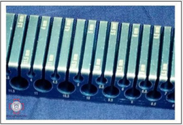
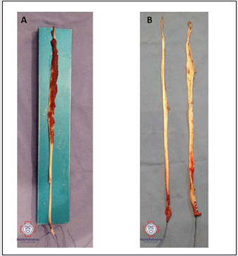
The tendons were prepared on a surgical table (Figure 3),
with removal of the muscle remnants, and then the thickness of
their quadruple shape was measured through a standardized
guide (Figure 4), whose holes have graduations every 05 mm. The
thickness was defined as the value following the measure in which
the quadruple graft was not able to cross longitudinally the hole of
the guide.
Figure 1: Steps for evaluating the maximum isometric extension (A) and knee flexion (B) forces, with the appropriate positioning of the dynamometer, rigid straps and examiner. The figures do not represent the angulation of the knee for performing the tests.
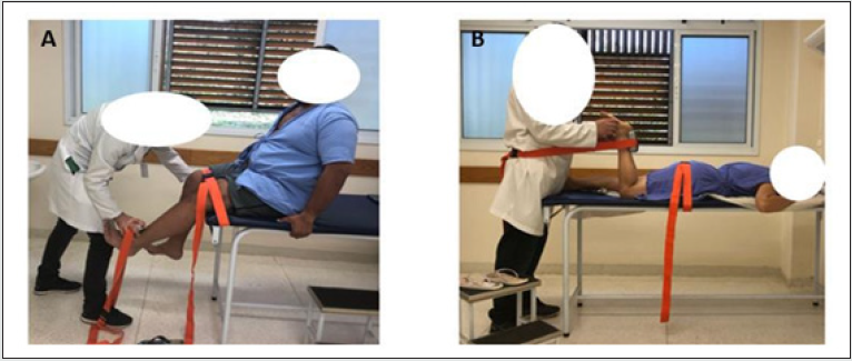
Figure 2: Intraoperative image during the removal of the knee flexor tendons, in the first stage of the anterior cruciate ligament reconstruction surgery.
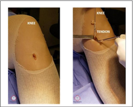
Figure 3: Intraoperative aspect of flexor tendons before (A) and after (B) removal of muscle remnants.
Statistical analysis
Data were processed in Excel® and SigmaStat®2.0 software
(GraphPad Software Jandel, SPSS, Chicago, IL, USA). The
Kolmogorov-Smirnov test was used to verify the normality of the
data distribution. Continuous variables with normal distribution
were presented as mean and standard deviation. The results were
organized in tables.
Student’s t-test was used to compare quantitative variables
regarding flexor and extensor torques according to gender.
Pearson’s correlation test was used to analyze the correlation of
the torques with the quantitative variables of the study, and the
multivariate analysis was performed using the linear regression
test with the ANOVA one-way test and the calculation of R2. The
linear regression test was also used for the prediction of a formula
for preoperative estimation of the diameter of flexor tendon grafts.
The level of statistical significance was set at 5% (p < 0.05).
Results
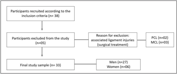
The study was attended by 38 amateur field soccer athletes.
The exclusion rate was 13% (Figure 5).
The study variables showed low variability coefficient, therefore
classified as homogeneous data. In the flexor torque analysis, the
male athletes presented means with statistically significant values
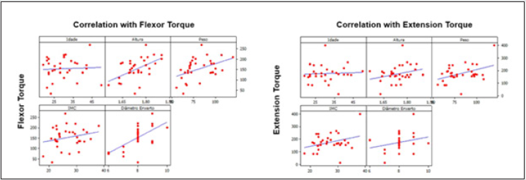
in relation to the female athletes (Table 2). Significant correlations
were observed between knee flexor torque and the height (p=0.002)
and weight (p=0.011) of the participants and also with the graft
diameter (p<0.001) (Table 3 and Figure 6).
Table 1: Characterization of the study sample, according to gender (M = male, F = female), age (years), dominant limb (D = right and E = left), height (m = meters), weight (Kg = kilograms), BMI (Body Mass Idex) and diameter of the graft (mm = millimeters).
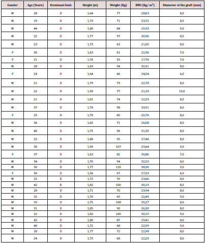
Table 3: Correlation of flexor and extensor torques with the quantitative variables of the study, according to the Pearson correlation test.

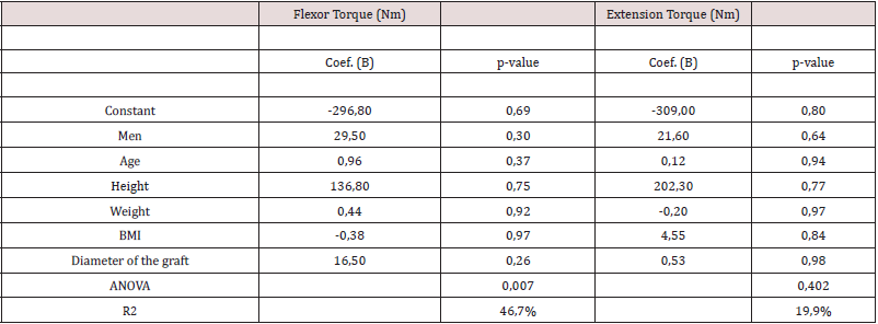
In the multivariate analysis, through the linear regression between the flexor and extension torques and the study variables, statistically significant results were observed for the flexor torque (p=0.007), but without statistical significance of the predictive variables (Table 4). The best statistical model to predict the diameter of the quadruple graft was from the flexor torque (Table 5), and with this the following equation was developed:
Graft Diameter= 6.57 + (0.01 x Flexor Torque)
Discussion
The main finding of this study was the positive and statistically
significant correlation between flexor tendon graft thickness and
isometric knee flexor torque in amateur soccer athletes. Through
this finding, this physical valence can be used as a parameter to
estimate the diameter of the hamstring’s tendons in knee ligament
reconstructions surgeries. In soccer players, regardless of their
performance level, the standard treatment for ACL injuries is
surgery. In this public, the choice of the most appropriate graft
remains controversial [23,24]. The existing consensus is that the
use of allografts should be avoided [25]. The use of hamstrings
tendons grafts, despite potentially causing knee flexion strength
deficit in specific tests [26,27] and also presenting biomechanical
disadvantages compared to bone/tendon grafts [28], is the
preferred graft for surgeons in practitioners of this sport [23,29].
Besides hamstrings tendons being one of the main autograft
options, in some situations they are also the priority of choice
[30,31], provided they present minimum diameters that guarantee
stability in the postoperative period. It is known that grafts in ACL
reconstruction surgery with diameters less than or equal to 8
mm are associated with higher rates of rerruptures [32]. For this
reason, different techniques have been developed to compensate
intraoperatively the unsatisfactory thickness of flexor tendons,
such as intrinsic adaptations to transform them into fivefold and
sixfold formats [33,34], solidarization with autografts or allografts
(hybrid graft) [35], and more recently, the preservation of muscle
remnants [36].
However, in some situations it is not possible to perform these
techniques, mainly when there is a scarcity of adequate instruments
or anatomic limitations of the graft itself. For reasons of this nature,
it is prudent that the knee surgeon has at hand, in a practical and reproducible way and prior to surgery, tools that provide a
guarantee of the average thickness of the flexor tendons. However,
up to now, there is no objective and commonly used method in
clinical practice to guide the surgeon in estimating the thickness
of the hamstring’s tendons preoperatively. Some imaging tests
have been studied with this objective [15,16]. Nuclear magnetic
resonance (NMR), considered the gold standard in diagnosing ACL
injury, can be used as an auxiliary measure in the preoperative
evaluation of tendon diameter. For this, one of the criteria with
good clinical accuracy is the measurement of tendon thickness at
its greatest diameter in the axial plane, with the medial epicondyle
of the femur as an anatomical reference [15]. Another possible
imaging method is ultrasonography (USG), which can be performed
based on anteroposterior and transverse diameters (measured
in mm) and cross-sectional area (mm²) [13,14]. However, unlike
MRI, USG is not a test of choice for the patient with knee ligament
injury and, in this case, would be performed only for the purpose of
assessing the thickness of the hamstring’s tendons. Moreover, the
results of USG with this purpose present conflicting results in the
literature [13,14] and, so far, it is not routinely used.
The relationship between epidemiological and anthropometric
parameters of easy practical execution, such as gender, age, weight,
height, lower limb length, body mass index, among others, and
hamstrings tendons thickness have also been studied [10,11,37,38].
However, among all these parameters, height is the only factor
that presents a significant association and reproducible results
[37,38], as observed in the current study, in which height presented
a positive and significant correlation with flexor torque. As it is
an easily evaluated parameter, even in a hospital environment, it
should be evaluated preoperatively as one of the ways to consider
the choice of graft in ACL reconstruction.
It is known that a physical training program with musculoskeletal
demands like soccer, based on aerobic and anaerobic overloads,
leads to gains not only in strength and muscle hypertrophy of the
segments worked, but also in hypertrophy of the tendinous unit, but
at a lower intensity [39-41]. This data from the literature justifies
the finding of a significant correlation between graft thickness and
knee flexor torque observed in the current study. Moreover, it is a
factor that corroborates the use of this biomechanical resource as
an adjuvant measure to predict the diameter of hamstrings tendons
in active populations.In this study, whose main objective was to
analyze the efficacy of a practical and additional resource to analyze
hamstrings tendons thickness in a specific population, we used the
isometric hand-held dynamometer, which is a tool well validated in
the literature for strength analysis [17,18,22] and has a much lower
cost than the isokinetic dynamometer.
The main precautions that must be taken when using this
portable instrument are the standardization of the test time, the
adequate positioning in the limb, the use of rigid belts, and joint
angulation. Of these, the biomechanical importance of defining
the lever arm, which is the measurement that will provide the
calculation of torque and whose size can change the peak force
produced, and the joint angulation, which should be optimized
according to the length x tension curve of the muscle grouping
analyzed, must be emphasized [19,20].
The main limitations of the study were its cross-sectional
design and gender bias, still common in studies with soccer
players [42,43]. Another limiting factor was the non-blinding of
the evaluators regarding the graft thickness because the procedure
was always performed and recorded by the surgeon and the same
evaluator. Moreover, the measurement of the graft thickness was
not performed continuously, but by intervals of 0.5 mm according
to the standardization of the specific guide. Further research with
this theme, in different populations, will be important for future
comparisons with the findings of the current study, as it is suggested
that soccer athletes have higher flexion and extension torques of
the knee in relation to the population in general.
Conclusion
Associated with the analysis of anthropometric characteristics, especially the patient’s height, knee flexor torque can be another tool for preoperative analysis of hamstrings tendons thickness during the surgical programming of ACL reconstruction.
References
- Mall NA, Chalmers PN, Moric M, Tanaka MJ, Cole BJ, et.al. (2014) Incidence and trends of anterior cruciate ligament reconstruction in the United States. Am J Sports Med 42(10): 2363-2370.
- Bram JT, Magee LC, Mehta NN, Patel NM, Ganley TJ, et.al. (2021) Anterior Cruciate Ligament Injury Incidence in Adolescent Athletes: A Systematic Review and Meta-analysis. The American Journal of Sports Medicine 49(7): 1962-1972.
- Beck NA, Lawrence JTR, Nordin JD, DeFor TA, Tompkins M et.al. (2017) ACL tears in school-aged children and adolescents over 20 years. Pediatrics 139(3): e20161877.
- Lynch TS, Parker RD, Patel RM, Andrish JT; MOON Group, Spindler KP, et.al. (2015) The Impact of the Multicenter Orthopaedic Outcomes Network (MOON) Research on Anterior Cruciate Ligament Reconstruction and Orthopaedic Practice. J Am Acad Orthop Surg 23(3): 154-163.
- Kaeding CC, Léger-St-Jean B, Magnussen RA (2017) Epidemiology and Diagnosis of Anterior Cruciate Ligament Injuries. Clin Sports Med 36(1): 1-8.
- Silvers-Granelli HJ, Bizzini M, Arundale A, Mandelbaum BR, Snyder-Mackler L, et.al. (2017) Does the FIFA 11+ Injury Prevention Program Reduce the Incidence of ACL Injury in Male Soccer Players? Clin Orthop Relat Res 475(10): 2447-2455.
- Diermeier T, Rothrauff BB, Engebretsen L, Lynch AD, Ayeni OR, (2020) Treatment after anterior cruciate ligament injury: Panther Symposium ACL Treatment Consensus Group. Knee Surg Sports Traumatol Arthrosc 28(8): 2390-2402.
- Magnussen RA, Lawrence JT, West RL, Toth AP, Taylor DC, et.al. (2012) Graft size and patient age are predictors of early revision after anterior cruciate ligament reconstruction with hamstring autograft. Arthroscopy 28(4): 526-531.
- Barrett GR, Luber K, Replogle W, Manley Jl (2010) Allograft anterior cruciate ligament reconstruction in the young, active patient: tegner activity level and failure rate. Arthroscopy 26(12): 1593-1601.
- Arnold MP, Calcei JG, Vogel N, Magnussen RA, Clatworthy M, et.al. (2021) ACL Study Group survey reveals the evolution of anterior cruciate ligament reconstruction graft choice over the past three decades. Knee Surg Sports Traumatol Arthrosc.
- Ho SWL, Tan TJL, Lee KT (2016) Role of anthropometric data in the prediction of 4-stranded hamstring graft size in anterior cruciate ligament reconstruction. Acta Orthop Belg 82: 72-77.
- Goyal S, Matias N, Pandey V, Acharya K (2016) Are pre-operative anthropometric parameters helpful in predicting length and thickness of quadrupled hamstring graft for ACL reconstruction in adults? A prospective study and literature review. Int Orthop 40(1): 173-181.
- Takenaga T, Yoshida M, Albers M, Nagai K, Nakamura T, et.al. (2019) Preoperative sonographic measurement can accurately predict quadrupled hamstring tendon graft diameter for AC reconstruction. Knee Surg Sports Traumatol Arthrosc 27(3): 797-804.
- Astur DC, Novaretti JV, Liggieri AC, Janovsky C, Nicolini AP, et.al. (2018) Ultrassonografia para avaliação do diâmetro dos tendões flexores do joelho: é possível predizer o tamanho do enxerto? Rev Bras Ortop 53(4): 404-409.
- Kupczik F, Tauscheck LOB, Schiavon MEG, Sbrissia B, Ernlund LSR, et.al. (2016) Predição do diâmetro do enxerto dos tendões flexores na reconstrução do ligamento cruzado anterior por meio da ressonância nuclear magné Rev Bras Ortop 51(4): 405-411.
- Camarda L, Grassedonio E, Albano D, Galia M, Midiri M, et.al. (2018) MRI evaluation to predict tendon size for knee ligament reconstruction. Muscles Ligaments Tendons J 7(3): 478-484.
- Whiteley R, Jacobsen P, Prior S, Skazalski C, Otten R, et.al. (2012) Correlation of isokinetic and novel hand-held dynamometry measures of knee flexion and extension strength testing. J Sci Med Sport 15(5): 444-450.
- Jackson SM, Cheng MS, Smith AR Jr, Kolber MJ (2017) Intrarater reliability of handheld dynamometry in measuring lower extremity isometric strength using a portable stabilization device. Musculoskelet Sci Pract 27: 137-141.
- Noorkoiv M, Nosaka K, Blazevich AJ (2014) Neuromuscular adaptations associated with knee joint angle-specific force change. Med Sci Sports Exerc 46(8): 1525-1537.
- Alegre LM, Ferri-Morales A, Rodriguez-Casares R, Aguado X (2014) Effects of isometric training on the knee extensor moment-angle relationship and vastus lateralis muscle architecture. Eur J Appl Physiol 114(11): 2437-2446.
- Jackson SM, Cheng MS, Smith AR Jr, Kolber MJ (2017) Intrarater reliability of handheld dynamometry in measuring lower extremity isometric strength using a portable stabilization device. Musculoskelet Sci Pract 27: 137-141.
- Mentiplay BF, Perraton LG, Bower KJ, Adair B, Pua YH, et.al. (2015) Assessment of Lower Limb Muscle Strength and Power Using Hand-Held and Fixed Dynamometry: A Reliability and Validity PloS One 10(10): e0140822.
- Martin-Alguacil JL, Arroyo-Morales M, Martín-Gomez JL, Monje-Cabrera IM, Abellán-Guillén JF, (2018) Strength recovery after anterior cruciate ligament reconstruction with quadriceps tendon versus hamstring tendon autografts in soccer players: A randomized controlled trial. Knee 25(4): 704-714.
- Britt E, Ouillette R, Edmonds E, Chambers H, Johnson K, et.al. (2020) The Challenges of Treating Female Soccer Players with ACL Injuries: Hamstring Versus Bone-Patellar Tendon-Bone Autograft. Orthop J Sports Med 8(11): 2325967120964884.
- Kaeding CC, Pedroza AD, Reinke EK, Huston LJ (2015) Risk factors and predictors of subsequent ACL injury in either knee after ACL reconstruction: prospective analysis of 2488 primary ACL reconstructions from the MOON cohort. Am J Sports Med 43(7): 1583-1590.
- Ardern CL, Webster KE, Taylor NF, Feller JÁ (2010) Hamstring strength recovery after hamstring tendon harvest for anterior cruciate ligament reconstruction: a comparison between graft types. Arthroscopy 26(4): 462- 469.
- Maletis GB, Inacio MC, Funahashi TT (2015) Risk factors associated with revision and contralateral anterior cruciate ligament reconstructions in the Kaiser Permanente ACLR registry. Am J Sports Med 43(3): 641- 647.
- Reinhardt KR, Hetsroni I, Marx RG (2010) Graft selection for anterior cruciate ligament reconstruction: a level I systematic review comparing failure rates and functional outcome. Orthop Clin N Am 41(2): 249- 262.
- Arliani GG, Pereira VL, Leão RG, Lara PS, Ejnisman B, et.al. (2019) Treatment of Anterior Cruciate Ligament Injuries in Professional Soccer Players by Orthopedic Surgeons. Rev Bras Ortop (Sao Paulo) 54(6): 703-708.
- Chen H, Liu H, Chen L (2020) Patellar Tendon Versus 4-Strand Semitendinosus and Gracilis Autografts for Anterior Cruciate Ligament Reconstruction: A Meta-analysis of Randomized Controlled Trials with Mid- to Long-Term Follow-Up. Arthroscopy 36(8): 2279-2291.e8
- Duchman KR, Lynch TS, Spindler KP (2017) Graft Selection in Anterior Cruciate Ligament Surgery: Who gets What and Why? Clin Sports Med 36(1): 25-33.
- Conte EJ, Hyatt AE, Gatt CJ Jr, Dhawan A (2014) Hamstring autograft size can be predicted and is a potential risk factor for anterior cruciate ligament reconstruction failure. Arthroscopy 30(7): 882-890.
- Nazari G, Barton KI, Bryant D, Getgood A, Brown CH Jr, et.al. (2020) Five- and six-strand hamstring grafts consistently produce appropriate graft diameters for anterior cruciate ligament reconstruction. Knee Surg Sports Traumatol Arthrosc.
- Brown CH Jr (2018) Editorial Commentary: How to Increase Hamstring Tendon Graft Size for Anterior Cruciate Ligament Reconstruction. Arthroscopy 34(9): 2641-2646.
- Kang H, Dong C, Wang F (2019) Small hamstring autograft is defined by a cut-off diameter of 7 mm and not recommended with allograft augmentation in single-bundle ACL reconstruction. Knee Surg Sports Traumatol Arthrosc 27(11): 3650-3659.
- Funchal LFZ, Ortiz R, Jimenez A, Funchal GDG, Cohen M, et.al. (2021) Remnant Muscle Preservation on Hamstring Tendon Autograft During ACL Reconstruction Promotes Volumetric Increase with Biological and Regenerative Potential. Orthopaedic Journal of Sports Medicine 9(3): 232596712199001.
- Pinheiro LF Jr, de Andrade MA, Teixeira LE, Bicalho LA, Lemos WG, et.al. (2011) Intra-operative four-stranded hamstring tendon graft diameter evaluation. Knee Surg Sports Traumatol Arthrosc 19(5): 811-815.
- Janssen RPA, Maria JF, Velden MJFV, Besselaar MV, Reijman M (2017) Prediction of length and diameter of hamstring tendon autografts for knee ligament surgery in Caucasians. Knee Surg Sports Traumatol Arthrosc 25(4): 1199-1204.
- Seynnes OR, Erskine RM, Maganaris CN, Longo S, Simoneau EM, et.al. (2009) Training-induced changes in structural and mechanical properties of the patellar tendon are related to muscle hypertrophy but not to strength gains. J Appl Physiol 107(2): 523-530.
- Magnusson SP, Hansen P, Kjaer M (2003) Tendon properties in relation to muscular activity and physical training. Scand J Med Sci Sports 13(4): 211-223.
- Reggiani C, Schiaffino S (2020) Muscle hypertrophy and muscle strength: dependent or independent variables? A provocative review. Eur J Transl Myol 30(3): 9311.
- Marom N, Dooley MS, Burger JA, Chang B, Coleman SH, et.al. (2020) Characteristics of Soccer Players Undergoing Primary Hip Arthroscopy for Femoroacetabular Impingement: A Sex- and Competitive Level–Specific Analysis. The American Journal of Sports Medicine 48(13): 3255-3264.
- Larruskain J, Lekue JA, Diaz N, Odriozola A, Gil SM (2018) A comparison of injuries in elite male and female football players: A five-season prospective study. Scand J Med Sci Sports 28(1): 237-245.

Top Editors
-

Mark E Smith
Bio chemistry
University of Texas Medical Branch, USA -

Lawrence A Presley
Department of Criminal Justice
Liberty University, USA -

Thomas W Miller
Department of Psychiatry
University of Kentucky, USA -

Gjumrakch Aliev
Department of Medicine
Gally International Biomedical Research & Consulting LLC, USA -

Christopher Bryant
Department of Urbanisation and Agricultural
Montreal university, USA -

Robert William Frare
Oral & Maxillofacial Pathology
New York University, USA -

Rudolph Modesto Navari
Gastroenterology and Hepatology
University of Alabama, UK -

Andrew Hague
Department of Medicine
Universities of Bradford, UK -

George Gregory Buttigieg
Maltese College of Obstetrics and Gynaecology, Europe -

Chen-Hsiung Yeh
Oncology
Circulogene Theranostics, England -
.png)
Emilio Bucio-Carrillo
Radiation Chemistry
National University of Mexico, USA -
.jpg)
Casey J Grenier
Analytical Chemistry
Wentworth Institute of Technology, USA -
Hany Atalah
Minimally Invasive Surgery
Mercer University school of Medicine, USA -

Abu-Hussein Muhamad
Pediatric Dentistry
University of Athens , Greece

The annual scholar awards from Lupine Publishers honor a selected number Read More...


