Lupine Publishers Group
Lupine Publishers
Menu
ISSN: 2638-6003
Research Article(ISSN: 2638-6003) 
Arthroscopic Fixation Of Tibial Spine Avulsion Fractures: Strangulation Technique Volume 4 - Issue 1
Neel M Bhavsar, Rameez A Musa, Sunil S Chodavadiya* , Trunal J Patel and Pankaj R Patel
- Department of Orthopaedics ,NHL Municipal Medical College, India
Received: March 29, 2020; Published: May 13, 2020
Corresponding author: Dr Sunil S Chodavadiya, Department of Orthopaedics , NHL Municipal Medical College, India
DOI: 10.32474/OSMOAJ.2020.04.000176
Abstract
Introduction: Tibial spine avulsion fracture is rare injury. Various methods have been described to treat these injuries. We are demonstrating the results of arthroscopic fixation of these fractures using strangulation technique.
Materials and Methods: 10 Patients with displaced tibial spine avulsion fractures without other associated ligament injuries were included in the study. Patients underwent arthroscopic fixation using strangulation technique. The postoperative results were analyzed using clinical tests, radiological evaluation and Lysholm score.
Observations and Results: We evaluated all 10 patients at final follow up. Radiographs showed that all fracture healed anatomically and all patients reported no symptoms of instability, such as giving – way episodes, clinical signs of anterior cruciate ligament deficiency were negative. Functional outcome assessed with help of Lhysolm score on each follow up. One patient had restricted range of motion in post op period.
Conclusion: Arthroscopic fixation of tibial spine avulsion fractures using strangulation technique is reliable technique, which gives excellent functional outcomes.
Keywords: Dr Sunil S Chodavadiya, Department of Orthopaedics , NHL Municipal Medical College, India
Introduction
Tibial spine fractures are relatively rare with an approximated
incidence of 3 per 100,000 per year.1The age group which is
more commonly involved in these fractures is 8-14 years of
children.2Because during these years of age strength of the
ligaments is more than the ossifying tibial eminence bone. Though
recently there has been increase in the incidence of these fractures
in adults3. Hayes et al found that 40% of tibial eminence fractures
reported in the literature occurred in adults4.
As tibial spine is the attachment site of ACL, insufficiency of the
ACL is associated with tibial spine avulsion fractures.5 In adults’
mode of injuries are usually high energy trauma like RTA, Sports
etc, so concomitant injuries to collateral ligaments and menisci
occurs more in adult age group5. The Meyers and Makeover
classification6, is the most commonly used classification of tibial
spine fractures. This injury produces disabilities in form of flexion
deformity, loss of extension and instability of the knee joint as
ACL is also involved, so it is important to fix this injury (especially
type 3 and 4).09,10,11It is also important to fix this injury with
prevention of native ACL because it has mechanoreceptors for
proprioception and neuromuscular control.12Various methods
have been described to fix this fracture which includes k-wire13,
cancellous screws14, Herbert screws14, staples16, stainless steel
wires17 , suture anchor18 , meniscal arrows19 , sutures20-24 or
combination25 of these methods and mini-open or arthroscopic
repair26 .
The purpose of this study is to evaluate the functional outcomes
of the arthroscopic repair of displaced tibial spine avulsion fractures
in adults using suture pull out technique using fibertape (Arthrex
,Napels). We hypothesized that arthroscopic fixation using suture
pull out technique in adults could give satisfactory functional
outcomes along with good knee stability and range of motion.
Materials and Methodology
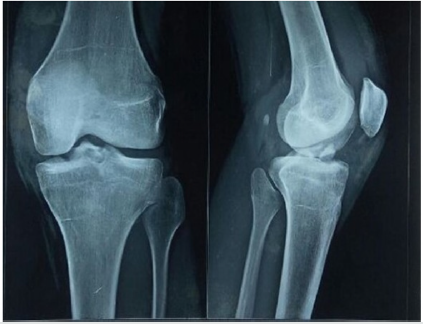
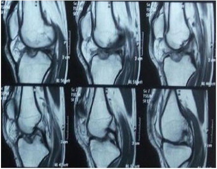
A study of 10 patients having displaced tibial spine avulsion fracture (Figure 1, 2) was done at our institute.
• Inclusion criteria were: age >18, patients willing to give
consent, closed injuries, type 2, 3 or 4 injury (according to
Meyer and Makeover classification)
• Exclusion criteria were: patient not willing to give consent,
open injuries, multiple ligament injuries
Preoperative work up included clinical examination, AP
and Lateral radiographs of Knee, MRI, and blood investigations.
Based on the radiological investigations fractures were classified
according to Meyer and Mckeever classification. Out of 10 patients
2 patients had type II, 1 patient had type IV and 7 patients had type
III injury. All patients were treated by arthroscopic suture pull-out
technique using suture tape.
Patient recovery was recorded post operatively which included
clinical examination, radiograph after 4 weeks and recording
functional outcome using Lysholm Knee score on each follow up.
Operative technique
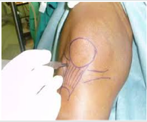
All patients were operated by same surgeon after 2 weeks of injury. Under spinal anesthesia patient kept in supine position with affected leg in 90 degree of flexion in leg holder and with tourniquet. Anteromedial and Anterolateral portals made (Figure 3) and diagnostic arthroscopy was done to rule out other associated injuries. Now avulsed fracture fragment identified and soft tissue around the avulsed fragment was debrided with help of shaver and soft tissue interposition along with transverse ligament was removed.
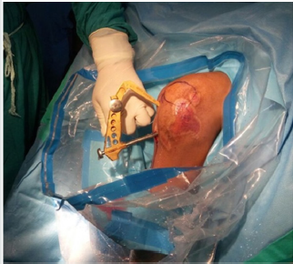
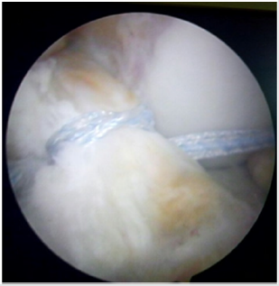
Reduction of the fragment is attempted with help of probe and site of the base of fracture fragment in the carter is confirmed. With help of ACL jig 2 parallel tunnels were made (Figure 4) from the anteromedial aspect of upper part of tibia exiting just at the level of tuberosity on either side of base of ACL attachment., Tunnels were made as close as possible to attachment of the ACL without damaging the structure. With help of suture lasso, PDS is passed around the base of ACL fragment and railroading of fiber tape was done. Base of the ACL fragment is strangulated (Figure 5) with fiber tape (Arthrex, Naples) in a crisscross manner with each end of the fiber tape passing through opposite tunnel. Reduction of the fragment was maintained with probe while tying the sutures.
Post Op Rehab
Patient was immobilized with long knee brace for 2 weeks and partial weight bearing was allowed. After 2 weeks gradual weight bearing walking and knee mobilization started along with passive knee range of motion exercises. After 4 weeks active knee range of motion exercises started with quadriceps stretching and strengthening exercises till next few months. After 6 months postoperatively patients were allowed to return for pre injury activity.
Post Op Evaluation
All patients were followed up monthly till 6 months and the yearly. On each monthly follow up clinical examination was done which included assessment of knee range of motion and laxity. Monthly AP and Lateral radiographs were taken to see the radiological union. Functional outcome assessment was done by Lysholm knee score on every monthly follow up.
Discussion
Tibial spine avulsion fractures are rare and occur mostly in age group 8 -14 years [1], though there is increase in the number of this injury in adults has been noted recently [2-4]. This injury is also associated with laxity of the ACL as it is the site of the attachment of the ACL [5]. Fixation of this type of fracture is necessary as they can be the cause of disabilities like loss of range of motion and stability of the knee joint [6-11]. And it is necessary to preserve the native ACL as it has mechanoreceptors for proprioception and neuromuscular control [12]. Various methods have been described to fix this fracture which includes k-wire [13], cancellous screws [14], Herbert screws [15], staples [16], stainless steel wires [17], suture anchor [18], meniscal arrows [19], sutures [20-24] or combination [25] of these methods and mini-open or arthroscopic repair [26]. Suture fixation is relatively better than screw fixation. As there is no need for second surgery for hardware removal, no impingement of the hardware in the notch. Suture fixation is better both, clinically and bio-mechanically [27-29]. the aim for fixation of the tibia spine avulsion fractures is to restore the knee stability and ACL competence. Studies have shown that laxity may present even after fixation in 10 % of patients [30]. In our study only 1 patient had post-operative complication in form of inability to full extension (arthrofibrosis), which was improved by regular high intensity physiotherapy. So out of 10 patients 9 patients had satisfactory functional outcomes after arthroscopic fixation by suture pull through technique. All 9 patients reached at the pre injury work level after 6 months of fixation. R Rajanish et al. [31] have done the similar method with the use of wire. But we have used tape as we believe that it causes less tissue compression due to larger surface area with more strength.
Outcomes and results
The study consisted 10 patients (9 male and 1 female). The average age of patients was 23.9 years (Ranging from 18 to 38 years). Mode of injury was sports in 4 patients and Road traffic accident was in 6 patients. Out of 10 patients, 1 patient had type IV, 2 patients had type II and 7 patients had type III injury according to Meyers and Makeover classification. Average time from injury to surgery was 13.6 days. Average radiological union time was 3.8 months. Average follow up time was 12.3 months (Range: 4 to 24) at final follow up functional outcome in form of average Lysholm knee score was 96.7 (Range: 88 to 100). No patient showed signs of ACL laxity at final follow up. All patients reached at pre-injury work level at around 6 months.
Conclusion
Arthroscopic suture pull-out fixation for tibial spine avulsion fracture results in excellent clinical and radiological outcomes without any significant complications. Use of tape leads to better compression and reduction of fracture fragment due to larger surface area.
References
- Hargrove R, Parsons S, Payne R (2004) Anterior tibial spine fracture - an easy fracture to miss. Accid Emerg Nurs 12(3): 173-175.
- Tolo VT (1998) Fractures and dislocations around the knee.In: Green NE, Swiontkowski MF, eds. Skeletal trauma in children. Philadelphia: W.B. Saunders 444-447.
- Ishibashi Y, Tsuda E, Sasaki T,et al. (2005) Magnetic resonance imaging aids in detecting concomitant injuries in patients with tibial spine fractures. Clin Orthop 434: 207-212.
- Hayes JM, Masear VR (1984) Avulsion fracture of the tibial eminence associated with severe medial ligamentous injury in an adolescent. A case report and literature review. Am J Sports Med 12(4): 330-333.
- McLennan JG (1982) The role of arthroscopic surgery in the treatment of fractures of the intercondylar eminence of the tibia. J Bone Joint Surg Br 64(4): 477-480.
- Meyers MH, McKeever FM (1959) Fracture of the intercondylar eminence of the tibia. J Bone Joint Surg Am 41(2): 209-222.
- Casalonga A, Bourelle S, Chalencon F, De Oliviera L, Gautheron V, et al. (2010) Tibial intercondylar eminence fractures in children: The long-term perspective. Orthop Traumatol Surg Res 96(5): 525-530.
- Zaricznyj B (1977) Avulsion fracture of the tibial eminence: Treatment by open reduction and pinning. J Bone Joint Surg Am 59(8): 1111-1114.
- Sullivan DJ, Dines DM, Hershon SJ, Rose HA (1989) Natural history of a type III fracture of the intercondylar eminence of the tibia in an adult. A case report. Am J Sports Med 17(1):132-133.
- Panni AS, Milano G, Tartarone M, Fabbriciani C (1998) Arthroscopic treatment of malunited and nonunited avulsion fractures of the anterior tibial spine. Arthroscopy 14(3): 233-240.
- Koukoulias NE, Germanou E, Lola D, Papavasiliou AV, Papastergiou SG, et al. (2012) Clinical outcome of arthroscopic suture fixation for tibial eminence fractures in adults. Arthroscopy 28(10): 1472-1480.
- Koukoulias NE, Germanou E, Lola D, Papavasiliou AV, Papastergiou SG et al. (2012) Clinical outcome of arthroscopic suture fixation for tibial eminence fractures in adults. Arthroscopy 28(10): 1472-1480.
- Furlan D, Pogorelić Z, Biocić M, Jurić I, Mestrović J, (2010) Pediatric tibial eminence fractures: arthroscopic treatment using K-wire. Scand J Surg 99(1): 38-44.
- Doral MN, Atay OA, Leblebicioğlu G, Tetik O (2001) Arthroscopic fixation of the fractures of the intercondylar eminence via transquadrici- pital tendinous portal. Knee Surg Sports Traumatol Arthrosc 9(6): 346-349.
- Wiegand N, Naumov I, Vámhidy L, Nöt LG (2014) Arthroscopic treatment of tibial spine fracture in children with a cannulated Herbert screw. Knee 21(2): 481-485.
- Kobayashi S, Terayama K (1994) Arthroscopic reduction and fixation of a completely displaced fracture of the intercondylar eminence of the tibia. Arthroscopy 10(2): 231-235.
- Oohashi Y (2001) A simple technique for arthroscopic suture fixation of displaced fracture of the intercondylar eminence of the tibia using folded surgical steels. Arthroscopy 17(9): 1007-1011.
- Vega JR, Irribarra LA, Baar AK, Iñiguez M, Salgado M, et al. (2008) Arthroscopic fixation of displaced tibial eminence fractures: a new growth plate-sparing method. Arthroscopy 24(11): 1239-1243.
- Wouters DB, de Graaf JS, Hemmer PH, Burgerhof JG, Kramer WL, et al. (2011) The arthroscopic treatment of displaced tibial spine fractures in children and adolescents using Meniscus Arrows®. Knee Surg Sports Traumatol Arthrosc 19(5): 736-739.
- Huang TW, Hsu KY, Cheng CY, Chen LH, Wang CJ, (2008) et al. Arthroscopic suture fixation of tibial eminence avulsion fractures. Arthroscopy 24(11): 1232-1238.
- Hunter RE, Willis JA (2004) Arthroscopic fixation of avulsion fractures of the tibial eminence: technique and outcome. Arthroscopy 20(2): 113-121.
- May JH, Levy BA, Guse D, Shah J, Stuart MJ, et al. (2011) ACL tibial spine avulsion: mid-term outcomes and rehabilitation. Orthope- dics 34(2): p. 89.
- Tudisco C, Giovarruscio R, Febo A, Savarese E, Bisicchia S, et al. (2010) Inter- condylar eminence avulsion fracture in children: long-term follow-up of 14 cases at the end of skeletal growth. J Pediatr Orthop B 19(5): 403-408.
- Wagih AM (2015) Arthroscopic treatment of avulsed tibial spine frac- tures using a transosseous sutures technique. Acta Orthop Belg 81(1):141-146.
- Gans I, Babatunde OM, Ganley TJ (2013) Hybrid fixation of tibial emi- nence fractures in skeletally immature patients. Arthrosc Tech 2(3): e237-e242.
- Accousti WK, Willis RB (2003) Tibial eminence fractures. Orthop Clin North Am 34(3): 365-375.
- Ahn JH, Yoo JC (2005) Clinical outcome of arthroscopic reduction and suture for displaced acute and chronic tibial spine fractures. Knee Surg Sports Traumatol Arthrosc 13(2): 116-121.
- Huang TW, Hsu KY, Cheng CY, Chen LH, Wang CJ, et al. (2008) Arthroscopic suture fixation of tibial eminence avulsion fractures. Arthroscopy 24(11): 1232-1238.
- Shook BJ, Frank HG, Freedman KB (2006) Arthroscopic suture fixation of tibial eminence fractures. Orthopedics 29(7): 577-581.
- Aderinto J, Walmsley P, Keating JF (2008) Fractures of the tibial spine: Epidemiology and outcome. Knee 15(3): 164-167.
- Rajanish R, Jaseel M, Murugan C, Kumaran C (2018) Arthroscopic tibial spine fracture fixation: Novel techniques. Journal of Orthopaedics 15(2): 372-374.

Top Editors
-

Mark E Smith
Bio chemistry
University of Texas Medical Branch, USA -

Lawrence A Presley
Department of Criminal Justice
Liberty University, USA -

Thomas W Miller
Department of Psychiatry
University of Kentucky, USA -

Gjumrakch Aliev
Department of Medicine
Gally International Biomedical Research & Consulting LLC, USA -

Christopher Bryant
Department of Urbanisation and Agricultural
Montreal university, USA -

Robert William Frare
Oral & Maxillofacial Pathology
New York University, USA -

Rudolph Modesto Navari
Gastroenterology and Hepatology
University of Alabama, UK -

Andrew Hague
Department of Medicine
Universities of Bradford, UK -

George Gregory Buttigieg
Maltese College of Obstetrics and Gynaecology, Europe -

Chen-Hsiung Yeh
Oncology
Circulogene Theranostics, England -
.png)
Emilio Bucio-Carrillo
Radiation Chemistry
National University of Mexico, USA -
.jpg)
Casey J Grenier
Analytical Chemistry
Wentworth Institute of Technology, USA -
Hany Atalah
Minimally Invasive Surgery
Mercer University school of Medicine, USA -

Abu-Hussein Muhamad
Pediatric Dentistry
University of Athens , Greece

The annual scholar awards from Lupine Publishers honor a selected number Read More...


