Lupine Publishers Group
Lupine Publishers
Case ReportOpen Access
Multiple Focal Choroidal Excavations in Association with Protein Rich Diet Volume 3 - Issue 1
Hakan Özdemir, Cansu Ekinci1*, Ahmet Elbay and Betul Tugcu2
- 1Department of Ophthalmology, University Faculty of Medicine, Turkey
- 2Bezmialem University Faculty of Medicine, Department of Ophthalmology, Turkey
Received:February 23, 2021; Published:March 8, 2021
Corresponding author: Cansu Ekinci, Bezmialem University Faculty of Medicine, Department of Ophthalmology, Adnan Menderes Bulv Vatan Cad 34093, İstanbul, Turkey
DOI: 10.32474/TOOAJ.2021.03.000158
Introduction
Choroidal excavation is a novel entity that is diagnosed with optical coherence tomography (OCT). In 1959, Klien, et al. [1] first described a concave-shaped chorioretinal abnormality with undifferentiated choriocapillaris and retinal pigment epithelium. Then in 2006, Jampol [2] defined similar changes with OCT and first used the term of focal choroidal excavation (FCE). In 2011, Margolis, et al. [3] described two FCE patterns, based on the shape of the lesions: nonconforming, if the photoreceptors are detached from retina pigment epithelium (RPE) and conforming, when the RPE follows the photoreceptor layer without any optically clear space between the outer retina and RPE. These two forms are also thought to convert to each other during the clinical process of the disease [4]. Although it is known to be mostly idiopathic, its relationship with central serous chorioretinopathy, choroidal neovascular membrane and inflammatory diseases such as multiple evanescent white dot syndrome and Vogt Koyanagi Harada disease has been reported [5]. In this report, we present a patient with multiple focal choroidal excavation following protein-rich diet.
Case
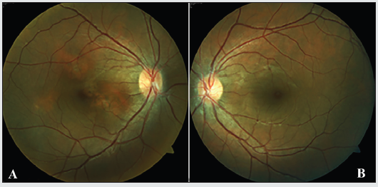
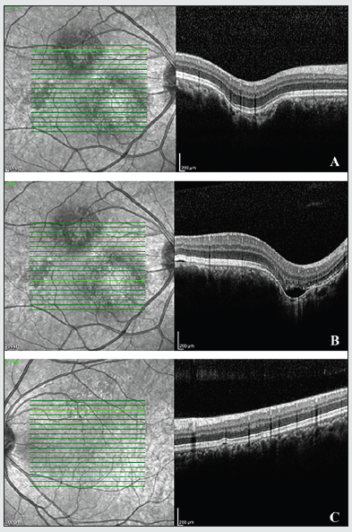
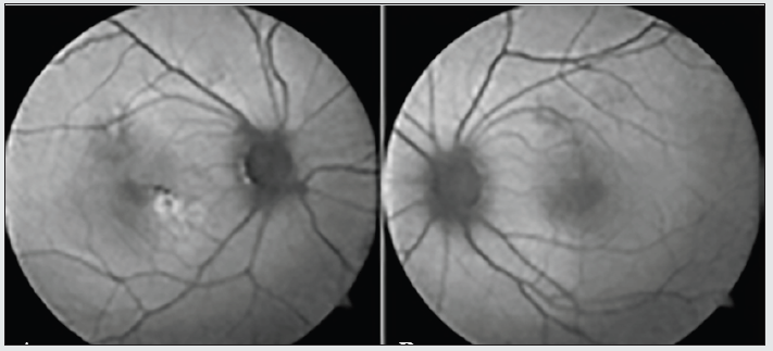
21-year-old male patient applied with blurred vision in his right eye since one week. He was on a protein rich diet since he was doing bodybuilding and had been using approximately 2g/kg/day protein powder since 3 months. His best corrected visual acuity was 0,6 in his right eye (RE) and 0,9 in his left eye (LE). There was no refractive disorder. Slit lamp examination and intraocular pressure were normal. Fundus examination revealed cavitary lesions in the macula of the RE (Figure 1a) and pigmentary changes above the macula of the LE (Figure 1b). OCT showed multiple conforming (Figure 2a) and non-conforming (Figure 2b) type of focal choroidal excavations with small retinal pigment epithelium detachments in the right eye and a small retinal pigment epithelium detachment in the left eye (Figure 2c). When evaluated with fundus autofluorescence (FAF), there have been hypo and hyperautofluorescent areas in the macula of the right eye in accordance with the pigmentary changes (Figure 3a) and hypoautofluorescent area above the macula of the left eye (Figure 3b). Fundus fluorescein angiography showed multiple hyperfluorescent window defects in the macula of the RE and hyperfluorecence consistent with window defect above macula of the LE. There was no ischemia, leakage, macular edema and all vascular structures were normal . Protein powder stopped and the patient was examined 6 months after the diagnosis. No changes in the findings were detected.
Figure 1: A and B reveals the color fundus photograph. In the right eye yellowish lesions and pigmentary changes around fovea exist.

Figure 2: Figure 2: Optical coherence tomography scans of the patient. In figure 2A, a conforming focal choroidal excavation and in figure 2B a non-conforming focal choroidal excavation are seen in the right eye. In figure 2C, a small retinal pigment epithelial detachment exists in the left eye..

Figure 3: Fundus autofluorescence (FAF) images of the patient. In figure 3A, in the right eye, hypo and hyper autofluorescent areas in macula, in figure 3B, in the left eye, hypoautofluorescent area above the macula were seen..

Discussion
Choroidal excavation is an idiopatic local pit or cupping of the choroid which is usually located near fovea. It has mostly no association with any accompanying systemic disease and condition, but concurrent choroidal disorders such as central serous chorioretinopathy, choroidal neovascular membrane and polypoidal choroidal vasculopathy are reported [2-6]. In our case, since the clinical complaints of the patient occured following protein rich diet, choroidal excavation and protein-rich diet are thought to have a causative relationship. Although it is mostly believed not to be associated with any systemic disease, ocular symptoms and findings of our patient occured following a systemic condition such as protein rich diet. Kheir, et al. [7] presented two cases of bilateral multiple serous pigment epithelial detachment following acute major weight loss and protein rich diet. They mentioned the interesting similarity between the Bruch’s membrane (BM) and glomerular basement membrane (GBM) suggesting that the protein rich diet may have effects on retina and choroid. In many individuals with membranoproliferative glomerulonephritis type 2, similar electron dens deposits as in the GBM were found in choriocapillaris-BM-retinal pigment epithelial interface [8]. According to this resemblance, protein-rich diet is suggested to be harmful for the BM. Damaged BM can not remain its barrier function for biomolecules and may lead to choroidal excavation. Additionally Coppo, et al. showed increased proteinuria in 14 patients having protein rich diet (1.6 g/kg/d) for 4 weeks. Our patient had been on a protein rich diet since 3 months and he had taken a fairly high amount of protein which could presumably affect BM and choroid [9] In our case, although there were multiple excavations in the right eye of the patient; RPE detachment and loss of visual acuity in the left eye, which might result in new excavations, suggest bilateral asymmetric involvement.
Conclusion
In this case report, since the pathogenesis of FCE is not well-known, we comment that the protein rich diet can damage Bruch’s membrane and may cause choroidal disorders such as excavations. Further examinations with longer term follow-ups and larger patient- groups are required to understand the precise pathogenesis.
Acknowledgement
The study was designed by HÖ and CE; data were collected and analyzed by CE and AE; data interpretation and manuscript preparation were undertaken by CE, AE, HÖ and BT. All authors approved the final version of the paper.
Conflict of Interest
No grant support or funding.
No conflict of interest.
References
- Klien BA (1959) The pathogenesis of some atypical colobomas of the choroid. Am J Ophthalmol 48: 597-607.
- Jampol LM, Shankle J, Schroeder R, Tornambe P, Spaide RF, et al. (2006) Diagnostic and therapeutic challenges. Retina 26(9): 1072-1076.
- Margolis R, Mukkalamala SK, Jampol LM, Spaide RF, Ober MD, et al. (2011) The expanded spectrum of focal choroidal excavation. Arch Ophthalmol 129(10): 1320-1325.
- Wakabayashi Y, Nishimura A, Higashide T, Ijiri S, Sugiyama K (2010) Unilateral choroidal excavation in the macula detected by optical coherence tomography: case report. Acta Ophthalmol 88(3): 87-91.
- Roy R, Saurabh K, Vyas C, Sharma P, Bansal A (2017) From fruste focal choroidal excavation in a case of serpiginous choroiditis. Clin Exp Ophthalmol 101(2): 302-304.
- Luk FO, Fok AC, Lee A, Liu AT, Lai TY (2015) Focal choroidal excavation in patients with central serous chorioretinopathy. Eye (Lond) 29(4): 453-459.
- Kheir V, Ambresin A, Mantel I (2017) Multiple serous pigment epithelial detachments in associated with major weight loss: case report and review of the literature. Ret Cases Brief Rep pp. 1-5.
- Appel GB, Cook HT, Hageman G (2005) Membranoproliferative glomerulonephritis type 2 (dense deposit disease: an update. J Am Soc Nephrol 16(5): 1392-1403.
- Coppo R, Amore A, Roccatello D (1988) Microalbuminuria in single kidney patients: Relationship with protein intake. Clin Nephrol 29(5): 219-228.


