Lupine Publishers Group
Lupine Publishers
Case ReportOpen Access
Micromechanical Properties of Individual Intraocular Lenses (IOL) Volume 3 - Issue 2
Felix Klein, Erika Koch and Norbert Hampp*
- University of Marburg, Department of Chemistry, Germany
Received:March 22, 2021; Published:April 9, 2021
Corresponding author: Norbert Hampp, University of Marburg, Department of Chemistry, Hans-Meerwein-Str. 4, 35032 Marburg, Germany
DOI: 10.32474/TOOAJ.2021.03.000160
Abstract
Mechanical properties can now be analyzed on single intraocular lenses (IOLs) after all machining steps ready for implantation employing micromechanical analytics. This is important because IOLs are implanted through a so-called shooter. Folding into the loader chamber and squeezing the IOL through the about 1 mm wide nozzle into the eye’s capsular bag means a lot of mechanical stress to the polymer. We compare hydrophilic and hydrophobic materials in terms of unfolding speed and time to complete unfolding and found significant differences. Hydrophilic materials seem to be better suited for implantation than hydrophobic ones. A 2D mapping of IOL shows that the mechanical properties are pretty constant over the optics’ full area. This is somewhat astonishing because the thicker middle part and the thinner outer rim part experienced quite different mechanical treatment. Micromechanical analytics are a great advance over the prior testing on larger material pieces which did not go through the lathing process.
Introduction & Results
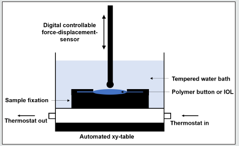
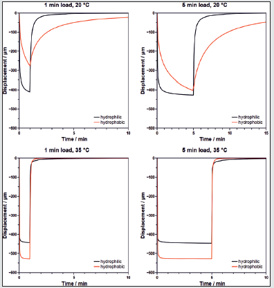
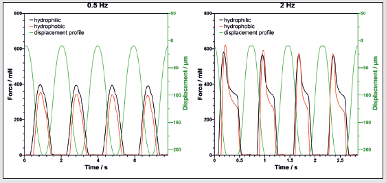
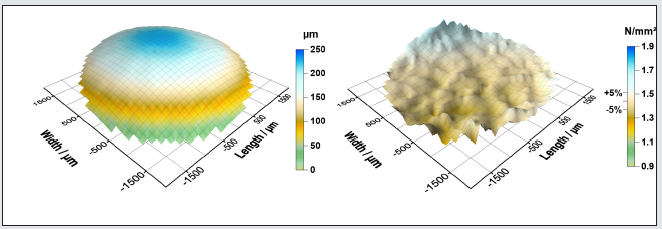
Cataract is an age-dependent disease where the natural eye lens becomes more and more cloudy [1]. In a short, often ambulant surgery, the natural lens is removed and replaced by an artificial polymeric intraocular lens (IOL) [2]. IOLs are the most used human implants. The IOL is applied by a syringe-like shooter through a small incision into the cornea and the capsular bag [3]. This requires that the IOL is tightly folded into the loader chamber of the shooter and then is injected into the capsular bag through an about 2 mm diameter nozzle (Figure 1) [4]. This means high mechanical stress to the IOL polymer. The mechanical properties of polymers for IOL manufacturing, e.g., tensile strength or dynamic mechanical analyses (DMA), are commonly tested on macroscopic samples with standardized test equipment [5,6]. We have developed a process to measure the micromechanical properties of IOL materials on buttons, i.e. small polymer discs where the IOLs are made from, or ready-made IOLs after all fabrication steps. For this process, a variable tip is attached at a displacement-force-sensor. We used a ‘LNP® nano touch’ (Ludwig Nano Präzision, Nordheim, Germany) with an absolute displacement range of ± 2 mm and a maximum test force of 1400 mN (Figure 2). The ‘LNP® nano touch’ is a socalled micro-indentor system optimized for the analysis of elastomers. It supports the mechanical testing following the norms for Shore AM, IRHD-M, and VLRH. Adjustable force and displacementdriven measurements, as well as dynamical mechanical analysis (DMA) are possible, too. The shooters are loaded at the beginning of the surgery and typically rest several minutes before use. During this time, the IOL stays folded in the shooter chamber. A pretty high force is applied. We used 925 mN in our tests, then left the polymers for 1 to 5 min before the force was released. The loading of the shooters is done at room temperature. During the unfolding inside the capsular bag, the temperature of the IOL raises to 35 °C, the temperature of the eye. This test comprises stiffness of the material and unfolding speed after high force loads. It is easy to see that there is significantly different behavior between hydrophobic and hydrophilic polymers. Hydrophobic polymers, here HRI-5814 (homemade), contain only very low water amounts, but hydrophilic materials, here H26R1 (Actiol, Germany), contain 26% water, making their behavior more complex [7,8]. Whereas hydrophobic polymers are highly elastic, hydrophilic ones behave more like sponges [9]. At higher loads, not only spring-like deformations are caused, but water is squeezed between different areas in the IOL. The longer the high load is applied to the polymer, the longer this water redistribution takes. The simulation of a high load is shown in Figure 3. Table 1 summarizes the time to 99 percent unfolding calculated from the displacement curves. The hydrophilic material shows a moderate temperature dependence. The unfolding time (> 99%) is in the range of 2-3 min at 35 °C. Final IOL positioning and unfolding fit well the surgery timescale. The hydrophobic material in contrast shows a strong temperature dependence. At room temperature, it unfolds very slowly, but at 35 °C quite a bit too fast and this is not optimal because this might cause damage to the capsular bag. The determination of elastic and viscoelastic properties of polymers is done by dynamical mechanical analysis (DMA) (Figure 4). At low frequency, the compression is proportional to the displacement, which means it is elastic. Retraction of the indenter and reduction of force is elastic only in the very first part. When the shoulder in the curve appears, we observe a viscoelastic expansion of the polymer. The shoulder means that the indenter is no longer in contact with the polymer because the retraction speed is higher than the expansion of the polymer. At higher frequency, this effect becomes much more striking. The hydrophilic materials show a distinct change from elastic to viscoelastic behavior. The speed of redistribution of water in the sponge-like polymer is responsible for this. The hydrophobic material shows a rather similar behavior, despite it is expected that it should have an almost fully elastic expansion. The combination of the LNP® nano touch sensor with a computer- controlled xy-stage allows to measure the elasticity (and some more parameters) with high resolution over the whole IOL surface. In our example, it is a 29 x 29 grid (Figure 5).
Figure 1: State-of-the-art shooter used for the implantation of intraocular lenses. The lens is folded in the loader chamber (left) and then is pushed by the blue piston through the nozzle into the capsular bag (middle), where it unfolds (right).

Figure 2: Experimental setup. The digital controllable force-displacement sensor can operate on submerged thermostated samples. The fixation of the samples is critical not to distort the measurements.

Figure 3: Comparison of a simulated high load (935 mN) on a hydrophilic and hydrophobic polymer button for 1 min and 5 min force loading. An r = 1.35 mm ruby indentor has been used.

Figure 4: Comparison of elastic and viscoelastic behavior at low (0.5 Hz) and high (2 Hz) dynamic frequencies at 35 °C. Displacement was between -10 μm and -210 μm referred to the surface of the sample (green line).

Figure 5: Topography (left) of the optics of the measured hydrophilic intraocular lens with the associated micromechanical map of the complex E module (right) measured at 35 °C. The median value is 1.44 ± 0.08 N/mm2. On average, the elasticity over the whole optic varies about 5%. For the 2D mapping of the IOL, an r = 0.2 mm steel indentor with a displacement-controlled program between -10 μm and -60 μm has been used.

Summary
Micromechanical measurements are a valuable tool to analyze ready-made IOLs. The lathing process, as well as the steam sterilization, mean considerable mechanical stress to the polymer. Hydrophilic polymers are not affected by this and have the same elasticity over the whole optical area despite the rather different thickness. Also, the unfolding time of the hydrophilic materials match more the desired time-scale. The unfolding times at room temperature, as well as at 35 °C, are in the minute range. The hydrophobic materials at room temperature are too slow and at 35 °C too fast.
References
- Bloemendal H, De Jong W, Jaenicke R, Lubsen N H, Slingsby C, et al. (2004) Ageing and vision: Structure, stability and function of lens crystallins. Prog Biophys Mol Biol 86(3): 407-485.
- Allen D, Vasavada A (2006) Cataract and surgery for cataract. BMJ 333(7559): 128-132.
- Linebarger EJ, Hardten DR, Shah GK, Lindstrom RL (1999) Phacoemulsification and modern cataract surgery. Surv Ophthalmol 44(2): 123-147.
- Allen D, Habib M, Steel D (2012) Final incision size after implantation of a hydrophobic acrylic aspheric intraocular lens: New motorized injector versus standard manual injector. J Cataract Refract Surg 38(2): 249-255.
- Swallowe GM (1999) Mechanical Properties and Testing of Polymers. Dordrecht: Springer Netherlands.
- Menard KP, Menard N (2017) Dynamic Mechanical Analysis. In: Encyclopedia of Analytical Chemistry. Chichester, UK: John Wiley & Sons Ltd 2017: 1-25.
- Werner L (2008) Biocompatibility of intraocular lens materials. Curr Opin Ophthalmol 19(1): 41-49.
- Bellucci R (2013) An Introduction to Intraocular Lenses: Material, Optics, Haptics, Design and Aberration. In: Cataract. Basel: Karger 3: 38-55.
- Clayton AB, Chirila TV, Lou X (1997) Hydrophilic sponges based on 2-hydroxyethyl methacrylate. V. Effect of crosslinking agent reactivity on mechanical properties. Polym Int 44(2): 201-207.


