Lupine Publishers Group
Lupine Publishers
Menu
ISSN: 2644-1209
Research Article(ISSN: 2644-1209) 
Automation in Vision Testing: Glaucoma Detection Software Volume 1 - Issue 4
Shilpa* and Gargi Khanna
- National institute of information technology, E&CED, Hamirpur, India
Received: May 26, 2018; Published: June 07, 2018
*Corresponding author: Shilpa, National institute of information technology, E&CED, Hamirpur, India
DOI: 10.32474/TOOAJ.2018.01.000117
Abstract
Vision testing is the phenomenon of detecting the vision problems which are generally undertaken to improve prognosis and disabilities. Apart from visual acuity measurement and refractive error problems, other eye related retinal disorders include diabetic retinopathy, age related muscular degeneration and glaucoma. Retinal imaging plays an important role in the detection of these disorders. Early detection of these disorders can prevent the permanent vision loss in patients. The aim of this research work is to implement a software algorithmic program in the field of ophthalmology for the detection of glaucoma. In the present work, Glaucoma detection is done by cup to disc ratio measurement to locate the damaged optic nerve head. DRIVE database is used to acquire retinal images. Further codes are implemented using MATLAB software to get the results. This software system can be utilized everywhere, especially in school health programs in Himachal Pradesh in urban as well as rural areas where it is difficult to carry high maintenance ophthalmic machines.
Keywords: Glaucoma; Retinal image; MATLAB
Introduction
Glaucoma has been nicknamed the “sneak thief of sight” because it mostly goes undetected and causes irreversible damage to the eye. Glaucoma is the term for a diverse group of eye diseases. It is a serious ocular, chronic, irreversible neurodegenerative disease. It is one of the common causes of blindness with about 79 million in the world likely to be afflicted with glaucoma by the year 2020. It is a significant cause of blindness in the world [1]. The incidence of glaucoma increases with age but the disease is more prevalent among individuals with a family history of glaucoma [2]. It can occur at any age and can lead to permanent vision loss and blindness. Glaucoma is a referred as a group of eye disorders that leads to optic nerve damage which is mostly associated with an elevated fluid pressure in the eye i.e. Intra-Ocular Pressure (IOP) [3]. Glaucoma is further classified as:
a) Open-angle glaucoma
b) Closed-angle glaucoma
c) normal-pressure
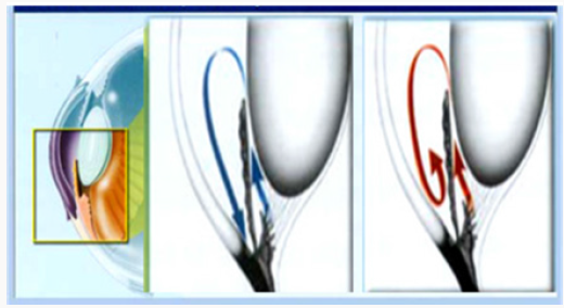
Glaucoma can only be treated if we know the elevations and fluctuations happening in aqueous flow. So, it is necessary to measure the intraocular pressure for accurate treatment of Glaucoma [4-8]. IOP is the pressure exerted by the ocular fluid called “aqueous humor” that fills the anterior chamber of the eye between the lens and cornea. Aqueous humor is produced by the eye to bathe and nourish its different parts. The aqueous humor normally flows out of the eye through various paths and chambers. When these paths get clogged, aqueous humor gets trapped in the eye as shown in Figure 1. This causes a pressure buildup and leads to high IOP. Normal IOP is in the range of 10-21 mmHg [4,5]. When pressure in the eye gets too high, the optic nerve which sends visual signals to the brain can get damaged. This in turn will not allow some signals from the eye to reach the brain. This leads to a reduction in vision and if not managed, may cause loss of peripheral vision and leads to blindness, which is not reversible unfortunately. In most glaucoma patients, the IOP increases above the normal range (>21mmHg) up to 60mmHg, when the aqueous outflow is lower than the inflow. High IOP is a major risk factor for glaucoma, but it can be treated if diagnosed early as well as it requires constant monitoring through medical scomapecialist, hence there is an urgent need to device some portable and easy method to diagnose Glaucoma. The Intra- Ocular Pressure (IOP) in the eye is measured by a instrument called as tonometer [3,4] Calculation of cup to disc ratio is one of the methods in assessment of damaged optic nerve head but it is quiet expensive method . With the introduction software algorithms, simple fundus image can be used for the detection of Cup-to-Disc ratio [6]. The proposed method provides the easy measurement of intraocular pressure for the treatment of Glaucoma [9].
Methodology
This research work is aimed at developing a smart solution to enable early detection of glaucoma using MATLAB R2017b software tool. Accurate and easy diagnosis of glaucoma through analysis of the neuro-retinal optic disc and cup is one of the crucial works. The proposed algorithm is designed to work in MATLAB Software. First step is to get the good quality Fundus images. For the present work the fundus images are taken from DRIVE [7] database which is freely available and provide number of fundus images captured using Cannon CRS non-mydriatic camera.
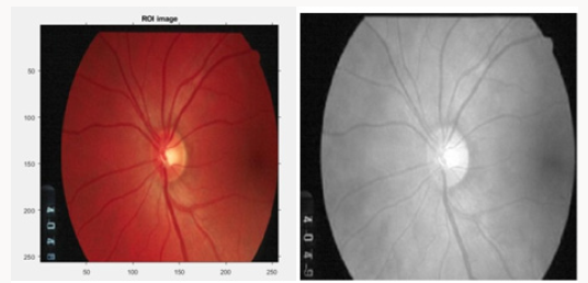
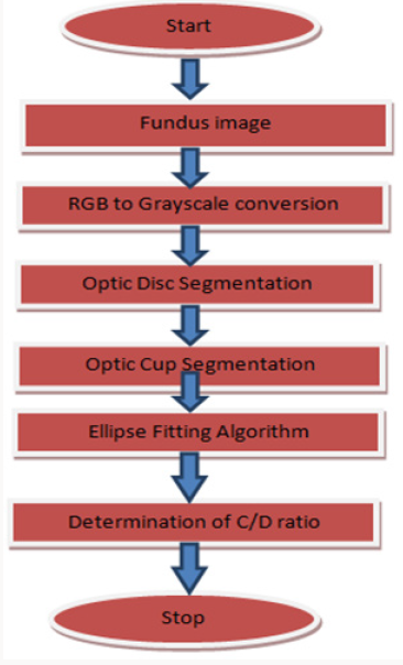
Step 1: Using MATLAB software, input fundus image is first resized according to the parameters then it is converted into gray scale image from RGB as shown in the Figure 2 next step includes optic disc and optic cup segmentation as described in the flow chart Figure 3.
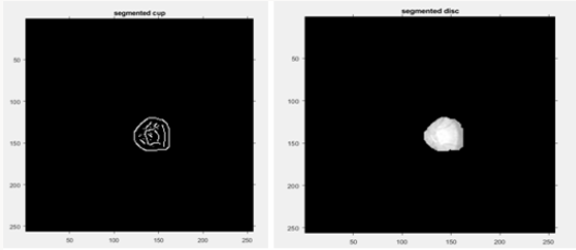
Step2: Disc segmentation-The retinal image is processed to detect the disc boundary using edge detection algorithm for image segmentation using canny as shown in Figure 4.
Figure 3: The patient’s second-month control, OCT image shows that the fluid is regressing after treatment.
Step3: Cup segmentation-For detecting cup boundary again color and edge analysis technique is applied (Threshold initialization based level set according to the user) as shown in Figure 4.
Step 4: After the cup boundary and Disc segmentations CDR has been detected, ellipse fitting algorithms which are already defined in MATLAB were employed to eliminate some of the cup boundary changes in curvature using convex hull algorithm.,/
The CDR is consequentially obtained based on the height of detected cup and disc. According to the CDR calculations it gives the final result. A CDR value that is greater than 0.56 indicates glaucoma is present in the patient and CDR value less than 0.5 is detected as no glaucoma.
Results
In this work the appearance characteristics of the Optic Nerve Head (ONH), which includes cupping or excavation of the optic disc, an enlargement of the optic cup-to-disc ratio (CDR) are identified. A CDR value that is greater than 0.56 indicates glaucoma is present in the patient and CDR value less than 0.5 is detected as no glaucoma. The algorithm designed is able to work on all glaucoma fundus images.
Summary and Conclusion
With this system, technology is available in a quite handy form; one can keep track of the glaucoma anywhere anytime. It can be used by anyone as a replacement in the conditions when patient is not able to visit the doctor frequently. In future this system can be advanced through some other techniques so that it can found its applicability in retinal imaging by smart phone itself so that this algorithm can work as a mobile application where patient can click the image and analyze it using this software Smartphone application and integration of imaging system can make a revolutionary changes in the sector of ophthalmology as the retinal imaging system are very costly and cannot be found easily in all eye care centers. It is reliable and simple to handle. Hence the Proper utilization and validation of smartphone application in the ophthalmology can lead the advancement in the health care sector and that will be beneficial for each section of society.
References
- A Mariotti, D Pascolini (2012) Global estimates of visual impairment. Br J Ophthalmol 96 (5): 614-618.
- World Glaucoma Association.
- Glaucoma Society of India.
- E Chihara (2008) “Assessment of True Intraocular Pressure: The Gap between Theory and Practical Data”. Elsevier Survey on Opthamology 53(3): 203-218.
- Aqueous Fluid Pathway.
- Cheng J, Liu J, Wong DWK, Yin F, Cheung C, et al. (2011) Automatic optic disc segmentation with peripapillary atrophy elimination. Int Conf IEEE Eng Med Biol Soc 2011: 6224-6227.
- DRIVE: Digital Retinal Images for Vessel Extraction.
- Kwon YH, Fingert JH, Kuehn MH (2009) Primary open-angle glaucoma. N Engl J Med 360(11): 1113-1124.
- Shilpa, Gargi Khanna (2017) Techniques and Automation in Eye Care and Vision Testing Using Smartphone. International Journal of Ophthalmology & Visual Science 2(4): 88-92.

Top Editors
-

Mark E Smith
Bio chemistry
University of Texas Medical Branch, USA -

Lawrence A Presley
Department of Criminal Justice
Liberty University, USA -

Thomas W Miller
Department of Psychiatry
University of Kentucky, USA -

Gjumrakch Aliev
Department of Medicine
Gally International Biomedical Research & Consulting LLC, USA -

Christopher Bryant
Department of Urbanisation and Agricultural
Montreal university, USA -

Robert William Frare
Oral & Maxillofacial Pathology
New York University, USA -

Rudolph Modesto Navari
Gastroenterology and Hepatology
University of Alabama, UK -

Andrew Hague
Department of Medicine
Universities of Bradford, UK -

George Gregory Buttigieg
Maltese College of Obstetrics and Gynaecology, Europe -

Chen-Hsiung Yeh
Oncology
Circulogene Theranostics, England -
.png)
Emilio Bucio-Carrillo
Radiation Chemistry
National University of Mexico, USA -
.jpg)
Casey J Grenier
Analytical Chemistry
Wentworth Institute of Technology, USA -
Hany Atalah
Minimally Invasive Surgery
Mercer University school of Medicine, USA -

Abu-Hussein Muhamad
Pediatric Dentistry
University of Athens , Greece

The annual scholar awards from Lupine Publishers honor a selected number Read More...


