Lupine Publishers Group
Lupine Publishers
Menu
ISSN: 2641-1725
Case Reporte(ISSN: 2641-1725) 
Epiplolic Apendagitis: A Diagnosis In Disuse Volume 5 - Issue 1
Pedro Nogarotto Cembranel*
- Medical Sciences Course, Health Sciences School, Faculdade Ceres (FACERES), Brazila
Received: March 05, 2020; Published: March 12, 2020
*Corresponding author: Pedro Nogarotto Cembranel, Medical Sciences Course, Health Sciences School, Faculdade Ceres (FACERES), São José do Rio Preto, SP, Brazil
DOI: 10.32474/LOJMS.2020.05.000204
Abstract
Introduction: Epiploic appendagitis (EA) is an unusual, benign and self-limited clinical condition. The diagnosis is made by abdominal computed tomography (CT) and the treatment is conservative. The wrong diagnosis can lead to hospitalizations, antibiotics and unnecessary surgical intervention.
Case report: Female patient, 38 years old, with abdominal pain in the left iliac fossa for 4 days. Laboratory exams with no abnormalities, and abdominal tomography (CT) showed smearing of the anti-messenteric border and thickening of the adjacent fascia. The diagnostic hypothesis of EA was made, analgesic and anti-inflammatory prescribed for home treatment, with complete remission of the condition in 7 days.
Discussion and conclusion of the case: EA is a rare entity with low incidence, however it should be considered as a diagnostic hypothesis when it comes to acute abdomen in the emergency. The diagnosis of early EA aims to avoid the use of medications and unnecessary surgical intervention.
Introduction
The omental appendages are projections of the outer surface of the colon, filled with fat, covered with serosa and projecting into the peritoneal cavity. Epiploic appendagitis (EA) is an unusual, benign and self-limiting clinical condition [1]. It results from the spontaneous venous torsion or thrombosis of the veins that drain the epploic appendages [2]. It manifests as acute abdominal pain. The diagnosis is made by computed tomography (CT) of the abdomen and the treatment is conservative. The wrong diagnosis can lead to hospitalizations, antibiotics and unnecessary surgical intervention [3].
Case Report
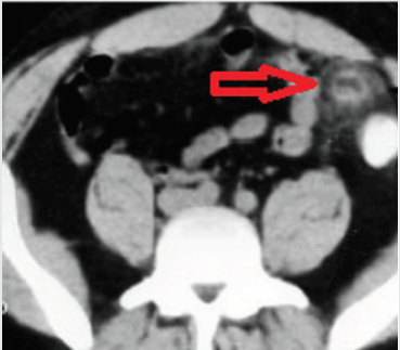
A 38-year-old female patient arrives at the emergency department complaining of continuous colic abdominal pain associated with vomiting and diarrhea for 4 days. She denies fever and urinary disorders. Upon examination, the abdomen was painful on palpation of the lower floor, especially in the left iliac fossa, with reduced hydro-air noises. Laboratory tests including blood count and urine tests were normal. Abdominal CT showed smearing of the anti-messenteric border and thickening of the adjacent fascia (Figure 1), with a diagnostic hypothesis of EA.
Prescribed analgesics and anti-inflammatory drugs for outpatient treatment with favorable evolution, with total remission of symptoms in 7 days.
Discussion
Approximately 50 to 100 epiploic resources are present
throughout the colon, with predominance in the transverse and
sigmoid colon, ranging from 0.5 to 5 cm [1]. EA is a benign clinical
condition, which occurs secondarily in spontaneous venous torsion
or thrombosis of the veins that drain the epploid appendages [1-2].
The usual clinic is for acute abdominal pain located in the
lower left quadrant, which may mimic acute abdomen, leading to
an incorrect diagnosis of appendicitis or acute diverticulitis. There
may be an increase in leukocytes in the blood and an increase in the
erythrocyte sedimentation rate, without urinary changes [4].
The diagnosis is made through abdominal CT, with a finding of
paracolic, oval mass, from 1 to 5 cm, with fat density, accompanied
by thickening of the peritoneal lining and attenuation of
periapendicular fat [5-6].
Treatment is conservative, on an outpatient basis and dispenses
with the use of antibiotics or surgical treatment. It consists of the
administration of analgesics and anti-inflammatory drugs, with
complete improvement of symptoms [7-8].
Conclusion
EA is a rare entity with low incidence, but it should be considered as a diagnostic hypothesis when it comes to acute abdomen in the emergency. The diagnosis of early EA aims to avoid the use of medications and unnecessary surgical intervention.
References
- Melo AS, Moreira LB, Pinheiro RA, Noro F, Alves JR, et al. (2002) Apendicite Epiplóica: Aspectos na Ultra-Sonografia e na Tomografia Computadorizada. Radiol Bras 35(3):171-174.
- Varela U, Fuentes MV, Rivadeneira R (2004) Procesos inflamatorios del tejido adiposo intraabdominal, causa no quirurgica de dolor abdominal agudo: hallazgos en tomografia computada. Rev Chil Radiol 10(1): 28-34
- Schnedl WJ, Krause R, Tafeit E, Tillich M, Wallner-Liebmann SJ, et al. (2011) Insights sobre apendagite epipló Nat Rev Gastroenterol Hepatol 8(1): 45-49.
- Vinson DR (1999) Epiploic appendagitis: a new diagnosis for the emergency physician. Two cases report and a review. J Emerg Med 17(5): 827-832.
- Subramaniam R (2006) Acute appendagitis: emergency presentation and computed tomographic appearances. Emergency Medicine Journal 23(10): e53.
- Singh AK, Gervais DA, Hahn PF, Rhea J, Mueller PR, et al. (2004) CT Appearance of Acute Appendagitis. American Journal of Roentgenology 183(5): 1303-1307.
- Sangha S, Soto JA, Becker JM, Farraye FA (2004) Case Report: Primary Epiploic Appendagitis: An Underappreciated Diagnosis. A Case Series and Review of the Literature. Dig Dis Sci 49(2): 347-350.
- Sand M, Gelos M, Bechara FG, Sand D, Wiese TH, et al. (2007) Epiploic appendagitis-clinical characteristics of an uncommon surgical diagnosis. BMC Surg 1: 7-11.

Top Editors
-

Mark E Smith
Bio chemistry
University of Texas Medical Branch, USA -

Lawrence A Presley
Department of Criminal Justice
Liberty University, USA -

Thomas W Miller
Department of Psychiatry
University of Kentucky, USA -

Gjumrakch Aliev
Department of Medicine
Gally International Biomedical Research & Consulting LLC, USA -

Christopher Bryant
Department of Urbanisation and Agricultural
Montreal university, USA -

Robert William Frare
Oral & Maxillofacial Pathology
New York University, USA -

Rudolph Modesto Navari
Gastroenterology and Hepatology
University of Alabama, UK -

Andrew Hague
Department of Medicine
Universities of Bradford, UK -

George Gregory Buttigieg
Maltese College of Obstetrics and Gynaecology, Europe -

Chen-Hsiung Yeh
Oncology
Circulogene Theranostics, England -
.png)
Emilio Bucio-Carrillo
Radiation Chemistry
National University of Mexico, USA -
.jpg)
Casey J Grenier
Analytical Chemistry
Wentworth Institute of Technology, USA -
Hany Atalah
Minimally Invasive Surgery
Mercer University school of Medicine, USA -

Abu-Hussein Muhamad
Pediatric Dentistry
University of Athens , Greece

The annual scholar awards from Lupine Publishers honor a selected number Read More...


