Lupine Publishers Group
Lupine Publishers
Menu
ISSN: 2641-1725
Case Report(ISSN: 2641-1725) 
Caudal Regression Syndrome: Case Report and Review Volume 4 - Issue 4
Sophie José1*, Patricia Zelaya2 and Cesar Trejo2
- 1Ginecóloga Obstetra. Jefe departamento de Ginecología y Obstetricia, Hospital General Atlántida, USA
- 2Médico General, UNAH, USA
Received: February 06, 2020; Published: February 11, 2020
*Corresponding author: Sophie José, Ginecóloga Obstetra. Jefe departamento de Ginecología y Obstetricia, Hospital General Atlántida. Honduras, USA
DOI: 10.32474/LOJMS.2020.04.000195
Abstract
Introduction: Caudal regression syndrome is a congenital malformation ranging from agenesis of the lumbosacral spine to the most severe cases of sirenomelia. The etiology of this syndrome is not well known. Obstetric ultrasonography is the diagnostic tool of choice. Surviving infants have usually a normal mental function and they require extensive urologic and orthopedic assistance.
Clinical case: A 38-year-old pregnant woman, an obstetric ultrasound performed at 29 weeks gestation reports a single fetus, alive, axial cuts of the spine shows the absence of the sacral and coccyx portion. Lower hypoplasic limbs, contracted and crossed in Buddha position. Therefore, it is concluded as a fetus with suspected caudal regression syndrome. At 38 weeks gestation where the baby was born, with hypoplasia of the lower hemibody, narrow hips, distal leg atrophy, lower limbs in flexoabduction, bilateral equine foot, motor paresis of lower limbs, permeable anus. A total column radiograph confirms the interruption of the distal column with the absence of a sacral column, without continuity with the hypoplasic pelvis.
Discussion and conclusion: The antenatal screening will probably offer the opportunity for better management of this condition. Ultrasound and fetal MRI can be used to make a prenatal diagnosis. Early detection and prompt treatment are very important to decrease the risk of complications and improve the prognosis. A multidisciplinary approach is the task.
Keywords: Caudal regression syndrome; Sacral agenesis; Maternal diabetes; Congenital malformation.
Introduction
Caudal regression syndrome is a congenital malformation ranging from agenesis of the lumbosacral spine to the most severe cases of sirenomelia. The etiology of this syndrome is not well known. Maternal diabetes, genetic predisposition, and vascular hypoperfusion have been suggested as possible causative factors. The degree of associated anomalies usually parallels the severity of the primary defect. Obstetric ultrasonography is the diagnostic tool of choice revealing the absent distal vertebrae of the fetal spine; magnetic resonance imaging is useful for better evaluation of fetal anatomy. Perinatal management depends mainly on gestational age at diagnosis and severity of the lesion. It should include genetic counseling and serial ultrasonography to assess interval growth and amniotic fluid volume. Surviving infants have usually a normal mental function and they require extensive urologic and orthopedic assistance. Their long-term morbidity consists mostly of neurogenic bladder dysfunction resulting in progressive renal damage and disabling neuromuscular deficits of the lower extremities. Neurosurgical and orthopedic intervention with physical rehabilitation is indicated to improve the quality of their lives.
Clinical Case
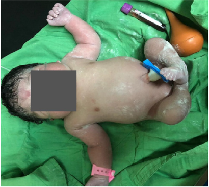
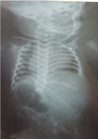
This is a 38-year-old woman with a gyneco-obstetric history of 4 pregnancies, delivery with death baby at 36 weeks gestation, miscarriage at the first trimester of pregnancy, then a cesarean section for a macrosomic fetus, newborn weight 5100 grams. The current pregnancy is a high-risk obstetric pregnancy. Goes on your first prenatal checkup at 12 weeks gestation, routine exams are requested that are within normal parameters. Pathological history of type 2 Diabetes mellitus whit more than 4 years of evolution treated with insulin with poor treatment adherence and poor metabolic control. Glycosylated hemoglobin (HbA1c) reports 8%. The obstetric ultrasound performed at 29 weeks gestation reports a single fetus, alive, with absence of right kidney, left kidney of dysplastic appearance. Urinary bladder with normal appearance, axial cuts of the spine shows the absence of the sacral and coccyx portion. Lower hypoplastic limbs, contracted and crossed in Buddha position. The estimated fetal weight was 1117 grams corresponding to intrauterine growth restriction for gestational age. Unable to see genitals. Without other structural alterations evident at the time of the study. Therefore, it is concluded as a fetus with suspected caudal regression syndrome. The cesarean section is scheduled at 38 weeks gestation where the female baby is born, Apgar 6/7, weight 2587 grams, size 47 cm, head circumference 34 cm. The physical examination of the neonate shows hypoplasia of the lower hemibody, soft tissue augmentation in the lumbosacral region, flattened buttocks with a short intergluteal cleft, external genitals of female appearance, narrow hips, distal leg atrophy, lower limbs in flexoabduction, bilateral equine foot, motor paresis of lower limbs, permeable anus. No abnormalities of the genitourinary tract, posterior intestine, cardiac and respiratory were found. Neurological development is preserved. (Figures 1 & 2) The newborn is undergoing imaging studies, a total column radiograph confirms the interruption of the distal column with the absence of a sacral column, without continuity with the hypoplasic pelvis. (Figure 3) Ultrasound of the abdomen that reports both kidneys of normal size and shape, preserving their renal parenchyma-cortex relationship, both with significant calicial ectasia. Right kidney measures 4.2 x 2.3 cm left kidney measures 4 x 2.3 cm. Newborn with stable vital signs is sent to a multidisciplinary approach with neurology, orthopedics, urology, rehabilitation.
Figure 1: Neonate with hypoplasia of the lower hemibody, distal leg atrophy, lower limbs in flexoabduction, bilateral equine foot.

Figure 2: Flattened buttocks with a short intergluteal cleft, external genitals of female appearance, narrow hips.

Figure 3: Frontal radiograph of the neonatal pelvis demonstrates the absence of sacrum, coccyx and hypoplasia of pelvic bones.

Discussion
Definition
Caudal regression syndrome (CRS) is a relative uncommon congenital anomaly. A rare congenital disorder that affects the development of the lower segment. CRS occurs approximately in 40000–100000 pregnancies. CRS, also known as caudal regression sequence, caudal dysplasia, caudal aplasia, femoral hypoplasia, phocomelic diabetic embryopathy, or sacral agenesis, is a spectrum of anomalies involving the caudal end of the trunk. CRS comprises various degrees of anomalous formation of the caudal trunk. The spectrum of disease can vary from isolated partial agenesis of the sacrococcygeal spine to the complete absence of sacral, lumbar, or lower thoracic vertebrae. Characteristic features include short intergluteal cleft, flattened buttocks, narrow hips, distal leg atrophy, and talipes deformities. Urinary and bowel dysfunction are nearly universal. Motor function is generally more affected than sensory function and is correlated with the level of spinal aplasia. In very mild cases, such as isolated coccygeal agenesis, patients may be asymptomatic. Associated anomalies are common, often involving abnormalities of the genitourinary tract, hindgut, cardiac, and respiratory system. Intelligence and mental function generally are preserved [1,2].
Etiology
The pathogenesis involves abnormal differentiation of the
developing spine, spinal cord, and caudal mesoderm. Although CRS
is rare in the general population, maternal hyperglycemia is thought
to play an important role, and the malformation is seen much more
commonly in infants of diabetic mothers [1].
In 1961 Duhamel explained the spectrum of CRS, of which
sirenomelia was thought to be the most severe form. Since then
numerous etiological theories have been proposed, but the
underlying mechanisms are still not clearly understood. Several
theories have been proposed to understand this anomaly. The first
hypothesis, based on the overall malformation of the caudal body,
postulates a primary defect in the generation of the mesoderm,
an embryologic insult to the caudal mesoderm, occurring
between 28 and 32 days of gestation. Interaction of genetic and
environmental factors (teratogenic agents, such as cadmium or
retinoic acid and vitamin A, maternal diabetes, insulin, embryonal
trauma, severe fluctuations in temperature, vitamin deficiencies,
lithium salts, radiation, stress, alcohol, amphetamines, and trypan
blue) during the 3rd gestational week might interfere with the
formation of notochord, resulting in abnormal development of
caudal structures. The second hypothesis, based on the aberrant
abdominal and vascular pattern of affected individuals, postulates
a primary vascular defect that leaves the caudal part of the embryo
hypoperfused, an aetiology based on the vascular theory associated
with the presence of an abnormal, single umbilical artery [2].
CRS is not specific to diabetes and only 1% of diabetic mother
babies may develop this type of malformation. Renshaw classified
the spectrum of CRS into 5 types based on the type of defect and
articulation between bones. Type 1 has total or partial unilateral
sacral agenesis. Type II has variable lumbar and total sacral agenesis
and the ilia articulates with the sides of the lowest vertebra. Type
III: has variable lumbar and total sacral agenesis and the caudal
end plate of the lowest vertebra rests above fused ilia or an iliac
amphiarthrosis. Type IV: has a fusion of soft tissues in both lower
limbs; and type V, also known as sirenomelia, has fused bones of
lower limbs [3].
Diagnosis
Classic features are suggestive. Prenatal diagnosis by ultrasound
is possible at 22 weeks of gestation, seen as a sudden interruption of
the spine due to the absence of vertebrae and a frog-like position of
the lower limbs. However, sirenomelic fetuses have been diagnosed
at 16- and 19-weeks’ gestation by the transvaginal route. The firsttrimester
diagnosis is difficult because of incomplete ossification
of the sacrum at that time. However, a short crown-rump length
and abnormal appearance of the yolk sac have been proposed
as early ultrasonographic signs of CRS. In a recent case report,
CRS was diagnosed antenatally after detection of a large nuchal
translucency, although the diagnosis was not confirmed until 16
weeks. Sonographic findings are variable, depending on the defect’s
extent and severity. Because the syndrome is not associated with
aneuploidy, a fetal karyotype is not warranted [4].
The morphological ultrasound is oriented towards the diagnosis
and helps to count the other frequently associated malformations.
Fetal magnetic resonance (MRI) makes it possible to confirm the
diagnosis and to determine the level of the terminal medullary
cone, which will be a major prognostic factor. The spinal cord is
usually dysplastic, and the terminal cone is too high [3,5].
Treatment
Prenatal diagnosis is important so that appropriate patient counseling can be provided, and postnatal interventions performed. The treatment depends upon clinical symptoms, including the degree of neurological deficits. The main goals of treatment include maintaining and improving renal, cardiac, pulmonary and GI function, preventing renal infection and achieving continence. Urinary incontinence is treated with anticholinergic agents, which decreases the detrusor muscle tone, increases the bladder capacity, and thus decreases intravesicular pressure and urinary frequency. Orthopedic intervention is necessary to correct the associated malformations. Physical therapy can help to prevent secondary deformities, skin ulcers and assists in improving quality of life. Surviving infants usually have normal mental function but do require extensive urologic and orthopedic assistance. Their longterm morbidity consists mostly of a neurogenic bladder dysfunction resulting in progressive renal damage and disabling neuromuscular deficits of the lower extremities [6].
Prognosis
Survival is the rule if the vital systems are unaffected or minimally affected. These patients have normal intelligence and therefore lead otherwise normal lives except for neuromuscular deficits of the lower limbs and sphincters. However, secondary neurogenic bladder leading to progressive renal damage and deterioration of renal function remains an important comorbid factor; therefore, extensive and long-term urologic attention is needed [7].
Conclusion
Caudal regression syndrome is a rare entity, characterized by sacrococcygeal dysgenesis with an abrupt termination of a blunt-ending spinal cord. The balance of glycemic figures in periconceptional women with diabetes, the antenatal screening will probably offer the opportunity for better management of this condition. Ultrasound and fetal MRI can be used to make a prenatal diagnosis, while MRI is the imaging modality of choice in adults. Early detection and prompt treatment are very important to decrease the risk of complications, and thus, to improve the prognosis. A multidisciplinary approach is the task. It should not be forgotten that the purpose of rehabilitation is not to correct all deformities but increase the functionality of everyday life.
Deficiencies
- Heuser CC, Hulinsky RS, JacksonGM (2018) Caudal Regression Syndrome. Obstetric Imaging: Fetal Diagnosis and Care 291-294e1.
- Isik Kaygusuz, E, Kurek Eken M, Sivrikoz ON, Cetiner H (2015) Sirenomelia: a review of embryogenic theories and discussion of the differences from caudal regression syndrome. The Journal of Maternal-Fetal & Neonatal Medicine 29(6): 949-953.
- Bouchahda H, Houda El Mhabrech, Hechmi Ben Hamouda, Sobhi Ghanmi, Rim Bouchahda, et al. (2017) Prenatal diagnosis of caudal regression syndrome and omphalocele in a fetus of a diabetic mother.Pan African Medical Journal27:128.
- Singh SK, SinghRD, SharmaA (2005) Caudal regression syndrome-case report and review of literature. Pediatric Surgery International 21(7):578-581.
- Negrete LM, Chung M, Carr SR, Tung GA (2015) In utero diagnosis of caudal regression syndrome. Radiology Case Reports10(1): 1049.
- Kumar Y, Nishant Gupta, Kusum Hooda, Pranav Sharma, Salil Sharma, et al. (2017) Caudal Regression Syndrome: A Case Series of a Rare Congenital Anomaly. Pol J Radiol 82: 188-192.
- Irem Bicakci, Selin Turan Turgut, Bekir Turgut, Afitap Icagasioglu, Zeliha Egilmez, et al. (2014) A case of caudal regression syndrome: walking or sitting? The Pan African Medical Journal 18:92.
References

Top Editors
-

Mark E Smith
Bio chemistry
University of Texas Medical Branch, USA -

Lawrence A Presley
Department of Criminal Justice
Liberty University, USA -

Thomas W Miller
Department of Psychiatry
University of Kentucky, USA -

Gjumrakch Aliev
Department of Medicine
Gally International Biomedical Research & Consulting LLC, USA -

Christopher Bryant
Department of Urbanisation and Agricultural
Montreal university, USA -

Robert William Frare
Oral & Maxillofacial Pathology
New York University, USA -

Rudolph Modesto Navari
Gastroenterology and Hepatology
University of Alabama, UK -

Andrew Hague
Department of Medicine
Universities of Bradford, UK -

George Gregory Buttigieg
Maltese College of Obstetrics and Gynaecology, Europe -

Chen-Hsiung Yeh
Oncology
Circulogene Theranostics, England -
.png)
Emilio Bucio-Carrillo
Radiation Chemistry
National University of Mexico, USA -
.jpg)
Casey J Grenier
Analytical Chemistry
Wentworth Institute of Technology, USA -
Hany Atalah
Minimally Invasive Surgery
Mercer University school of Medicine, USA -

Abu-Hussein Muhamad
Pediatric Dentistry
University of Athens , Greece

The annual scholar awards from Lupine Publishers honor a selected number Read More...


