Lupine Publishers Group
Lupine Publishers
Menu
ISSN: 2641-6921
Research Article(ISSN: 2641-6921) 
Microstructure And Composition Can Significantly Affect The Properties Of Tissue-Engineered Materials Containing Living Cells Volume 5 - Issue 1
Jianming Wang1*, Meng Li2, Zhizhong Shen2, Xiaorui Tian1 and Shengbo Sang2
- 1General Hospital of TISCO, North Street, Xinghualing District, China
- 2Taiyuan University of Technology, China
Received: January 15, 2022; Published: January 24, 2022
*Corresponding author: Jianming Wang, & Shengbo Sang, General Hospital of TISCO, North Street, Xinghualing District, Taiyuan 030024, China, Taiyuan University of Technology, Taiyuan 030024, China
DOI: 10.32474/MAMS.2022.05.000203
Opinion
Bioengineering synthetic materials to replace some specific human functions is the inevitable direction of development in the field of materials in the near future. Synthetic materials containing living cells will have a broad application prospect in skin replacement. For example, the market scale of wound repair (avoiding donor site injury, promoting healing, and improving healing quality), such as in vitro validation of beauty products will be 10 billion US dollars. Up to now, in the research of skin substitute materials, many research teams around the world have released a lot of research results, but they still have not fully developed an artificial composite material that can replace human skin. At the same time, they have not developed skin organ products that can solve wound coverage and skin function substitution. Our previous research combined with the research progress of other teams, found the material characteristics of other teams in simulating skin structure, and made improvements. Our research found that GelMA-PEGDA co-network hydrogel to prepare tissue-engineered skin with RRs structure. We have found that 10%GelMA-2%PEGDA hydrogels showed the adequate bioactivity, excellent structural support, suitable degradation rate and high mechanical stability mechanical stability [1]. The advantages of mold one-time forming gelma are low cost, low equipment requirements, low technical requirements, and fast manufacturing speed. It can simulate the net ridge structure between dermis and epidermis, so as to induce the close connection between them; [2].
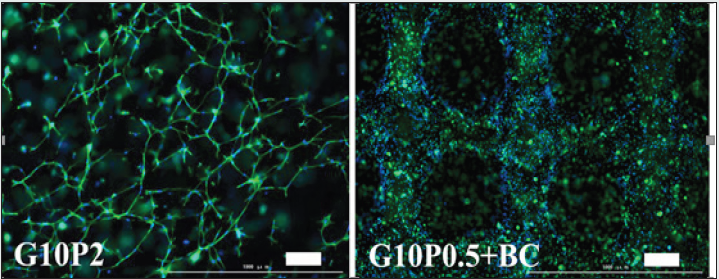
Figure 1: HSFs contained within G10P2 hydrogels and G10P0.5+BC at day 5 stained with DAPI for nuclei (blue) and FITCphalloidin for F-actin (green). Scale bar: 200 μm.

The disadvantage is that the looseness of the internal structure
of the material is completely formed by the cavity after crosslinking
and water loss after the material solidification, as shown in Figure
1 and a film layer with few pores will be formed on the material
surface due to molecular tension, which may partially lead to
unfavorable fluid exchange and cell migration and growth. The
current research of our team is based on the deficiencies found in
the previous study to further improve the appropriate raw material
ratio to make it suitable for biological ink as 3D biological printing.
The raw material ratio of biological ink in this study is 10%
gelma-0.5% pegda-0.1% BC (BC, bacterial cell). It uses 3D printing
technology to maintain the network ridge structure on the material
surface, The internal structure of the material is improved by
printing and forming, so that the internal structure is also a threedimensional
network structure, which can facilitate the liquid
exchange around the cells and the migration and growth of cells in
the three-dimensional space [3,4].
The preliminary research results suggest that the expected
effect has indeed been achieved. Its in vitro experiment shows that,
as shown in the figure, the statistical result of cell proliferation in
3D printing material is about three times higher than that of mold
forming material under the same cell inoculation density and
culture time. In vivo validation: We conducted in vivo experiments
after inoculation of material printing cells. The results showed that
the proliferation of two main cells constituting the skin, epidermal
cells and fibroblasts, was faster than that of mold forming
materials, and the wound healing speed was also faster. The key
was that the healing quality could be significantly different at the
time point of 2 weeks, The specific results will be introduced in
the next treatise. We all know that time is life in clinical rescue. If a
material can achieve in vitro proliferation in the shortest time and
meet the transplantation requirements, it will greatly increase its
practicability for clinical application, and has a very broad prospect
in clinical transformation application. The same cell inoculation
density has great differences in the proliferation rate of different
materials, which shows that the structure, composition, and ratio
of materials have a significant impact on the cell proliferation in
materials. The two materials currently developed by our team
have their own advantages and disadvantages. How to avoid the
disadvantages and combine the advantages of the two schemes will
be the problem to be solved in the next research.
References
- Zhizhong S, Yanyan C, Meng L, Yayun Y, Rong C , et al. (2021) Construction of tissue-engineered skin with rete ridges using co-network hydrogels of gelatin methacrylated and poly(ethylene glycol) diacrylate Mater Sci Eng C Mater Biol Appl 129: 112360.
- Clement AL, Moutinho Jr, Pins GD (2013) Micropatterned dermal-epidermal regeneration matrices create functional niches that enhance epidermal morphogenesis. Acta Biomater 912: 9474–9484,
- Zhou X, Tenaglio S, Esworthy T, Hann SY, Cui H, et al. (2020) Three-Dimensional Printing Biologically Inspired DNA-Based Gradient Scaffolds for Cartilage Tissue Regeneration. ACS Appl Mater Interfaces (29): 33219-33228.
- Vila A, Torras N, Castano AG, Garcia-Diaz M, Comelles J, et al. (2020) Hydrogel co-networks of gelatine methacrylate and poly (ethylene glycol) diacrylate sustain 3D functional in vitro models of intestinal mucosa. Bio fabrication 12(2): 025008.

Top Editors
-

Mark E Smith
Bio chemistry
University of Texas Medical Branch, USA -

Lawrence A Presley
Department of Criminal Justice
Liberty University, USA -

Thomas W Miller
Department of Psychiatry
University of Kentucky, USA -

Gjumrakch Aliev
Department of Medicine
Gally International Biomedical Research & Consulting LLC, USA -

Christopher Bryant
Department of Urbanisation and Agricultural
Montreal university, USA -

Robert William Frare
Oral & Maxillofacial Pathology
New York University, USA -

Rudolph Modesto Navari
Gastroenterology and Hepatology
University of Alabama, UK -

Andrew Hague
Department of Medicine
Universities of Bradford, UK -

George Gregory Buttigieg
Maltese College of Obstetrics and Gynaecology, Europe -

Chen-Hsiung Yeh
Oncology
Circulogene Theranostics, England -
.png)
Emilio Bucio-Carrillo
Radiation Chemistry
National University of Mexico, USA -
.jpg)
Casey J Grenier
Analytical Chemistry
Wentworth Institute of Technology, USA -
Hany Atalah
Minimally Invasive Surgery
Mercer University school of Medicine, USA -

Abu-Hussein Muhamad
Pediatric Dentistry
University of Athens , Greece

The annual scholar awards from Lupine Publishers honor a selected number Read More...


